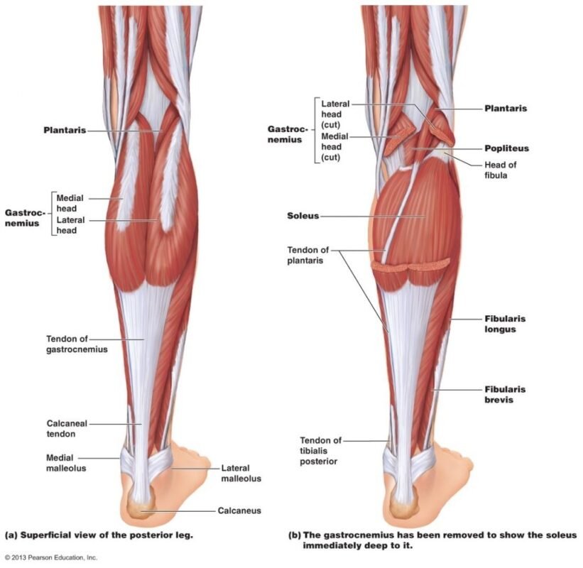
Plantaris Muscle is a small muscle that typically originates at the lateral supracondylar line of the femur and the knee joint capsule, from where it continues distally, forming a long and slender tendon.
The plantaris muscle consists of a small, thin muscle belly, and a long thin tendon that forms part of the posterior superficial compartment of the calf. Together with the gastrocnemius, and soleus, they are collectively referred to as the triceps surae muscle. The muscle originates from the lateral supracondylar line of the femur just superior and medial to the lateral head of the gastrocnemius muscle as well as from the oblique popliteal ligament in the posterior aspect of the knee.[rx] The muscle ranges from 7 to 13 cm long varying highly in both size and form when present.[rx]

Origin and Insertion of Plantaris Muscle
- Plantaris is a long, slender muscle that consists of a short, fusiform belly (7-10 cm) and a long, thin tendon extending inferiorly.
- It originates from the inferior end of the lateral supracondylar line of the femur, just superior to the lateral head of the gastrocnemius muscle.
- The attachment often extends onto the oblique popliteal ligament. Its tendon then travels anteromedially along the medial border of the gastrocnemius.
- The plantaris tendon inserts onto the posterior surface of the calcaneus, medial to the calcaneal tendon (common tendon of the soleus and gastrocnemius muscles, also known as Achilles’ tendon). Sometimes, plantar might join the calcaneal tendon, or merge with the flexor retinaculum of the ankle or leg fascia.
Nerve Supply of Plantaris Muscle
- Lumber and sacral plexus (formed from the anterior rami of spinal nerves L4, L5, S1–4)
- The plantaris muscle is innervated by the tibial nerve, a branch of the sciatic nerve in the sacral plexus. Signaling for contraction begins in the frontal lobe of the brain with the pre-central gyrus (primary motor cortex). Upper motor neurons synapse with lower motor neurons at the anterior horn of the spinal cord in the sacral plexus (formed from the anterior rami of spinal nerves L4, L5, S1–4).
- The lower motor neuron fibers continue down the sciatic nerve and then diverge into the tibial and common fibular nerves. The tibial nerve runs medially at the knee joint. When the tibial nerve receives an action potential, the plantaris muscle contracts, providing weak plantar flexion of the foot and weak flexion of the knee.[rx]
Blood Supply of Plantaris Muscle
Plantaris has a dual blood supply.
- Superficially it receives blood from the lateral sural artery, a branch of the popliteal artery.
- Its deep surface is supplied by the superior lateral genicular artery, which also stems from the popliteal artery.
- The distal part of the plantaris tendon receives blood from the calcaneal branches of the posterior tibial artery.
The Function of Plantaris Muscle
- Plantaris acts weakly to plantarflex the foot and flex the knee. It is considered a vestigial muscle and can be used as a tendon graft in reconstructive orthopedic surgery.
- The plantaris acts to weakly plantarflex the ankle joint and flex the knee joint.
- The plantaris muscle may also provide proprioceptive feedback information to the central nervous system regarding the position of the foot. The unusually high density of proprioceptive receptor end organs supports this notion.[rx]
- Its motor function is so minimal that its long tendon can readily be harvested for reconstruction elsewhere with little functional deficit. Often mistaken for a nerve by new medical students (and thus called the “freshman nerve”), the muscle was useful to other primates for grasping with their feet.[rx]
Primary Actions of the Plantaris
- The plantaris muscle is not a prime mover and does not have a primary action but assists with the actions of other muscles at the knee and ankle joints.

Secondary Actions of the Plantaris
Assists with flexion of the knee
Agonists
- Biceps Femoris
- Semitendinosus
- Semimembranosus
Antagonists
- Vastus Lateralis
- Vastus Medialis
- Vastus Intermedius
- Rectus Femoris
Assists with plantarflexion of the foot at the ankle
Agonists
- Gastrocnemius
- Soleus
Antagonists
- Tibialis Anterior