Coccydynia Causes Symptoms Coccydynia means the inflammation of the tailbone (coccyx or bony area located deep between the buttocks above the anus) is referred to as coccydynia. Coccydynia is associated with pain and tenderness at the tip of the tailbone between the buttocks. Sitting often worsens coccyx pain.
Another Names of Coccydynia
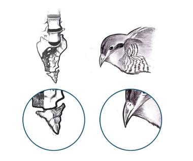
Coccygodynia (also referred to as coccydynia, coccalgia, coccygalgia, or coccygeal pain) is a painful syndrome affecting the tailbone (coccygeal) region. The word coccyx is derived from the Greek word kokkyx (“cuckoo”), on the basis of this structure’s resemblance to the shape of a cuckoo’s beak (see the image below).
Coccygodynia is a rare condition but can be highly unpleasant when it does occur. Patients’ chief complaint is pain, which typically is triggered by or occurs while sitting on hard surfaces. The pain often varies and sometimes is aggravated by arising from the sitting position.
Direct trauma to the tailbone is the most common cause of coccydynia, and usually leads to inflammation surrounding the coccyx, which contributes to pain and discomfort.
There are many cases reported in which pain begins with no identifiable origin (called idiopathic coccydynia
The above factors may result from an injury to the coccyx, or may develop as idiopathic coccydynia.
Causes of Coccydynia
A diagnosis of coccydynia will usually identify one of the following underlying causes of pain
- Local trauma – A direct injury to the coccyx is probably the most common cause of coccydynia. A fall on the tailbone can inflame the ligaments and injure the coccyx or the coccygeal attachment to the sacrum. Coccygeal trauma usually results in a bruised bone, but may also result in a fracture or dislocation either in the front or back of the coccyx.
- Repetitive stress – Activities that put prolonged pressure on the tailbone, such as horseback riding and sitting on hard surfaces for long periods of time, may cause the onset of coccyx pain. Tailbone pain from these causes usually is not permanent, but if inflammation and symptoms are not managed, the pain may become chronic and cause long-term altered mobility of the sacrococcygeal joint.
- Childbirth – During delivery, the baby’s head passes over the top of the coccyx, and the pressure against the coccyx can sometimes result in injury to the coccygeal structures (the disc, ligaments, and bones). While uncommon, the pressure can also cause a fracture in the coccyx.
- Tumor or infection – Rarely, coccydynia can be caused by a nearby tumor or infection that puts pressure on the coccyx.
- Referred coccyx pain – In rare cases pain will be referred to the coccyx from elsewhere in the spine or pelvis, such as a lumbar herniated disc or degenerative lumbar disc.
- Obesity – Pelvic rotation, including movement of the coccyx, is usually lessened in individuals who are overweight, leading to more continual stress being placed on the coccyx and increasing the chances of developing coccyx pain. One study found that a Body Mass Index (BMI) of more than 27.4 in women and 29.4 in men increases the risk for coccydynia following repetitive stress or a one-time injury.
- Gender – Women have a higher chance of developing coccydynia than men, due to a wider pelvic angle as well as trauma to the coccyx endured during childbirth.
If pain is mild or moderate, it may not be necessary to identify the exact cause of coccydynia. In some cases, however, coccyx pain is severe or of a serious origin, so it is important to have a general idea why pain has developed so that it can be treated most effectively.
What are the symptoms of Coccydynia?
Because coccydynia strictly refers to inflammation in or near the tailbone, symptoms are highly localized. An individual experiencing coccydynia may encounter-
- Pain in the coccygeal region (tip of the tailbone)
- Local tenderness
- The pain can range from mild to severe. It is usually worse when
- Sitting down
- Moving from sitting to a standing position
- When touched or when some pressure falls on the coccyx
- Worsens with constipation, feels better after bowel movement
- Some people can tolerate sitting in the same position for only a few minutes. They feel restless after a few minutes. They have to get up and move around to get relieved of the pain
- Pain makes everyday activities difficult Ex. Driving, bending over, sitting down etc.
- Sitting on a soft surface may be more painful than sitting on something hard. Sitting on a soft surface places most of your weight on your coccyx rather than on the hard bones below your pelvis.
- Difficulty in sitting and leaning against the buttocks
Other symptoms of coccydynia
- Backache
- Shooting pain down the legs
- Pain before or when stools are passed
- Pain during sex (aggravates at or after sex)
- Painful buttocks and hips
- Increased pain during monthly periods in women
- Difficulty to sleep comfortably at night, you may need to keep changing the positions while you sleep
- Symptoms usually improve with relief of pressure when standing or walking
- Pain in the tailbone area that worsens when sitting or leaning against something
- Tenderness and redness in the affected area
- Discomfort during bowel movements
- Difficulty standing up from a resting position and performing other everyday activities, such as driving or bending down
- Bruising near the coccyx
- Some low back pain
- Deep, intense aches in the tailbone region
- Sharp pangs in the coccyx area
Test for coccygeal pain
- A local anaesthesia is injected into the coccygeal area. If the pain is related to the coccyx, it reduces and there is immediate relief.
- If this anaesthetic test is positive then a dynamic (sit/stand) X-ray or MRI scan show whether the coccyx dislocates when the patient sits
Other investigations
- Stool guaiac test – should be done for occult test to assess for Gastro-intestinal pathology
- Dynamic radiographs – obtained in both sitting and standing positions may be more useful than static
- X-rays – because they allow for measurement of the saggital rotation of the pelvis and the coccygeal angle of incidence. A comparison of sitting and standing films will yield radiographic abnormalities in up to 70% of symptomatic coccydynia
- MRI and technetium Tc-99m bone scans – may demonstrate inflammation of sacrococcygeal area indicative of coccygeal hypermobility
- Provocative testing – of the coccyx such as pressing on the region with a blunted needle to elicit pain and pain relief with injection of local anaesthesia under fluoroscopic guidance may be helpful in diagnosis
- Basically the diagnosis of coccydynia is done by a health care professional by taking a thorough medical history from the patient and completing a physical examination
- X-ray of the sacrum and coccyx – to rule out an obvious fracture or a large tumour
- An MRI scan – to rule out infection or spinal tumour as a cause of pain
- Bone scans and CT scans – give little information. They are generally not done. Typically all imaging studies will be negative.
- A thorough inspection / palpation of this area – is needed to detect any abnormal masses or abscesses (infections)
- A lateral X-ray of the coccyx – is taken to help detect any significant coccygeal pathology such as a fracture
Differential Diagnosis
Coccyx Fracture (broken tailbone)
It presents with –
- Pain that increases in severity when sitting or getting up from a chair or when experiencing a bowel movement
- Provoked pain over the tailbone
- Nausea
- Bruising or swelling in the tailbone area
Sacro-coccygeal dislocation
- It is a rare injury
- Presents with pain in the sacro-coccygeal area generally followed by a fall or injury
- On examination, a step will be felt in the continuity of sacrum and coccyx
- The tip of the coccyx will not be palpable
- Per rectal examination reveals a small bump on running the finger along the sacrococcygeal curvature
- Plain radiographs of sacrococcygeal region – reveals anterior dislocation of coccyx over sacrum
Intracoccygeal dislocation – Here there is dislocation of one coccygeal segment from the other
Intrapelvic malignancy and / or metastatic lesions ,Ischial bursitis / Ischiogluteal bursitis
- Ischial bursa (small sac filled with lubricating fluid) is located in the upper buttock area
- Inflammation in this bursa is called Ischial bursitis
- It causes dull pain in this area which is most noticeable when climbing uphill
- The pain sometimes occurs after prolonged sitting on hard surfaces
- It is also called as ‘weaver’s bottom’ or ‘tailor’s bottom’
Sacro-iliac joint pain or Sacroiliac joint dysfunction
- It presents with pain in sacroiliac joint
- Pain is experienced often in the lower back or at the back of the hips
- Pain is sometimes present in the groins and thighs
- Pain is typically worse with standing and walking and improves while lying down
- Stiffness and burning sensation can be felt in the pelvis
References


