Muscles of Anterior Compartment of Forearm (Flexors of Forearm)
Superficial layer
Pronator teres
-
Function: Pronation of radio-ulnar joint
-
Origin: Coronoid process and medial epicondyle of humerus
-
Insertion: Lateral surface of the radius
-
Innervation: Median nerve (C6, C7)
Flexor carpi radialis
-
Function: Flexion and adduction at the wrist
-
Origin: Medial epicondyle of humerus
-
Insertion: Base of second metacarpal
-
Innervation: Median nerve (C6, C7)
Palmaris longus
-
Function: Flexion at the wrist, tensing of the palmar aponeurosis
-
Origin: Medial epicondyle of humerus
-
Insertion: Flexor retinaculum
-
Innervation: Median nerve (C7, C8)
Flexor carpi ulnaris
-
Function: Flexion and adduction at the wrist
-
Origin: Medial epicondyle of humerus and olecranon
-
Insertion: Pisiform, hook of hamate and fifth metacarpal
-
Innervation: Median nerve (C7, C8)
Intermediate Layer
Flexor digitorum superficialis
-
Function: Flexion of the proximal interphalangeal joint of the second, third, fourth, and fifth finger. Also has a weaker flexion action on the metacarpophalangeal joints of the same fingers
-
Origin: Medial epicondyle, coronoid process, and anterior radius
-
Insertion: Second, third, fourth, and fifth middle phalanges
-
Innervation: Median nerve (C7, C8, T1)
Deep Layer
Flexor digitorum profundus
-
Function: Flexion of the distal interphalangeal joint of the second, third, fourth, and fifth finger
-
Origin: Medial and the anterior surface of the proximal ulna and interosseous membrane
-
Insertion: Second, third, fourth, and fifth distal phalanges
-
Innervation: Ulnar nerve (C8, T1) for the medial part, anterior interosseous nerve (C8,T1) for the lateral
Flexor pollicis longus
-
Function: Flexion of the interphalangeal joint of the thumb
-
Origin: Anterior aspect of radius as well as interosseous membrane
-
Insertion: Base of distal phalanx of thumb
-
Innervation: Anterior interosseous nerve (C7, C8)
Pronator quadratus
-
Function: Pronator of the forearm
-
Origin: Anterior aspect of distal ulna
-
Insertion: Anterior aspect of the distal radius
-
Innervation: Anterior interosseous nerve (C7, C8)
Brachioradialis
-
Function: Weak flexor of the forearm
-
Origin: Proximal supracondylar ridge on the humerus
-
Insertion: Lateral surface of the distal end of radius
-
Innervation: Radial nerve (C5, C6, C7)
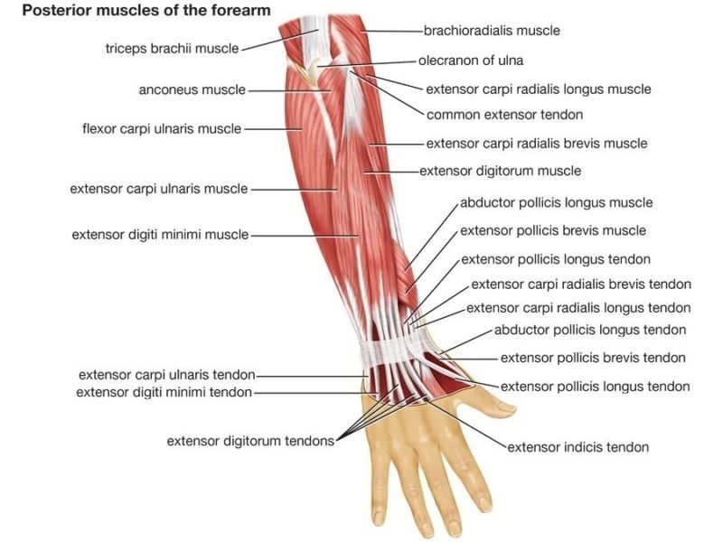
Muscles of Posterior Compartment of Forearm
Superficial
Extensor carpi radialis longus
-
Function: Extension and abduction of the wrist
-
Origin: Proximal supracondylar ridge on the humerus
-
Insertion: Dorsal base of the second metacarpal
-
Innervation: Radial nerve (C6, C7)
Extensor carpi radialis brevis
-
Function: Extension and abduction of the wrist
-
Origin: Lateral epicondyle of humerus
-
Insertion: Dorsal base of the third metacarpal
-
Innervation: Deep branch of the radial nerve (C7, C8)
Extensor digitorum
-
Function: Extension of the proximal interphalangeal joint of the second, third, fourth, and fifth fingers. Also has a weaker extension action on the metacarpophalangeal joints of the same fingers
-
Origin: Lateral epicondyle of humerus
-
Insertion: Extensor expansions on the dorsal aspect of second, third, fourth, and fifth middle and distal phalanges
-
Innervation: Posterior interosseous nerve (C7, C8)
Extensor digit minimi
-
Function: Extension of the little finger at the metacarpophalangeal joint and interphalangeal joint
-
Origin: Lateral epicondyle of humerus
-
Insertion: Extensor expansion on the dorsal aspect of fifth phalanx
-
Innervation: Posterior interosseous nerve (C7, C8)
Extensor carpi ulnaris
-
Function: Extension and adduction of the wrist
-
Origin: Lateral epicondyle of the humerus and posterior ulna
-
Insertion: Fifth metacarpal base
-
Innervation: Posterior interosseous nerve (C7, C8)
Deep Layer
Extensor indices
-
Function: Extension of the index finger
-
Origin: Dorsal surface of distal ulna and interosseous membrane
-
Insertion: Extensor expansion of second finger
-
Innervation: Posterior interosseous nerve (C7, C8)
Supinator
-
Function: Supination of the forearm
-
Origin: Lateral epicondyle and supinator crest of ulna
-
Insertion: Lateral surface of radius
-
Innervation: Deep branch of radial nerve (C7, C8)
Abductor policies longus
-
Function: Abduction of the thumb by acting on the carpometacarpal joint and the metacarpophalangeal joint
-
Origin: Dorsal aspects of the proximal radius, ulna, and interosseous membrane
-
Insertion: Base of the first metacarpal
-
Innervation: Posterior interosseous nerve (C7, C8)
Extensor pollicus longus
-
Function: Extension of the thumb by acting on the carpometacarpal joint, the metacarpophalangeal joint, and the interphalangeal joint.
-
Origin: Dorsal aspects of the middle ulna and interosseous membrane
-
Insertion: Distal phalanx of 1st finger
-
Innervation: Posterior interosseous nerve (C7, C8)
Extensor pollicus brevis
-
Function: Extension of the thumb by acting on the carpometacarpal joint and the metacarpophalangeal joint
-
Origin: Dorsal aspects of middle radius and interosseous membrane
-
Insertion: Distal phalanx of 1st finger
-
innervation: Posterior interosseous nerve (C7, C8)
Intrinsic Muscles of Hand
Thenar muscles
Opponents policies
-
Function: Opposition of the thumb
-
Origin: Flexor retinaculum and tubercle of trapezium
-
Insertion: Lateral aspect of first metacarpal
-
Innervation: Recurrent branch of median nerve (C8, T1)
Abductor policies Brevis
-
Function: Abduction of the thumb at the metacarpophalangeal joint
-
Origin: Flexor retinaculum and tubercle of scaphoid
-
Insertion: Lateral aspect of proximal phalanx of first finger
-
Innervation: Recurrent branch of median nerve (C8, T1)
Flexor pollicus brevis
-
Function: Flexion of the thumb at the metacarpophalangeal joint
-
Origin: Flexor retinaculum and tubercle of trapezium
-
Insertion: Lateral aspect of proximal phalanx of first finger
-
Innervation: Recurrent branch of median nerve (C8, T1)
Adductor Compartment
Adductor pollicus
-
Function: Adduction of the thumb
-
Origin: Second, third metacarpal, and capitate
-
Insertion: Proximal phalanx and extensor expansion of 1st finger
-
Innervation: Deep branch of ulnar nerve (C8, T1)
Hypothenar Muscles
Abductor digiti minimi
-
Function: Abduction of the little finger at the metacarpophalangeal joint
-
Origin: Pisiform
-
Insertion: Medial aspect of proximal phalanx of fifth finger
-
Innervation: Deep branch of ulnar nerve (C8, T1)
Flexor digiti minimi brevis
-
Function: Flexion of the little finger at the metacarpophalangeal joint
-
Origin: Flexor retinaculum and hook of hamate
-
Insertion: Medial aspect of proximal phalanx of fifth finger
-
Innervation: Deep branch of ulnar nerve (C8, T1)
Opponens digiti minimi
-
Function: Opposition of the little finger
-
Origin: Flexor retinaculum and hook of hamate
-
Insertion: Medial aspect of fifth metacarpal
-
Innervation: Deep branch of ulnar nerve (C8, T1)
Short Muscles
Lubricants
-
Function: Flexion of the metacarpophalangeal joints with extension of the interphalangeal joints
-
Origin: Arise from tendons of flexor digitorum profundus. First 2 are unipennate, and the third and fourth are bipennate
-
Insertion: Extensor expansions of second, third, fourth, and fifth finger
-
Innervation: Median nerve (C8, T1) for the lateral 2 lumbricals, deep branch of ulnar nerve (C8, T1) for the medial 2 lumbricals
Dorsal interossei
-
Function: Abduction of the second, third, and fourth finger away from the axial line
-
Origin: Adjacent metacarpals
-
Insertion: Extensor expansions and proximal phalanges of the second, third, and fourth fingers
-
Innervation: Deep branch of ulnar nerve (C8, T1)
Palmar interossei
-
Function: Adduction of the second, third, and fourth finger towards the axial line
-
Origin: Palmar surfaces of second, fourth, and fifth metacarpals
-
Insertion: Extensor expansions and proximal phalanges of the second, fourth, and fifth fingers
-
Innervation: Deep branch of ulnar nerve (C8, T1)
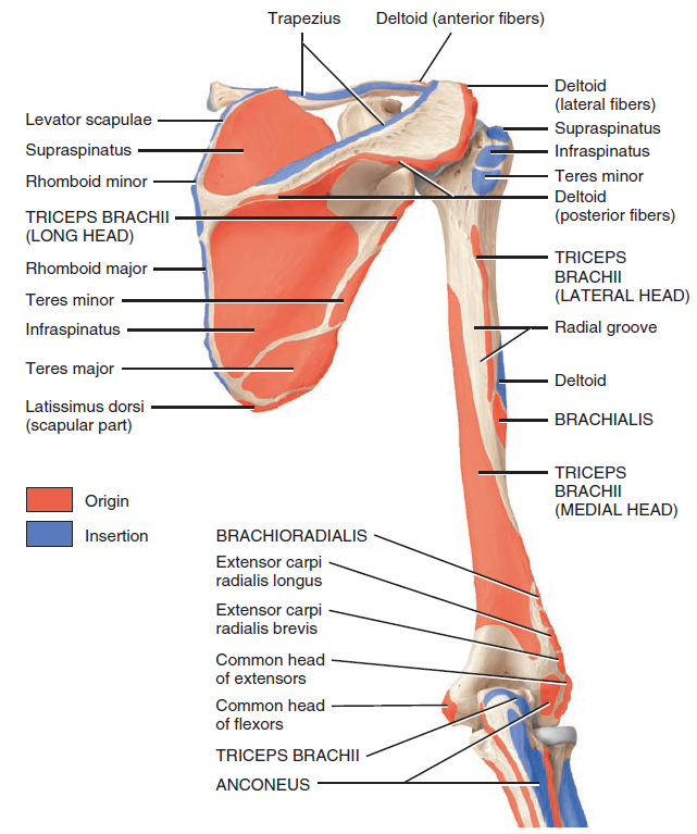
Surgical Considerations
The deltoid is a significant factor when considering the anterior surgical approach to gain access to the shoulder joint. Some of these technical procedures include, but are not limited to the following:
-
Open Bankart repair/capsular reconstructions – indicated in the setting of recurrent anterior (or other directional) instability of the shoulder
-
Shoulder arthroplasty – indicated for cases of post-traumatic deformity, advanced degenerative arthritis, and/or avascular necrosis includes hemiarthroplasty, total shoulder arthroplasty (TSA), reverse total shoulder arthroplasty (TSA)
-
Rotator cuff repair contemporary – indications remain somewhat controversial although most of these procedures are now being performed arthroscopically popular approaches (as opposed to the deltopectoral approach) include the mini-open approach (lateral deltoid-splitting approach)
References