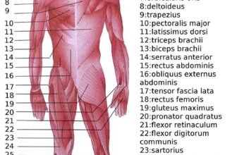
The shoulder composed of the clavicle, scapula, and humerus, is an intricately designed combination of four joints, the Glenohumeral (GH) Joint, the Acromioclavicular (AC) Joint and the Sternoclavicular (SC) Joint, and a “floating joint”, known as the Scapulothoracic (ST) joint.
Structure
The shoulder consists of a ball-and-socket joint formed by the humerus and scapula and their surrounding structures – ligaments, muscles, tendons – which support the bones and maintain the relationship of one to another. These supporting structures attach to the clavicle, humerus, and scapula, the latter providing the glenoid cavity, acromion and coracoid processes. The main joint of the shoulder is the shoulder joint (or glenohumeral joint), between the humerus and the glenoid process of the scapular.[rx] The acromioclavicular joint and sternoclavicular joint also play a role in shoulder movements.[rx] White hyaline cartilage on the ends of the bones (called articular cartilage) allows the bones to glide and move on each other, and the joint space is surrounded by a synovial membrane. Around the joint space are muscles – the rotator cuff, which directly surrounds and attaches to the shoulder joint – and other muscles that help provide stability and facilitate movement.
Two filmy sac-like structures called bursae permit smooth gliding between bone, muscle, and tendon. They cushion and protect the rotator cuff from the bony arch of the acromion.[rx]
The glenoid labrum is the second kind of cartilage in the shoulder which is distinctly different from the articular cartilage. This cartilage is more fibrous or rigid than the cartilage on the ends of the ball and socket. Also, this cartilage is also found only around the socket where it is attached.[rx]
Nerve supply and passage
The skin around the shoulder is supplied by C2-C4 (upper), and C7 and T2 (lower area). The brachial plexus emerges as nerve roots from the cervical vertebrae C5-T1. Branches of the plexus, in particular from C5-C6, supply the majority of the muscles of the shoulder.[rx]
Blood vessels
The subclavian artery arises from the brachiocephalic trunk on the right and directly from the aorta from the left. This becomes the axillary artery as it passes beyond the first rib. The axillary artery also supplies blood to the arm and is one of the major sources of blood to the shoulder region. The other major sources are the transverse cervical artery and the suprascapular artery, both branches of the thyrocervical trunk which itself is a branch of the subclavian artery.[rx] The blood vessels form a network (anastomosis) behind the shoulder that helps to supply blood to the arm even when the axillary artery is compromised. The axillary artery supplies blood to the arm and is one of the major sources of blood for the shoulder region.
Function
The muscles and joints of the shoulder allow it to move through a remarkable range of motion, making it one of the most mobile joints in the human body. The shoulder can abduct, adduct, rotate, be raised in front of and behind the torso and move through a full 360° in the sagittal plane. This tremendous range of motion also makes the shoulder extremely unstable, far more prone to dislocation and injury than other joints[rx]
The following describes the terms used for different movement of the shoulder:[rx]
| Name | Description | Muscles |
|---|---|---|
| Scapular retraction[rx] (aka scapular adduction) | The scapula is moved posteriorly and medially along the back, moving the arm and shoulder joint posteriorly. Retracting both scapulae gives a sensation of “squeezing the shoulder blades together.” | rhomboideus major, minor, and trapezius |
| Scapular protraction[rx] (aka scapular abduction) | The opposite motion of scapular retraction. The scapula is moved anteriorly and laterally along the back, moving the arm and shoulder joint anteriorly. If both scapulae are protracted, the scapulae are separated and the pectoralis major muscles are squeezed together.[rx] | serratus anterior (prime mover), pectoralis minor and major |
| Scapular elevation[rx] | The scapula is raised in a shrugging motion. | levator scapulae, the upper fibers of the trapezius |
| Scapular depression[12] | The scapula is lowered from elevation. The scapulae may be depressed so that the angle formed by the neck and shoulders is obtuse, giving the appearance of “slumped” shoulders. | pectoralis minor, lower fibers of the trapezius, subclavius, latissimus dorsi |
| Arm abduction[rx] | Arm abduction occurs when the arms are held at the sides, parallel to the length of the torso, and are then raised in the plane of the torso. This movement may be broken down into two parts: True abduction of the arm, which takes the humerus from parallel to the spine to perpendicular; and upward rotation of the scapula, which raises the humerus above the shoulders until it points straight upwards. | True abduction: supraspinatus (first 15 degrees), deltoid; Upward rotation: trapezius, serratus anterior |
| Arm adduction[rx] | Arm adduction is the opposite motion of arm abduction. It can be broken down into two parts: downward rotation of the scapula and true adduction of the arm. | Downward rotation: pectoralis minor, pectoralis major, subclavius, latissimus dorsi (same as scapular depression, with pec major replacing lower fibers of trapezius); True Adduction: latissimus dorsi, subscapularis, teres major, infraspinatus, teres minor, pectoralis major, long head of triceps, coracobrachialis. |
| Arm flexion[rx] | The humerus is rotated out of the plane of the torso so that it points forward (anteriorly). | pectoralis major, coracobrachialis, biceps brachii, anterior fibers of the deltoid. |
| Arm extension[rx] | The humerus is rotated out of the plane of the torso so that it points backward (posteriorly) | latissimus dorsi and teres major, long head of triceps, posterior fibers of the deltoid |
| Medial rotation of the arm[rx] | Medial rotation of the arm is most easily observed when the elbow is held at a 90-degree angle and the fingers are extended so they are parallel to the ground. Medial rotation occurs when the arm is rotated at the shoulder so that the fingers change from pointing straight forward to pointing across the body. | subscapularis, latissimus dorsi, teres major, pectoralis major, anterior fibers of deltoid |
| Lateral rotation of the arm[rx] | The opposite of medial rotation of the arm. | infraspinatus and teres minor, posterior fibers of deltoid |
| Arm circumduction[rx] | Movement of the shoulder in a circular motion so that if the elbow and fingers are fully extended the subject draws a circle in the air lateral to the body. In circumduction, the arm is not lifted above parallel to the ground so that “circle” that is drawn is flattened on top. | pectoralis major, subscapularis, coracobrachialis, biceps brachii, supraspinatus, deltoid, latissimus dorsi, teres major and minor, infraspinatus, long head of triceps |
Clavicle
The clavicle or collar bone is a long, curved bone on the upper portion of the shoulder that connects with the scapula and the sternum.
Key Points
- The clavicle is a long, doubly curved bone that connects the arm to the body, located directly above the first rib. It acts as a strut to keep the scapula in place so the arm can hang freely.
- The clavicle is an attachment point for several muscles.
- Structurally, the clavicle can be divided into three parts: medial end, lateral end, and shaft.
- There are sex differences in clavicle shape—female clavicles are shorter and thinner than male clavicles.
Key Terms
- acromion: The outermost point of the shoulder blade.
The clavicle, or collarbone, is a slender s-shaped bone that extends between the sternum and the scapula and is located directly above the first rib. It functions to attach the upper arm to the trunk and provides support to allow free movement around the shoulder.

Left Clavicle: The left clavicle, viewed from above. Muscle attachment sites (pectoralis major, subclavius muscle, deltoid, and sterno-hyoid) are highlighted.
Medially the clavicle is quadrangular in shape and articulates with the manubrium of the sternum forming the sternoclavicular joint. Laterally, the clavicle is flattened and attaches to the acromion process of the scapula forming the acromioclavicular joint.
The shaft of the clavicle acts as the origin and attachment point for numerous muscles and ligaments. At the medial end of the shaft the pectoralis major originates from the anterior surface, the posterior surface gives origin to the sternohyoid muscle and the superior surface the sternocleidomastoid muscle.
The costoclavicular ligament attaches to the inferio≠r surface. Laterally the deltoid muscle originates from the anterior surface and the trapezius muscle attaches to the posterior surface at the trapezoid line. Adjacent to this is the conoid tubercle which is an attachment point for the conoid ligament.
The clavicle in males is typically thicker and longer than a female’s clavicle to account for the larger muscle mass operating through it.
Scapula
The scapula, or shoulder bone, is a flat, triangular bone that connects to the humerus and the clavicle.
Key Points
The scapula articulates with the humerus and the clavicle.
The scapula is flat and triangular.
The scapula articulates with the humerus at the glenoid fossa and the clavicle at the acromion process.
The scapula provides attachment sites for many muscles including the pectoralis minor, coracobrachialis, serratus anterior, triceps brachii, biceps brachii, and the subscapularis.
The scapula has two main surfaces: the costal (front facing) surface and the dorsal (rear facing) surface.
Key Terms
acromion: The outermost point of the shoulder blade.
glenoid: A shallow depression in a bone, especially in the scapula.
The scapula, or shoulder blade, is a flat, triangular bone located to the posterior of the shoulder. The scapula articulates with the clavicle through the acromion process, a large projection located superiorly on the scapula forming the acromioclavicular joint. The scapula also articulates with the humerus of the upper arm to form the shoulder joint, or glenohumeral joint, at the glenoid cavity.
Due to its flat nature, the scapula presents two surfaces and three borders; the front-facing costal surface and the rear-facing dorsal surface, as well as the superior, lateral, and medial borders.

Costal surface: Costal surface of the left scapula. The subscapular fossia for subscapularis, serratus, pector minor regions are highlighted.
The serratus anterior originates from the costal surface, which also provides an attachment for the subscapularis muscle. The dorsal surface gives origin to the supraspinatus and infraspinatus muscles, and inferiorly to the teres minor and major. It is divided by a ridge-like structure called the spine of the scapula, from which the deltoid and trapezius muscles originate.

Dorsal surface: Dorsal surface of the left scapula.
The lateral border is the thickest border of the scapula and extends downwards from the glenoid cavity. Immediately below the glenoid cavity is the infraglenoid tuberosity, which is the origin for the long head of the triceps brachii.
Immediately above the glenoid cavity is the supraglenoid tubercle and its associated hook-like coracoid process, from which the long and short heads of the biceps brachii originate.
Four muscles attach to the medial border of the scapula. To the anterior side the serratus anterior attaches, whilst posteriorly the levator scapulae and rhomboids minor and major attach.


