
The urinary bladder is a complex organ that stores urine temporarily. It’s made up of several parts, including:
The urinary bladder is a temporary storage reservoir for urine. It is located in the pelvic cavity, posterior to the symphysis pubis, and below the parietal peritoneum. The size and shape of the urinary bladder varies with the amount of urine it contains and with the pressure it receives from surrounding organs.

The inner lining of the urinary bladder is a mucous membrane of transitional epithelium that is continuous with that in the ureters. When the bladder is empty, the mucosa has numerous folds called rugae. The rugae and transitional epithelium allow the bladder to expand as it fills.
The second layer in the walls is the submucosa, which supports the mucous membrane. It is composed of connective tissue with elastic fibers.
The next layer is the muscularis, which is composed of smooth muscle. The smooth muscle fibers are interwoven in all directions and, collectively, these are called the detrusor muscle. Contraction of this muscle expels urine from the bladder. On the superior surface, the outer layer of the bladder wall is parietal peritoneum. In all other regions, the outer layer is fibrous connective tissue.
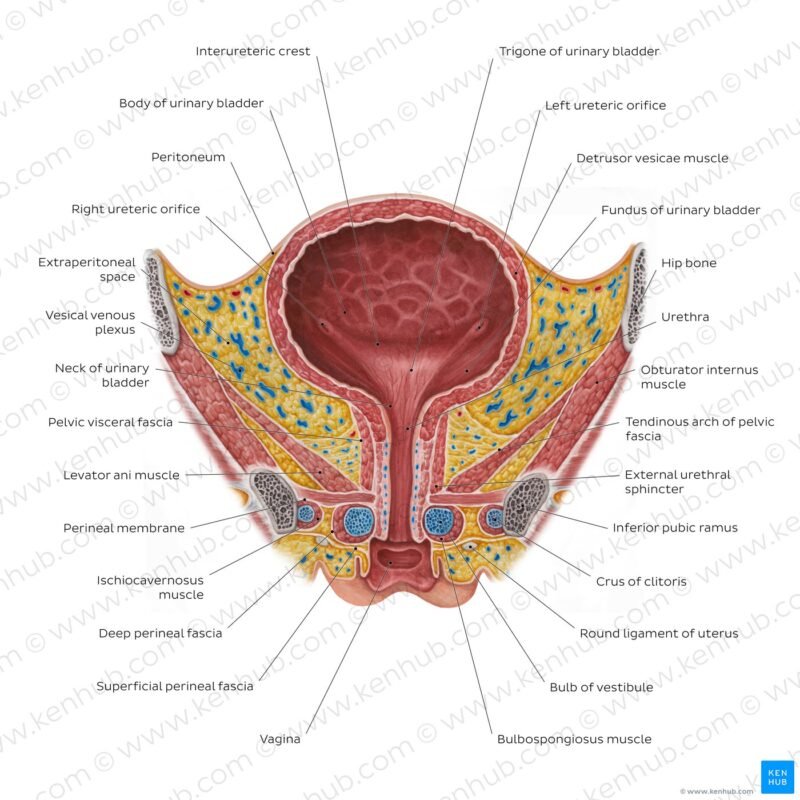
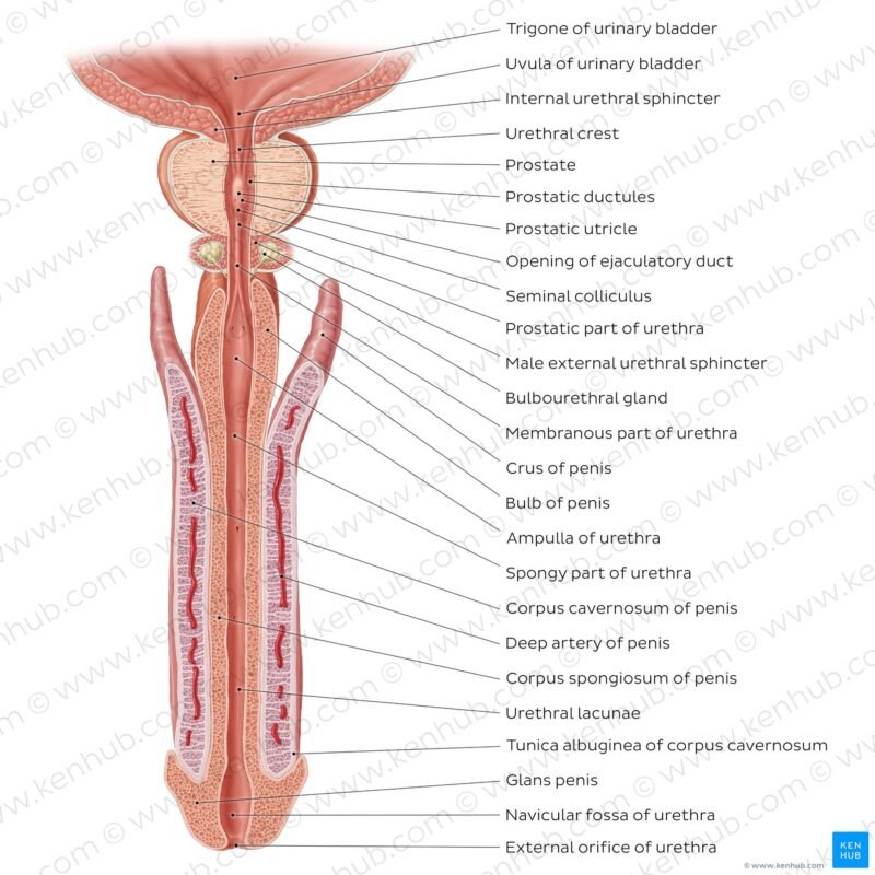
There is a triangular area, called the trigone, formed by three openings in the floor of the urinary bladder. Two of the openings are from the ureters and form the base of the trigone. Small flaps of mucosa cover these openings and act as valves that allow urine to enter the bladder but prevent it from backing up from the bladder into the ureters. The third opening, at the apex of the trigone, is the opening into the urethra. A band of the detrusor muscle encircles this opening to form the internal urethral sphincter.
- Layers: The bladder has multiple layers, including:
- Urothelium: The layer of cells that lines the inside of the bladder
- Lamina propria: A connective tissue layer that surrounds the urothelium
- Detrusor muscle: The thick, smooth muscle layer that contracts to expel urine
- Fatty connective tissue: The layer that covers the outside of the bladder and separates it from other organs
- Urothelium: The layer of cells that lines the inside of the bladder
- Parts: The bladder has four main parts:
- Dome: The top-front part of the bladder that points toward the abdominal wall
- Body: The central part of the bladder that stores urine
- Fundus: The bottom-back part of the bladder, also known as the base
- Neck: The narrow group of muscles at the base of the bladder that connects to the urethra
- Dome: The top-front part of the bladder that points toward the abdominal wall
- Trigone: A triangular area at the base of the bladder where the ureters connect
- Sphincter muscles: Two circular muscles that close tightly to prevent urine from leaking
- Nerves: The bladder receives sensory and motor supply from the nervous system, which alerts the person when it’s time to urinate
Four parts make up the structure (anatomy) of the bladder:
- Dome. The dome, or apex, is the top-front part of your bladder. It points toward your abdominal wall.
- Base. The base is the bottom-back part of your bladder, also referred to as the fundus.
- Body. The bladder body makes up the area between the dome and the base.
- Neck. The bladder neck is along the base of your bladder. It’s a narrow group of muscles that connect to your urethra.
Disclaimer: Each person’s journey is unique, treatment plan, life style, food habit, hormonal condition, immune system, chronic disease condition, geological location, weather and previous medical history is also unique. So always seek the best advice from a qualified medical professional or health care provider before trying any treatments to ensure to find out the best plan for you. This guide is for general information and educational purposes only. Regular check-ups and awareness can help to manage and prevent complications associated with these diseases conditions. If you or someone are suffering from this disease condition bookmark this website or share with someone who might find it useful! Boost your knowledge and stay ahead in your health journey. We always try to ensure that the content is regularly updated to reflect the latest medical research and treatment options. Thank you for giving your valuable time to read the article.


