Scoliosis is a spinal deformity consisting of lateral curvature and rotation of the vertebrae. Scoliosis is defined as a deviation of the normal vertical line of the spine, consisting of a lateral curvature with rotation of the vertebrae within the curve. Typically, for scoliosis to be considered, there should be at least 10° of spinal angulation on the posterior-anterior radiograph associated with vertebral rotation (rx).The causes of scoliosis vary and are classified broadly as congenital, neuromuscular, syndrome-related, idiopathic and spinal curvature due to secondary reasons. Congenital scoliosis is due to a vertebral abnormality causing the mechanical deviation of the normal spinal alignment. Scoliosis can be due to neurological conditions (eg, cerebral palsy or paralysis), muscular abnormalities (eg, Duchenne muscular dystrophy) or other syndromes (eg, Marfan syndrome and neurofibromatosis). Occasionally, significant lateral deviation of the spine can occur with little or no rotation of the spine and without bony abnormalities. In these cases, the ‘scoliosis’ can be the result of pain, spinal cord abnormalities, tumors (both intraspinal and extraspinal) and infection.
Scoliosis is a spinal disorder where the spine curves sideways in an “S” or “C” shape rather than remaining straight. This curvature can vary in degree and may affect body symmetry, posture, and sometimes lead to discomfort or pain. Scoliosis is most common in children and adolescents, although it can affect adults as well.
Medical literature often has more specific names or terms for scoliosis:
- Kyphoscoliosis: a combination of outward and lateral spine curvature
- Dextroscoliosis: the curvature of the spine to the right
- Rotoscoliosis (rotatory): the curvature of the vertebral column turned on its axis
- Levoconvex: the curvature of the spine to the left
- Thoracolumbar: curvature related to both the thoracic and lumbar regions of the spine
- Scoliosis can be classified into several types based on the cause and age of onset:
Idiopathic Scoliosis:
The most common type, especially in adolescents. Its cause is unknown but may have a genetic component.Adolescent Idiopathic Scoliosis (AIS): Occurs during the growth spurt just before puberty.
Adult Idiopathic Scoliosis: May continue from adolescent cases or develop later in life.
Congenital Scoliosis:
Develops from abnormalities in the vertebrae at birth due to improper spinal formation.Neuromuscular Scoliosis:
Occurs in individuals with neuromuscular conditions such as cerebral palsy, muscular dystrophy, or spinal muscular atrophy.Degenerative Scoliosis (or Adult Scoliosis):
Associated with aging changes in the spine, including disc degeneration and arthritis.Syndromic Scoliosis:
Associated with specific syndromes such as Marfan syndrome or Ehlers-Danlos syndrome.
As stated above, idiopathic scoliosis and its subtypes comprise over 80% of all scoliosis patients. However, there are three other main types of scoliosis:
- Functional: In this type of scoliosis, the spine is normal, but an abnormal curve develops because of a problem somewhere else in the body. This could be caused by one leg being shorter than the other, carrying heavy loads that cause unequal weight-bearing, or by muscle spasms in the back.
- Neuromuscular: In this type of scoliosis, there is a problem when the bones of the spine are formed. Either the bones of the spine fail to form completely or they fail to separate from each other during fetal development. This type of congenital scoliosis develops in people with other disorders, including birth defects, muscular dystrophy, cerebral palsy, or Marfan syndrome (an inherited connective tissue disease). People with these conditions often develop a long C-shaped curve and have weak muscles that are unable to hold them up straight. If the curve is present at birth, it is called congenital scoliosis. This type of scoliosis is often much more severe (severe scoliosis) and needs more aggressive treatment than other forms of scoliosis.
- Degenerative: Unlike the other forms of scoliosis that are found in children and teens, degenerative scoliosis occurs in older adults. It is caused by changes in the spine due to arthritis known as spondylosis. Weakening of the normal ligaments and other soft tissues of the spine combined with abnormal bone spurs can lead to an abnormal curvature of the spine. The spine can also be affected by osteoporosis, vertebral compression fractures, and disc degeneration.
There are other potential causes of scoliosis, including spine tumors such as osteoid osteoma. This is a benign tumor that can occur in the spine and cause pain. The pain causes people to lean to the opposite side to reduce the amount of pressure applied to the tumor. This can lead to a spinal deformity. In addition, researchers suggest that genetics (hereditary), muscle disorders, and/or abnormal fibrillin metabolism may play a role in causing or contributing to scoliosis development.
Causes of Scoliosis
While the exact origin of scoliosis is often unclear—especially in idiopathic cases—numerous factors may play a role. Here are 20 potential causes or contributing factors:
Idiopathic Factors:
No known cause; genetics may play a role.Congenital Vertebral Anomalies:
Improper formation of vertebrae during fetal development.Cerebral Palsy:
Neuromuscular disorder affecting muscle tone and posture.Muscular Dystrophy:
Progressive muscle weakness can lead to spinal imbalance.Spinal Muscular Atrophy:
A genetic disorder affecting motor neurons.Connective Tissue Disorders:
Conditions like Marfan syndrome or Ehlers-Danlos syndrome.Genetic Predisposition:
A family history of scoliosis increases the risk.Growth Imbalances:
Asymmetrical growth of the spinal components during adolescence.Injury or Trauma:
Spinal injuries or fractures may result in curvature.Inflammatory Conditions:
Infections or inflammation can disrupt normal spine development.Tumors or Spinal Cord Abnormalities:
Abnormal growths impacting spinal alignment.Degenerative Changes:
Wear and tear or arthritis in older adults.Osteoporosis:
Weakened bones that can lead to vertebral deformities.Postural Habits:
Consistently poor posture may contribute over time.Muscular Imbalance:
Uneven strength or tension in the back muscles.Hormonal Influences:
Hormonal changes during growth spurts.Nutritional Imbalances:
Poor nutrition affecting bone development.Excessive Physical Stress:
Repetitive strain from certain sports or activities.Environmental Factors:
Influences that are still under study.Inflammatory Arthritis:
Conditions such as rheumatoid arthritis affecting the spine.
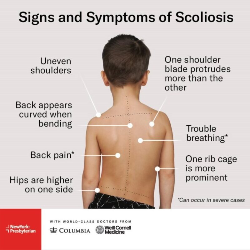
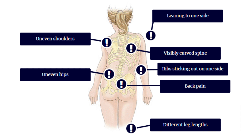
Symptoms of Scoliosis
Scoliosis symptoms can range from subtle to noticeable. Here are 20 signs and symptoms to look out for:
Visible Lateral Curve of the Spine:
Noticeable curve when viewed from behind.Uneven Shoulders:
One shoulder may appear higher than the other.Asymmetrical Waistline:
The waist may seem uneven or twisted.Prominent Shoulder Blade:
One shoulder blade may stick out.Uneven Hips:
One hip may be higher or more prominent.Back Pain:
May range from mild to severe.Stiffness in the Back:
Reduced flexibility and movement.Fatigue:
Increased tiredness due to muscle strain.Height Loss:
A reduction in height over time due to spinal curvature.Rib Prominence:
One or more ribs may stick out.Head Tilt:
The head may tilt to one side.Muscle Imbalance:
Differences in muscle tone and development.Nerve Compression:
Tingling or numbness in the arms or legs.Difficulty Breathing:
Severe curves can impact lung capacity.Uneven Skin Creases:
Lines along the back may not match on both sides.Postural Imbalance:
General asymmetry when standing or walking.Restricted Range of Motion:
Difficulty in bending or twisting.Muscle Spasms:
Involuntary contractions in the back muscles.Shoulder or Neck Pain:
Discomfort that extends upward.Changes in Gait:
Altered walking patterns to compensate for pain or imbalance.
Diagnostic Tests for Scoliosis
Proper diagnosis of scoliosis involves several tests and evaluations. Here are 20 diagnostic methods that health professionals may use:
Physical Examination:
A general check for posture and symmetry.Forward Bend Test:
The patient bends forward to highlight spinal curvature.Adam’s Forward Bend Test:
A specific test to assess asymmetry.X-Ray Imaging:
Standard method to view the spine in detail.Cobb Angle Measurement:
Determines the degree of curvature on X-rays.Lateral X-Rays:
Side views that assess the depth of the curve.Magnetic Resonance Imaging (MRI):
Evaluates soft tissues, spinal cord, and nerves.Computed Tomography (CT) Scan:
Provides detailed cross-sectional images of the vertebrae.Bone Scan:
Detects abnormal bone activity or growths.Spinal Flexibility Assessment:
Tests to see how much the spine can move.Neurological Examination:
Checks nerve function and detects any compression.Scoliometer Reading:
A device that measures the angle of trunk rotation.Surface Topography:
Uses optical methods to map the back’s shape.3D Imaging Techniques:
Provides a three-dimensional view of the spine.Ultrasound Imaging:
Occasionally used in children for preliminary assessment.Genetic Testing:
In cases of congenital or syndromic scoliosis.Pulmonary Function Tests:
Assess lung capacity if the curvature might affect breathing.Blood Tests:
Rule out infections or inflammatory conditions.Postural Analysis Software:
Digital tools to analyze posture and spinal alignment.Dynamic X-Rays:
Taken during movement to evaluate spinal function.
Non-Pharmacological Treatments for Scoliosis
Managing scoliosis often involves non-drug interventions. Here are 30 non-pharmacological treatment options that may be recommended:
Physical Therapy:
Customized exercise programs to strengthen the back.Schroth Method:
A specialized exercise program to correct posture.Pilates:
Helps improve core strength and alignment.Yoga:
Promotes flexibility and balance.Swimming:
A low-impact activity that strengthens the muscles.Postural Training:
Techniques to improve everyday posture.Orthotic Bracing:
Use of a brace to slow curve progression.Chiropractic Care:
Manual adjustments that may relieve discomfort.Massage Therapy:
Relieves muscle tension and improves circulation.Acupuncture:
Traditional therapy to help manage pain.Core Strengthening Exercises:
Focused workouts that build trunk stability.Stretching Routines:
Daily stretches to improve spinal flexibility.Aerobic Exercises:
Activities like walking or cycling to boost overall fitness.Strength Training:
Targeted exercises to balance muscle groups.Electrical Stimulation:
Techniques to reduce muscle spasm and pain.Hydrotherapy:
Exercises performed in water to decrease stress on joints.Breathing Exercises:
Improve respiratory function, especially if lung capacity is affected.Flexibility Training:
Exercises focusing on increasing range of motion.Occupational Therapy:
Tailored activities to improve daily function.Balance Exercises:
Improve coordination and prevent falls.Posture Correction Strategies:
Practical tips for maintaining a healthy spine.Ergonomic Adjustments:
Modifications in the home or workplace (furniture, seating) to support posture.Pilates Ball Exercises:
Use of stability balls to enhance core strength.Feldenkrais Method:
A movement method to improve body awareness.Alexander Technique:
Focuses on posture and movement efficiency.Cognitive Behavioral Therapy (CBT):
Helps manage chronic pain and improve quality of life.Biofeedback:
Techniques that help control bodily functions.Nutritional Support:
Diet changes to support bone and muscle health.Lifestyle Modifications:
Overall changes to support long-term spinal health.Relaxation Techniques:
Practices such as meditation to manage stress and pain.
Medications for Scoliosis
While there is no drug to cure scoliosis, medications are often used to manage associated pain, inflammation, and discomfort. Here are 20 drugs that may be used in the management of scoliosis-related symptoms:
Ibuprofen:
A common nonsteroidal anti-inflammatory drug (NSAID) for pain and inflammation.Naproxen:
Another NSAID used for pain relief.Acetaminophen:
A pain reliever that can help ease discomfort.Diclofenac:
An NSAID useful for reducing inflammation.Celecoxib:
A selective COX-2 inhibitor for pain management.Aspirin:
Occasionally used for its anti-inflammatory effects.Cyclobenzaprine:
A muscle relaxant that can ease muscle spasms.Gabapentin:
Often prescribed for nerve-related pain.Pregabalin:
Used for neuropathic pain and muscle discomfort.Corticosteroids:
Short-term use to decrease severe inflammation.Tramadol:
A moderate opioid for more intense pain.Codeine:
An opioid sometimes prescribed for pain management.Methocarbamol:
A muscle relaxant aiding in the reduction of muscle tension.Naproxen Sodium:
A formulation of naproxen for pain relief.Meloxicam:
An NSAID that offers once-daily dosing.Etodolac:
Another NSAID option for reducing pain.Indomethacin:
Used occasionally for its strong anti-inflammatory properties.Buprenorphine:
In select cases, used for severe pain management.Extended-Release Gabapentin:
Provides sustained relief for neuropathic symptoms.Duloxetine (SNRI):
Helps manage chronic pain and improve mood.
Note: Always consult with a healthcare provider before starting any medication, as these drugs are used off-label for symptom management in scoliosis.
Prevention of Scoliosis
While not all cases of scoliosis can be prevented, early detection and healthy habits can help reduce the risk of curve progression. Consider these 10 preventive measures:
Early Screening and Detection:
Regular check-ups in schools or during pediatric visits.Routine Medical Examinations:
Early diagnosis can lead to better outcomes.Good Posture:
Encourage proper sitting, standing, and sleeping positions.Regular Exercise:
Maintain a balanced exercise routine that strengthens the core.Core Strengthening Routines:
Help support the spine and reduce the risk of imbalance.Proper Backpack Use:
Distribute weight evenly by using backpacks with padded straps.Weight Management:
Avoid excessive weight that may stress the spine.Ergonomic Furniture:
Use chairs and beds that support good posture.Avoid Heavy Loads on One Side:
Distribute weight evenly when carrying items.Educational Awareness:
Educate children and caregivers about the importance of early signs.
When to See a Doctor
It is important to consult a healthcare provider if you notice any of the following signs:
A visible curve in the spine or uneven shoulders and hips.
Persistent back pain or discomfort that does not improve with rest.
Changes in posture or difficulty with mobility.
Shortness of breath or changes in breathing patterns.
Numbness, tingling, or weakness in the arms or legs.
Rapid progression of spinal curvature in children or adolescents.
Any concerning changes during routine health check-ups.
Early diagnosis can lead to better management and treatment outcomes.
Physiotherapy Treatment
While scoliosis does not usually require immediate medical attention or endanger one’s life, it can greatly impede the quality of life due to immobility difficulties, pain and fatigue. After a spinal cord injury, one must be keenly aware of scoliosis. Most people with a spinal cord injury experience some paralysis of their torso muscles, which leads to difficulty in holding the backbone in its natural position.
Many people with spinal cord injuries have bad posture because of this, which leads to scoliosis as the years’ progress. This, however, does not to be inevitable. Scoliosis can be prevented after an SCI. And if you do get scoliosis, there are many options available to treat it. Here are the options available for people with paralysis to prevent, maintain and correct scoliosis.
Annual X-Rays
It is important to get annual X-rays of your back to monitor any curves that may be forming. You’ll want to find an orthopedic surgeon familiar with scoliosis, or a rehab medicine doctor who can monitor your scoliosis, and make sure that it isn’t getting worse. The main goal is to ensure that no scoliosis occurs, but if it does, you’ll want a doctor who can monitor the curvature and to make sure it is not endangering any important organs, such as your lungs.
Stay Active
Moving as much as possible if you have movement in your upper body can help a lot in preventing scoliosis post-injury. It is also important to do a daily range of motion exercises to keep your back in the natural position as often as possible.
Maintain a Healthy Weight
Eating healthy and maintaining a healthy weight post-injury can go a long way in preventing and maintaining scoliosis. Eating healthy can minimize the symptoms of scoliosis as well. Also, making sure you’re getting enough Vitamin D, can also make a huge difference in preventing osteoporosis, which can lead to scoliosis. To monitor osteoporosis, annual DEXA scans are also suggested. Talk to your doctor about getting one.
Stretch Your Back
Keeping your back long and limber can make a huge impact when it comes to preventing and minimizing the effects of scoliosis. Muscle memory is a real thing and is still something you can tap into. By stretching your back daily, this will force your back into its natural position. Massage of the curve and muscles affected can also help if done regularly.
Scoliosis Specific Exercise
Scoliosis specific exercises have been found to improve treatment outcomes when utilized in addition to bracing and other standards of care.[rx] Scoliosis specific exercises include methods such as Schroth[rx] which specifically aim to correct aesthetic differences and strengthened muscles and connective tissue that may have atrophied as a result of scoliosis and asymmetric posture. The Schroth method is a three-dimensional exercise approach aiming to improve spinal curvature and vertebral rotational angles. A physical therapist guides the patient through breathing techniques and exercises to strengthen weak muscle sand and relieve overactive ones. In addition to spinal realignment, the Schroth method improves self-image, quality of life, and lumbar extensor strength.[rx] Schroth exercises and other scoliosis specific exercises should be utilized in conjunction with bracing and other standards of care,[rx] and be performed under the guidance of a trained professional to ensure the exercises are effective and target the individual’s curve pattern so that the correct muscles are strengthened. Strengthening spinal muscles is a crucial preventive measure. This is because the muscles in the back are essential when it comes to supporting the spinal column and maintaining the spine’s proper shape. Exercises that will help improve the strength of the muscles in the back include rows and leg and arm extensions.[rx] Elastic resistance exercise may also be able to sustain the progression of spinal curvature.[rx] This type of exercise is able to sustain progression by equalizing the strength of the torso muscles found on each side of the body.
Self-care
Disability caused by scoliosis, as well as physical limitations during recovery from treatment-related surgery, often affects an individual’s ability to perform self-care activities.[rx] One of the first treatments of scoliosis is the attempt to prevent further curvature of the spine. Depending on the size of the curvature, this is typically done in one of three ways: bracing, surgery, or postural positioning through customized cushioning.[rx][rx][rx] Stopping the progression of the scoliosis can prevent the loss of function in many activities of daily living by maintaining range of motion, preventing deformity of the rib cage, and reducing pain during activities such as bending or lifting.
Occupational therapists are often involved in the process of selection and fabrication of customized cushions. These individualized postural supports are used to maintain the current spinal curvature, or they can be adjusted to assist in the correction of the curvature. This type of treatment can help to maintain mobility for a wheelchair user by preventing the deformity of the rib cage and maintaining an active range of motion in the arms.[rx]
For other self-care activities (such as dressing, bathing, grooming, personal hygiene, and feeding), several strategies can be used as a part of occupational therapy treatment. Environmental adaptations for bathing could include a bath bench, grab bars installed in the shower area, or a handheld shower nozzle.[rx] For activities such as dressing and grooming, various assistive devices and strategies can be used to promote independence. An occupational therapist may recommend a long-handled reacher that can be used to assist self-dressing by allowing a person to avoid painful movements such as bending over; a long-handled shoehorn can be used for putting on and removing shoes. Problems with activities such as cutting meat and eating can be addressed by using specialized cutlery, kitchen utensils, or dishes.
Productivity
Productive activities include paid or unpaid work, household chores, school, and play.[rx] Recent studies in healthcare have led to the development of a variety of treatments to assist in the management of scoliosis thereby maximizing productivity for people of all ages. Assistive technology has undergone dramatic changes over the past 20 years; the availability and quality of the technology has improved greatly.[rx] As a result of using assistive technology, functional changes may range from improvements in abilities, performance in daily activities, participation levels, and quality of life.[rx]
A common assistive technology intervention is specialized seating and postural control. For children with poor postural control, a comfortable seating system that provides them with the support needed to maintain a sitting position can be essential for raising their overall level of well-being.[rx] A child’s well-being in a productive sense involves the ability to participate in classroom and play activities.[rx] Specialized wheelchair seating has been identified as the most common prescription in the management of scoliosis in teenagers with muscular dystrophy.[rx]
With comfortable wheelchair seating, teenagers are able to participate in classroom activities for longer periods with less fatigue. By tilting the seating position 20° forward (toward the thighs), seating pressure is significantly redistributed, so sitting is more comfortable. If an office worker with scoliosis can sit for longer periods, increased work output is likely to occur and could improve quality of life. Tall, forward-sloping seats or front parts of seats, and when possible with tall desk with the opposite slope, can, in general, reduce pains and the need of bending significantly while working or studying, and that is particularly important with braced, fragile, or tender backs. An open hip angle can benefit the used lung volume and respiration.[rx][rx]
For those not using a wheelchair, bracing may be used to treat scoliosis. Lifestyle changes are made to compensate for the proper use of spine braces.
Leisure
Physical symptoms such as chest pains, back pains, shortness of breath, and limited spinal movement can hamper or preclude participation in leisure activities of a physical nature. The occupational therapist’s role is to facilitate participation by helping the patient manage these symptoms.
Bracing is a common strategy recommended by an occupational therapist, in particular, for individuals engaging in sports and exercise.[rx] An OT is responsible for educating an individual on the advantages and disadvantages of different braces, proper ways to wear the brace, and the day-to-day care of the brace.
To help a person manage heart and lung symptoms, such as shortness of breath or chest pains, an occupational therapist can teach the individual energy conservation techniques.[rx] This includes scheduling routine breaks during the activity, as suitable for the individual. For example, an occupational therapist can recommend that a swimmer take breaks between laps to conserve energy. Adapting or modifying the exercise or sport is another way a person with scoliosis can do it.[rx] Adapting the activity may change the difficulty of the sport or exercise. For example, it might mean taking breaks throughout an exercise. If a person with scoliosis is unable to participate in a sport or exercise, an OT can help the individual explore other physical activities that are suitable to his/her interests and capabilities. An occupational therapist and the person with scoliosis can explore enjoyable and meaningful participation in the sport/exercise in another capacity, such as coaching or refereeing.
Bracing
Bracing is most effective when the patient has bone growth remaining (is skeletally immature) and should aim to both prevent progression of the curve (prevent progression to surgery), as well as reduce the scoliosis curve. Reduction of the curve is important as the natural history of idiopathic scoliosis suggests it can continue to progress at a rate ~1 degree per year in adulthood,[rx] while the treatment results of bracing have been shown to hold over >15 years.[rx] In some cases with juveniles, bracing has reduced curves significantly, going from a 40 degrees (of the curve, mentioned in length above.) out of the brace to 18 degrees in it. Braces are sometimes prescribed for adults to relieve pain related to scoliosis. Bracing involves fitting the patient with a device that covers the torso; in some cases, it extends to the neck. The most commonly used brace is a TLSO, such as a Cheneau type brace, a corset-like appliance that fits from armpits to hips and is custom-made from fiberglass or plastic. It is worn upwards of 18–23 hours a day, depending on the doctor’s prescription, and applies pressure on the curves in the spine. The effectiveness of the brace depends not only on brace design and orthotist skill; patient compliance; and amount of wear per day, but also the “stiffness” of the spine resulting from a shortened spinal cord[rx][rx] and/or nerve tension.[rx] as evidence by the force necessary (mean force ~121 lbs) to physically correct scoliosis during spinal surgery[rx] The typical use of braces for idiopathic scoliosis is to prevent progression to surgical range as well as reduce the scoliotic curve of the spine as spinal fusion surgery can reduce mobility due to fusion of the vertebrate while potentially increasing pain long term.[rx] For non-idiopathic scoliosis (ie. neuromuscular, congenital, etc.) and those with additional comorbidities (ie. Marfans Syndrome) spinal surgery may be required due to structural changes in the spine.
Indications for Scoliosis Bracing: Scoliosis professionals determine the proper bracing method for a patient after a complete clinical evaluation. The patient’s growth potential, age, maturity, and scoliosis (Cobb angle, rotation, and sagittal profile) are also considered. Immature patients who present with Cobb angles less than 20 degrees should be closely monitored and proactively treated based on their risk of progression[rx] as surgery can be prevented with early intervention of conservative treatment.[rx] Immature patients who present with Cobb angles of 20 degrees to 29 degrees should be braced according to the risk of progression by considering age, Cobb angle increase over a six-month period, Risser sign, and clinical presentation. Immature patients who present with Cobb angles greater than 30 degrees should be braced. However, these are guidelines and not every patient will fit into this table. For example, an immature patient with a 17-degree Cobb angle and significant thoracic rotation or flatback could be considered for nighttime bracing. On the opposite end of the growth spectrum, a 29-degree Cobb angle and a Risser sign three or four might not need to be braced because there is reduced potential for progression.[rx]
Surgery is indicated by the Society on Scoliosis Orthopaedic and Rehabilitation Treatment (SOSORT) at 45 degrees to 50 degrees[rx] and by the Scoliosis Research Society (SRS) at a Cobb angle of 45 degrees.[rx] SOSORT uses the 45-degree to 50-degree threshold as a result of the well-documented, plus or minus five degrees measurement error that can occur while measuring Cobb angles.
Scoliosis braces are usually comfortable for the patient, especially when it is well designed and fit; also after the 7- to 10-day break-in period. A well fit and functioning scoliosis brace provides comfort when it is supporting the deformity and redirecting the body into a more corrected and normal physiological position.[rx]
The Scoliosis Research Society’s recommendations for bracing include curves progressing to larger than 25°, curves presenting between 30 and 45°, Risser sign 0, 1, or 2 (an X-ray measurement of a pelvic growth area), and less than six months from the onset of menses in girls.[rx]
Progressive scolioses exceeding 25° Cobb angle in the pubertal growth spurt should be treated with a pattern-specific brace like the Chêneau brace and its derivatives, with an average brace-wearing time of 16 hours/day (23 hours/day assures the best possible result).
The latest standard of brace construction is with CAD/CAM technology. With the help of this technology, it has been possible to standardize the pattern-specific brace treatment. Severe mistakes in brace construction are largely ruled out with the help of these systems. This technology also eliminates the need to make a plaster cast for brace construction. The measurements can be taken in any place and are simple (and not comparable to plastering). Available CAD/CAM braces include the Regnier-Chêneau brace, the Rigo-System-Chêneau-brace (RSC brace), the Silicon Valley Brace, and the Gensingen brace; braces can and should be customized to fit the individual’s curve pattern and reduce the curve as much as possible as immediate in-brace correction has been shown to be associated with better treatment outcomes.[rx][rx] Many patients prefer the “Chêneau light” brace as it has good in-brace corrections reported in international literature and is easier to wear than other braces in use today.[rx][rx] However, this brace is not available for all curve patterns.
Prior to 2013 the efficacy of bracing has not been definitively demonstrated in randomised clinical studies, with more limited studies giving inconsistent conclusions.[rx] In 2013 the Bracing in Adolescent Idiopathic Scoliosis Trial (BrAIST) published results establishing benefits of bracing in adolescents with idiopathic scoliosis. In the randomized cohort, 72% in the group instructed to wear a brace for 18 hours per day against 48% in the observation group sustained curve progression to under 50 degrees, the proxy used for not requiring surgery. Additionally results suggested that the more a patient wore the brace, the better the result.[rx][rx]
Casting
In progressive infantile and sometimes juvenile scoliosis, a plaster jacket applied early may be used instead of a brace. It has been proven possible[rx] to permanently correct cases of infantile idiopathic scoliosis by applying a series of plaster casts (EDF: elongation, derotation, flexion) on a specialized frame under corrective traction, which helps to “mould” the infant’s soft bones and work with their growth spurts. This method was pioneered by UK scoliosis specialist Min Mehta.[rx] EDF casting is now the only clinically known nonsurgical method of complete correction in progressive infantile scoliosis. Complete correction may be obtained for curves less than 50° if the treatment begins before the second year of life.[rx][rx]
Surgery
Surgery is usually recommended by orthopedists for curves with a high likelihood of progression (i.e., greater than 45 to 50° of magnitude), curves that would be cosmetically unacceptable as an adult, curves in patients with spina bifida and cerebral palsy that interfere with sitting and care, and curves that affect physiological functions such as breathing.[
Surgery for scoliosis is performed by a surgeon specializing in spine surgery. For various reasons, it is usually impossible to completely straighten a scoliotic spine, but in most cases, significant corrections are achieved.
The two main types of surgery are:
- Anterior fusion: This surgical approach is through an incision at the side of the chest wall.
- Posterior fusion: This surgical approach is through an incision on the back and involves the use of metal instrumentation to correct the curve.
One or both of these surgical procedures may be needed. The surgery may be done in one or two stages and, on average, takes four to eight hours. A Cochrane review could not draw conclusions on how effective surgical interventions were when compared to non-surgical interventions in patients with adolescent idiopathic scoliosis.[rx]
Spinal fusion with instrumentation
Spinal fusion is the most widely performed surgery for scoliosis. In this procedure, bone [either harvested from elsewhere in the body (autograft) or from a donor (allograft)] is grafted to the vertebrae so when they heal, they form one solid bone mass and the vertebral column becomes rigid. This prevents worsening of the curve, at the expense of some spinal movement. This can be performed from the anterior (front) aspect of the spine by entering the thoracic or abdominal cavities, or more commonly, performed from the back (posterior). A combination may be used in more severe cases, though the modern pedicle screw system has largely negated the need for this.[rx]
In recent years all-screw systems have become the gold-standard technique for adolescent idiopathic scoliosis. Pedicle screws achieve better fixation of the vertebral column and have better biomechanical properties than previous techniques, so enabling greater correction of the curve in all planes.[rx]
Pedicle screw-only posterior spinal fusion may improve major curve correction at two years among patients with adolescent idiopathic scoliosis (AIS)[rx] as compared to hybrid instrumentation (proximal hooks with distal pedicle screws) (65% versus 46%) according to a retrospective, matched-cohort study.[rx] The prospective cohorts were matched to the retrospective cohorts according to patient age, fusion levels, Lenke curve type, and operative method. The two groups were not significantly different in regard to age, Lenke AIS curve type, or Riser grade. The numbers of fused vertebrae were significantly different (11.7±1.6 for pedicle screw versus 13.0±1.2 for hybrid group). This study’s results may be biased due to the pedicle screw group’s being analyzed prospectively versus retrospective analysis of the hybrid instrumentation group.
In general, modern spinal fusions have good outcomes with high degrees of correction and low rates of failure and infection. However a systematic review of PubMed papers in 2008 concluded “Scoliosis surgery has a varying but high rate of complications”, although the non-standardised data on complications was difficult to assess and was incomplete.[rx] Patients with fused spines and permanent implants tend to have normal lives with unrestricted activities when they are younger; it remains to be seen whether those that have been treated with the newer surgical techniques develop problems as they age.[rx]
Thoracoplasty
A complementary surgical procedure a surgeon may recommend is called thoracoplasty (also called costoplasty). This is a procedure to reduce the rib hump that affects most scoliosis patients with a thoracic curve. A rib hump is evidence of some rotational deformity to the spine. Thoracoplasty may also be performed to obtain bone grafts from the ribs instead of the pelvis, regardless of whether a rib hump is present. Thoracoplasty can be performed as part of a spinal fusion or as a separate surgery, entirely.
Thoracoplasty is the removal (or resection) of typically four to six segments of adjacent ribs that protrude. Each segment is one to two inches long. The surgeon decides which ribs to resect based on either their prominence or their likelihood to be realigned by correction of the curvature alone. The ribs grow back straight.
Thoracoplasty has risks, such as increased pain in the rib area during recovery or reduced pulmonary function (10–15% is typical) following surgery. This impairment can last anywhere from a few months to two years. Because thoracoplasty may lengthen the duration of surgery, patients may also lose more blood or develop complications from the prolonged anesthesia. A more significant, though far less common, risk is the surgeon might inadvertently puncture the pleura, a protective coating over the lungs. This could cause blood or air to drain into the chest cavity, hemothorax or pneumothorax, respectively.[rx]
Surgery without fusion for growing children
Implants that aim to delay spinal fusion and to allow more spinal growth in young children is the gold standard for surgical treatment of early onset scoliosis. Surgery without fusion can be divided into three principles: distraction of the entire spine, compression of the short segment of spine, and guided-growth techniques. Distraction-based systems include Vertical, Expandable Prosthetic Titanium Ribs (VEPTR) & growing rods. The concept uses distraction to create additional soft-tissue space in-between the vertebrae, for the bone to later grow into. Its universal application was thrusted through the use of traditional growth rods which required repeated invasive surgeries every 6–12 months for the sustenance of growth, via distraction. Nowadays developed countries only use MAGEC (MAGnetic Expansion Control) rods to non-invasively lengthen the spine. In contrast, developing and under-developed countries still use traditional growing rods, which require invasive surgery every 6–12 months, because of high initial cost associated with procurement of MAGEC rods. Compression-based system include tethering using a flexible rope-like implant and are relatively new to receive FDA approval. Guided-growth technique include SHILLA (named after a hotel in Korea, where the concept was initiated). SHILLA has the advantage of being one-time surgery and is technologically less demanding compared with MAGEC rod. However, there are still two major disadvantages of using SHILLA: loss of correction and need for osteotomies.[rx][rx]
The failure of most of these standalone techniques has shown that the concept of “one size fits all” is not applicable for the surgical management of EOS. Therefore, newer concepts employing two or more of the above philosophies, i.e. various combinations of distraction-based, guided-growth, and compression-based approaches might be more suitable and biomechanically-speaking, a more optimal surgical intervention. One such combination currently used for surgery includes active apex correction (APC). It is a hybrid of guided-growth and compression-based management of deformity. The technique simply consists of replacing the apical fusion (of traditional SHILLA) with unilateral compression (via pedicle screws or any other means) on the convex side. The latest clinical results presented by spine researchers Aakash Agarwal and Alaaedldin Azmi Ahmad on APC shows good clinical results with no economic barrier to use the technology.[rx][rx][rxx]
Complications
The risk of undergoing surgery for scoliosis was estimated in 2008 to be varying, but with a high rate of complications. Possible complications may be inflammation of the soft tissue or deep inflammatory processes, breathing impairments, bleeding and nerve injuries. It is not yet clear what to expect from spine surgery in the long term.[rx][rx] Taking into account that signs and symptoms of spinal deformity cannot be changed by surgical intervention, surgery remains primarily a cosmetic indication, only especially in patients with adolescent idiopathic scoliosis, the most common form of scoliosis never exceeding 80°.[rx][rx] However, the cosmetic effects of surgery are not necessarily stable.[rx]
For spinal fusion surgery on AIS cases, with instrumentation attached using pedicle screws, complication rates were reported in 2011 as transient neurological injuries between 0% to 1.5%, a pedicle fracture rate of 0.24%, screw malposition assessed by radiography at 1.5%, 6% when assessed by CT scans though these patients were asymptomatic not requiring screw revision, and screw loosening noted in 0.76% of patients.[rx]
For surgery without fusion in growing children, substantial percentage of patients undergoing SHILLA technique experience loss of correction via crankshafting or adding-on (eg, distal migration). In addition, the need for osteotomies on the concave side has the potential of severe complications. For MAGEC rods, higher distraction magnitude resulted in the generation of higher distraction forces, and this in combination with off-axis loading (exemplified by “growth marks”) result in wear and breakage of MAGEC rod’s components.[rx][rx]
After-surgery care
Pain medication
In the event of surgery to correct scoliosis, pain medications and anesthesia will be administered. Before the surgery, the patient will receive anesthesia. With adults, the anesthesia will be administered through an IV in the antecubital region of the arm. With young children, however, the child will be asked to breathe in nitrous oxide, or laughing gas. Because needles can be frightening for a young child, the nitrous oxide will put them to sleep so the anesthesiologist can then insert the IV in order to give them the anesthesia. After the surgery, the patient will most likely be given morphine. Until the patient is ready to take the medicine by mouth, an IV will be giving them their medication. Morphine is the most common pain medicine used after scoliosis surgery, and is often administered through a patient-controlled analgesia (PCA) system. The PCA system allows the patient to push a button when they are feeling pain, and the PCA will emit the drugs into the IV and then into the body. To prevent overdoses, there is a limit on the number of times a patient can push the button. If a patient pushes the button too much at once, the PCA will reject the request.[rx]
Bowel and bladder function
For the patient’s bladder control, a catheter will be inserted so that a patient can urinate without having to move. A catheter is inserted because the patient will not have much free movement to be able to get up and walk to the bathroom. The most common type of catheter used after major surgeries is an indwelling Foley catheter. The indwelling Foley catheter is most often put in the urethra, with a tube leading into a drainage bag. Once the catheter is inserted into the urethra, a balloon is blown up inside the bladder in order to keep it from falling out. The balloon allows the catheter to remain inside the urethra until the patient is able to get up and go to the bathroom on their own.[rx] The drainage bag is connected to the side of the bed, and must be changed or emptied out once it is full.
Bowel control can vary from patient to patient. The combination of no food, very little fluids, and a lot of prescription drugs has the potential to cause many patients to become constipated. The body is used to a normal diet, and used to excreting waste in a system. Interrupting the system can cause bowel problems. This constipation can be resolved in a couple of ways. The first way, and the most common way, is to administer a rectal suppository. A rectal suppository is administered through the anus, and into the rectum. They are bullet-shaped and contain medicine that will help the patient’s bowels get back on track. Once the suppository is inserted, it is designed to melt off the wax-like case, and put the medicine in the body.[rx] If the suppository does not work, a laxative may be continued at home to keep the bowels in full function.
Diet
When first returning home after surgery, a nutritional diet is necessary in order to keep the body operating correctly. Junk food is not a good idea, as the grease and sugar can irregulate the bowels. Fruit, vegetables, and juices will be a vital part in the diet.[rx] Food and drink will be limited for the patient after surgery. Because the bowels are not fully active because of anesthetic, clear water and ice may be the only acceptable thing to ingest. After the digestive tract is back up to speed, soft food and drink like pudding, soup broth, and orange juice are acceptable.[rx] Very dark urine with a strong odor means that the person is most likely dehydrated and needs more fluids. In order for the urine to become a pale or clear color, the patient will need to drink a lot of water. Juices such as prune juice are a healthy option and prune juice also helps with constipation, a common factor after surgery. When it comes to food, whole grains should be added into the diet. Whole grains can be broken down easily by the body whereas processed grains and flour cannot be broken down easily. Processed grains and flour also add to constipation.[rx]
Disclaimer: Each person’s journey is unique, treatment plan, life style, food habit, hormonal condition, immune system, chronic disease condition, geological location, weather and previous medical history is also unique. So always seek the best advice from a qualified medical professional or health care provider before trying any treatments to ensure to find out the best plan for you. This guide is for general information and educational purposes only. Regular check-ups and awareness can help to manage and prevent complications associated with these diseases conditions. If you or someone are suffering from this disease condition bookmark this website or share with someone who might find it useful! Boost your knowledge and stay ahead in your health journey. We always try to ensure that the content is regularly updated to reflect the latest medical research and treatment options. Thank you for giving your valuable time to read the article.
The article is written by Team Rxharun and reviewed by the Rx Editorial Board Members
Last Update: April 16, 2025.


