Extensors Muscles of Arm; Types, Anatomy, Functions Extensors Muscles of Arm/Upper Limb Muscle comprises many muscles that are organized into anatomical compartments. These muscles act on the various joints of the hand, arm, and shoulder, maintaining tone, providing stability and allowing precise fluid movement. Axioappendicular groups of muscles arise from the axial skeleton to act upon the pectoral girdle. Scapulohumeral muscles originate from the scapula and insert into the proximal humerus. Included in this category are the rotator cuff muscles which provide stability to the glenohumeral joint. In the arm, the muscles of the anterior compartment are involved in flexion of the forearm, and the posterior comprises of the forearm extensors. Similarly, the anterior compartment of the forearm contains the flexors of the hand and the posterior has extensors. The hand is divided into the thenar, the hypothenar, the adductor compartment, as well as the short muscles of the hand.[rx][rx][rx][rx][rx] The upper extremity (UE) is comprised of its associated muscles, nerves, and vessels, organized into anatomical compartments.[rx] The muscles cross joints to provide tone, maintain dynamic joint stability, and perform dynamic functions of the entire extremity. In addition, the arteries and veins provide nourishment and remove waste, and the nerves provide the motor and sensory innervations.[rx] Types of Muscle of Upper Limb Muscle Pectoral Anterior Axioappendicular Muscles (Thoracoappendicular Muscles) Pectoral muscles lie in the chest and exert force through the shoulder to move the upper arm. Three pectoral muscles interact with the shoulder. Pectoralis major The pectoralis major is a large, fan-shaped muscle covering the chest. It is comprised of clavicular and sternocostal regions. Function – flexion, adduction, medial rotation of the humerus. Origin – clavicular head: medial clavicle anteriorly, sternocostal head: anterior sternum and costal cartilages of ribs 1 to 6 as well as an external oblique aponeurosis Insertion: the lateral edge of the intertrabecular groove of the humerus Innervation: medial pectoral nerve (C8, T1) lateral pectoral nerve (C5, C6, C7) of brachial plexus Attachments: The clavicular region originates from the clavicle and the sternocostal region originates from the sternum and the fascia of the oblique muscles of the abdomen. Both attach to the humerus. Actions: Adducts and rotates the upper arm. Pectoralis minor The pectoralis minor muscle is smaller and lies beneath the pectoralis major. Function: Depression of the shoulder, protraction of the scapula Origin: Third, fourth, fifth ribs close to their respective costal cartilages Insertion: Coracoid process Innervation: Medial pectoral nerve (C8, T1) Attachments: The pectoralis minor originates from the third to fifth ribs and attaches to the scapula. Actions: Supports and depresses the scapula. Subclavius Function: Depression and stabilization of the clavicle Origin: First rib medially Insertion: Middle of the clavicle, inferiorly Innervation: Nerve to subclavius (C5, C6) Serratus anterior The serratus anterior is located in the lateral wall of the chest. Function: Protraction of scapula, rotation of the scapula Origin: Lateral first to the eighth rib Insertion: anterior scapula, medially Innervation: long thoracic nerve (C5, C6, C7) Attachments: The muscle is formed of several strips originating from the second to eight ribs, each of which attaches to the scapula. Actions: Supports the scapula allowing for elevation of the upper arm. Posterior Axioappendicular Muscles Superficial layer Latissimus dorsi The latissimus dorsi originates from the lower back and covers a wide area. Function: Adduction, medial rotation, an extension of humerus Origin: Spinous processes of seventh to 12th thoracic vertebrae, iliac crest, thoracolumbar fascia, and inferior third and fourth rib Insertion: Intertubercular groove of humerus Innervation: Thoracodorsal nerve (C5,C6,C7) Attachments: The latissimus dorsi originates from the lower spine and ribs and the upper pelvis and fascia of the deep trunk muscles. The muscle converges into a tendon attaching to the humerus. Actions: Extends, adducts, and medially rotates the upper arm. Trapezius The trapezius is the most superficial muscle of the back and forms a broad flat triangle. Function: Elevation, depression, and retraction of the scapula, rotation of glenoid cavity Origin: Superior nuchal line, nuchal ligament, occipital protuberance, spinous processes of C7- T12 Insertion: Spine of scapula, acromion, and lateral clavicle Innervation: CN XI Actions: The superior region supports the arm and elevates and rotates the scapula, the intermediate region retracts the scapula, and the inferior region rotates and depresses the scapula. Attachments: The trapezius originates from the skull and spine of the upper back and neck. It attaches to the clavicle and scapula. Deep Layer Levator scapulae Function: Adduction, medial rotation, an extension of humerus Origin: Transverse processes of C1 through C4 vertebrae Insertion: Scapula at its medial border Innervation: Thoracodorsal nerve (C5, C6, C7) Rhomboid major Function: Retraction of scapula and depression of glenoid cavity Origin: Spinous processes of T2 through T5 vertebrae Insertion: Inferior aspect of medial scapula Innervation: Dorsal scapular nerve (C4, C5) Rhomboid minor Function: Retraction of scapula and depression of glenoid cavity Origin: Nuchal ligament as well as spines of C7 and T1 vertebrae Insertion: Superior aspect of medial scapula Innervation: Dorsal scapular nerve (C4, C5) Three deep muscles lie below the superficial muscles of the shoulder Levator Scapulae – A small, strap-like muscle that joins the neck to the scapula. Attachments: Originates from the side of the spine in the neck and attaches to the scapula. Actions: Elevates the scapula. Rhomboid Major – Sits inferiorly to the levator scapulae. Attachments: Originates from the spine in the upper back and attaches to the scapula in an inferior position to the levator scapulae attachment. Actions: Retracts and rotates the scapula. Rhomboid Minor – Sits between the levator scapulae and rhomboid major, with which it is paired in action and function. It retracts and rotates the scapula. Scapulohumeral (Intrinsic Shoulder Muscles) Location of the deltoid muscles Highlighted in orange, the deltoids cover the rounding of the shoulder joint. Intrinsic muscles originate from the scapula or clavicle and attach to the humerus. There are six intrinsic muscles, four of which form the rotator cuff.
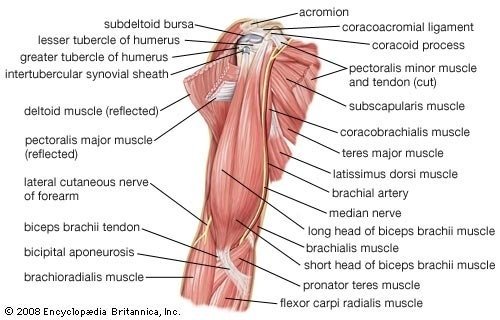
 Deltoid Muscle The deltoid muscle is a triangular muscle that covers the shoulder. The action of the muscle is complex, with the components acting in opposing and separate ways during the course of a contraction. Attachments: The deltoid muscle originates from the scapula and clavicle and attaches to the lateral surface of the humerus. Actions: The anterior region assists the pectoralis major during transverse flexion of the shoulder and acts weakly in strict transverse flexion. The lateral region assists in shoulder flexion when the shoulder is rotating, although it also assists the transverse abduction of the shoulder. The posterior region is the hyperextension of the shoulder, contributing to the transverse extension. Function: Anterior part: flexion and medial rotation of the arm, middle part: the abduction of arm, posterior part: extension and lateral rotation of the arm Origin: Lateral clavicle, acromion and scapular spine Insertion: Deltoid tuberosity Innervation: Axillary nerve (C5, C6) Teres major The teres major is a thick flattened muscle connecting the lower scapula with the humerus. Function: Adduction and medial rotation of the arm Origin: Posterior surface of scapula at its inferior angle Insertion: Intertubercular groove on its medial aspect Innervation: Lower scapular nerve (C5, C6) Attachments: Originates from the posterior of the scapula and attaches to the humerus. Actions: Adducts the shoulder and assists in rotation of the arm. Supraspinatus Function: Initiation of arm abduction Origin: Posterior scapula, superior to the scapular spine Insertion: Superior aspect of the greater tubercle Innervation: Suprascapular nerve (C5, C6) Part of rotator cuff muscles Infraspinatus Function: Lateral rotation of arm Origin: Posterior scapula, inferior to the scapular spine Insertion: Greater tubercle of the humerus, between the supraspinatus and teres minor insertion Innervation: Suprascapular nerve (C5, C6) Part of rotator cuff muscles Teres minor Function: Lateral rotation of arm Origin: Posterior surface of scapula at its inferior angle Insertion: Inferior aspect of the greater tubercle Innervation: Axillary nerve (C5, C6) Part of rotator cuff muscles Subscapularis Function: Adduction and medial rotation of the arm Origin: Anterior aspect of the scapula Insertion: Lesser tubercle of the humerus Innervation: Subscapular nerves (C5, C6, C7) Part of rotator cuff muscles Rotator cuff muscles: supraspinatus, infraspinatus, teres minor, subscapularis Muscles of Anterior Compartment of Arm (Flexors of Arm) Biceps brachii –The biceps brachii is a two-headed muscle. Although the majority of the muscle mass is located anteriorly to the humerus, it has no attachment to the bone itself. Function: Major flexion of forearm, supination of forearm, resists dislocation of shoulder Origin: Short head originates from the coracoid process. The long head is from the supraglenoid tubercle of scapula Insertion: Radial tuberosity and forearm fascia (as bicipital aponeurosis) Innervation: Musculocutaneous nerve (C5, C6) Attachments: Both heads originate from the scapula and attach via the bicipital aponeurosis to the fascia of the forearm. Action: Supination of the forearm. It also flexes the arm at the elbow and at the shoulder. Brachialis – The brachialis muscle lies within the distal region of the biceps brachii. Attachments: Originates from the humerus and attaches to the ulna. Action: Flexing of the arm at the elbow. Function: Flexion of forearm Origin: Distal anterior humerus Insertion: Coronoid process and ulnar tuberosity Innervation: musculocutaneous nerve (C5, C6, C7 small contribution) Coracobrachialis – The coracobrachialis lies within the two heads of the biceps brachii. Attachments: Originates from the scapula and attaches to the humerus. Action: Flexing of the arm at the shoulder, and weak adduction Function: Flexion and adduction of arm Origin: Coracoid process Insertion: Middle of the humerus, on its medial aspect Innervation: Musculocutaneous nerve (C5, C6, C7) Muscles of Posterior Compartment of Arm (Extensors of Arm) Triceps brachii Function: Major extensor of the forearm, resists dislocation of the shoulder Origin: Lateral head: above the radial groove, medial head: below the radial groove, long head: infraglenoid tubercle of the scapula Insertion: Olecranon process of ulna and forearm fascia Innervation: Radial nerve (C6,C7,C8) Anconeus- The anconeus is located in the superficial region of the forearm posterior compartment and is blended with the triceps brachii. Function: Extension of the forearm, stabilization of elbow joint Origin: Lateral epicondyle of humerus Insertion: Olecranon process and posterior ulna Innervation: Radial nerve (C7, C8, T1) Attachments: Originates from the humerus and attaches to the ulna. Actions: Moves the ulna during pronation and extends the forearm at the elbow. Muscles of Anterior Compartment of Forearm (Flexors of Forearm) Superficial layer Pronator Teres – A rectangular muscle located in the superficial region of the anterior compartment. Attachments: The pronator teres has two origins, one on the proximal end of the humerus and one of the distal end of the ulna. It attaches to the mid-region of the radius. Function: Pronation of radio-ulnar joint and Pronates the forearm. Origin: Coronoid process and medial epicondyle of humerus Insertion: Lateral surface of the radius Innervation: Median nerve (C6, C7) Pronator Quadratus – A square-shaped muscle located adjacent to the wrist in the deep region of the anterior compartment. Attachments: Originates from the ulna and attaches to the radius. Action: Pronates the forearm. Posterior Superficial Layer The superficial layer of the posterior forearm contains seven muscles. The posterior compartment of the forearm is split into superficial and deep regions. Anconeus – The anconeus is located in the superficial region of the forearm posterior compartment and is blended with the triceps brachii. Attachments: Originates from the humerus and attaches to the ulna. Action: Moves the ulna during pronation and extends the forearm at the elbow. Brachioradialis – The brachioradialis is located in the superficial region of the forearm posterior compartment,The brachioradialis is located in the superficial region of the forearm posterior compartment Attachments: Originates from the humerus and attaches to the distal end of the radius. Action: Flexes the forearm at the elbow. Attachments: Originates from the humerus and attaches to the distal end of the radius. Actions: Flexes the forearm at the elbow. Supinator – The supinator is located in the deep region of the forearm posterior compartment. Attachments: The supinator has two heads: one originating from the humerus, the other from the ulna. Together they attach to the radius. Action: Supinates the forearm. Flexor carpi radialis Function: Flexion and adduction at the wrist Origin: Medial epicondyle of humerus Insertion: Base of the second metacarpal Innervation: Median nerve (C6, C7) Palmaris longus Function: Flexion at the wrist, tensing of the palmar aponeurosis Origin: Medial epicondyle of humerus Insertion: Flexor retinaculum Innervation: Median nerve (C7, C8) Flexor carpi ulnaris Function: Flexion and adduction at the wrist Origin: Medial epicondyle of humerus and olecranon Insertion: Pisiform, hook of hamate and fifth metacarpal Innervation: Median nerve (C7, C8) Extensor Carpi Radialis Longus and Brevis – A pair of muscles located on the side of the forearm, allowing them to control the extension and abduction of the wrist. Attachments: Both originate from the humerus and attach to the base of the hand. Actions: Extend and abduct the wrist. Extensor Digitorum – The extensor digitorum is the main extensor of the fingers. Attachments: Originates from the humerus, splitting into four tendons at the wrist which travel through the carpal tunnel and attach to the digits. Actions: Extends fingers. Extensor Digiti Minimi – Originates from the extensor digitorum. In some people, these muscles cannot be individually defined. Attachments: Originates from the humerus and attaches to the little finger. Actions: Extends the little finger, and contributes to extension at the wrist. Extensor Carpi Ulnaris – Located on the other side of the forearm to the extensor carpi radialis longus and brevis, it performs a similar role. Attachments: Originates from the humerus and attaches to the base of the hand. Actions: Extension and adduction of the wrist. Intermediate Layer Flexor digitorum superficialis Function: Flexion of the proximal interphalangeal joint of the second, third, fourth, and fifth finger. Also has a weaker flexion action on the metacarpophalangeal joints of the same fingers Origin: Medial epicondyle, coronoid process, and anterior radius Insertion: Second, third, fourth, and fifth middle phalanges Innervation: Median nerve (C7, C8, T1) Deep Layer Flexor digitorum profundus Function: Flexion of the distal interphalangeal joint of the second, third, fourth, and fifth finger Origin: Medial and the anterior surface of the proximal ulna and interosseous membrane Insertion: Second, third, fourth, and fifth distal phalanges Innervation: Ulnar nerve (C8, T1) for the medial part, anterior interosseous nerve (C8,T1) for the lateral Flexor pollicis longus Function: Flexion of the interphalangeal joint of the thumb Origin: Anterior aspect of radius as well as interosseous membrane Insertion: Base of distal phalanx of thumb Innervation: Anterior interosseous nerve (C7, C8) Pronator quadratus Function: Pronator of the forearm Origin: Anterior aspect of the distal ulna Insertion: Anterior aspect of the distal radius Innervation: Anterior interosseous nerve (C7, C8) Brachioradialis Function: Weak flexor of the forearm Origin: Proximal supracondylar ridge on the humerus Insertion: Lateral surface of distal end of radius Innervation: Radial nerve (C5, C6, C7) Muscles of Posterior Compartment of Forearm Superficial Extensor carpi radialis longus Function: Extension and abduction of the wrist Origin: Proximal supracondylar ridge on humerus Insertion: Dorsal base of second metacarpal Innervation: Radial nerve (C6, C7) Extensor carpi radialis brevis Function: Extension and abduction of the wrist Origin: Lateral epicondyle of humerus Insertion: Dorsal base of the third metacarpal Innervation: Deep branch of the radial nerve (C7, C8) Extensor digitorum Function: Extension of the proximal interphalangeal joint of the second, third, fourth, and fifth finger. Also has a weaker extension action on the metacarpophalangeal joints of the same fingers Origin: Lateral epicondyle of humerus Insertion: Extensor expansions on the dorsal aspect of second, third, fourth, and fifth middle and distal phalanges Innervation: Posterior interosseous nerve (C7, C8) Extensor digit minimi Function: Extension of the little finger at the metacarpophalangeal joint and interphalangeal joint Origin: Lateral epicondyle of humerus Insertion: Extensor expansion on the dorsal aspect of the fifth phalanx Innervation: Posterior interosseous nerve (C7, C8) Extensor carpi ulnaris Function: Extension and adduction of the wrist Origin: Lateral epicondyle of the humerus and posterior ulna Insertion: Fifth metacarpal base Innervation: Posterior interosseous nerve (C7, C8) Muscles of the Forearm As with the upper arm, the forearm is split into the anterior and posterior compartment. Each contains many more muscles than described below due to the requirement for more complex movements in the wrist and hand. Superficial Layer Three muscles are located in the superficial layer of the anterior compartment of the forearm. Flexor Carpi Ulnaris – A long muscle originating near the elbow and passing through into the wrist. Attachments: Originates from the humerus and ulna and attaches to one of the carpal bones in the wrist. Actions: Flexion and adduction at the wrist. Palmaris Longus – A long muscle originating near the elbow and passing through into the wrist. Attachments: Originates from the humerus and attaches to the base of the hand. Actions: Flexion at the wrist. Flexor Carpi Radialis – A long muscle originating near the elbow and passing through into the wrist. Attachments: Originates from the humerus and attaches to the base of the digits. Actions: Flexion and abduction at the wrist. Pronator Teres – A rectangular muscle. Attachments: The pronator teres has two origins, one on the proximal end of the humerus and one of the distal end of the ulna. It attaches to the mid region of the radius. Actions: Pronates the forearm. Deep Layer There are four muscles in the deep layer of the posterior compartment of the forearm. Supinator – The supinator is located in the deep region of the forearm posterior compartment. Attachments: The supinator has two heads: one originates from the humerus, the other from the ulna. Together they attach to the radius. Actions: Supinates the forearm. Abductor Pollicis Longus – The abductor pollicis longus is situated immediately distal to the supinator muscle. Attachments: Originates from the radius and ulna attaching to the base of the thumb. Actions: Abducts the thumb. Extensor Pollicis Brevis – The extensor pollicis Brevis is located below the abductor pollicis longus. Attachments: Originates from radius and attaches to the base of the thumb. Actions: Extends the thumb. Extensor Indices Proprius – This muscle allows the index finger to be independent of the other fingers during extension. Attachments: Originates from the ulna and attaches to the index finger. Actions: Extends the index finger. Intermediate Layer There is just one muscle in the intermediate layer of the anterior compartment of the forearm. Flexor Digitorum Superficialis – Lying below the superficial region, the flexor digitorum superficialis is a key muscle controlling wrist and finger flex. Attachments: Originates from the humerus and the radius, splitting into four tendons at the wrist which travel through the carpal tunnel and attach to the fingers. Actions: Flexes fingers and wrist. Deep Layer There are three muscles in the deep layer of the anterior compartment of the forearm. Flexor Digitorum Profundus – A long muscle originating near the elbow and passing through into the wrist, lying adjacent to the flexor pollicis longus. Attachments: Originates from the ulna, splitting into four tendons at the wrist which travel through the carpal tunnel and attach distally to the fingers. Actions: Flexes the wrist and the most distal regions of the fingers. Flexor Pollicis Longus – A long muscle originating near the elbow and passing through into the wrist, lying adjacent to the flexor digitorum profundus. Attachments: Originates from the radius and attaches to the base of the thumb. Actions: Flexes the thumb. Pronator quadratus – A square-shaped muscle located adjacent to the wrist. Attachments: Originates from the ulna and attaches to the radius. Actions: Pronates the forearm. Deep Layer Extensor indices Function: Extension of the index finger Origin: Dorsal surface of the distal ulna and interosseous membrane Insertion: Extensor expansion of the second finger Innervation: Posterior interosseous nerve (C7, C8) Supinator Function: Supination of the forearm Origin: Lateral epicondyle and supinator crest of the ulna Insertion: Lateral surface of radius Innervation: Deep branch of the radial nerve (C7, C8) Abductor policies longus Function: Abduction of the thumb by acting on the carpometacarpal joint and the metacarpophalangeal joint Origin: Dorsal aspects of the proximal radius, ulna, and interosseous membrane Insertion: Base of the first metacarpal Innervation: Posterior interosseous nerve (C7, C8) Extensor policies longus Function: Extension of the thumb by acting on the carpometacarpal joint, the metacarpophalangeal joint, and the interphalangeal joint. Origin: Dorsal aspects of the middle ulna and interosseous membrane Insertion: Distal phalanx of 1st finger Innervation: Posterior interosseous nerve (C7, C8) Extensor policies Brevis Function: Extension of the thumb by acting on the carpometacarpal joint and the metacarpophalangeal joint Origin: Dorsal aspects of middle radius and interosseous membrane Insertion: Distal phalanx of 1st finger innervation: Posterior interosseous nerve (C7, C8) Intrinsic Muscles of Hand Thenar muscles The thenar muscles are three short muscles located at the base of the thumb and responsible for its fine movement. Opponents policies The opponent’s pollicis is the largest and deepest-lying of the thenar muscles. Function: Opposition of the thumb Origin: Flexor retinaculum and tubercle of trapezium Insertion: Lateral aspect of the first metacarpal Innervation: Recurrent branch of the median nerve (C8, T1) Attachments: Originates from the wrist and attaches to the thumb. Actions: Rotates the thumb towards the palm, producing opposition and improving grip. Abductor policies Brevis Located anteriorly to the opponens pollicis and proximal to the flexor pollicis brevis. Function: Abduction of the thumb at the metacarpophalangeal joint, Abducts the thumb. Origin: Flexor retinaculum and tubercle of the scaphoid Insertion: Lateral aspect of proximal phalanx of the first finger Innervation: Recurrent branch of the median nerve (C8, T1) Attachments: Originates from the wrist and attaches to the thumb. Flexor policies Brevis The smallest and most distal of the thenar muscles. Function: Flexion of the thumb at the metacarpophalangeal joint Origin: Flexor retinaculum and tubercle of trapezium Insertion: Lateral aspect of proximal phalanx of the first finger Actions: Flexes the thumb.Innervation: Recurrent branch of the median nerve (C8, T1) Attachments: Originates from the wrist and attaches to the thumb Adductor Compartment Adductor policies Function: Adduction of the thumb Origin: Second, third metacarpal, and capitate Insertion: Proximal phalanx and extensor expansion of 1st finger Innervation: Deep branch of ulnar nerve (C8, T1) Hypothenar Muscles Hypothenar Muscles The hypothenar muscles are located at the base of the little finger. Their naming, function, and organization are similar to those of the thenar muscles. Abductor digiti minimi The most superficial of the hypothenar muscles. Function: Abduction of the little finger at the metacarpophalangeal joint Origin: Pisiform Insertion: Medial aspect of proximal phalanx of the fifth finger Innervation: Deep branch of the ulnar nerve (C8, T1) Attachments: Originates from the wrist and attaches to the little finger. Actions: Abducts the little finger. Flexor digiti Minimi Brevis Located laterally to the digiti minimi. Function: Flexion of the little finger at the metacarpophalangeal joint Origin: Flexor retinaculum and hook of hamate Insertion: Medial aspect of proximal phalanx of the fifth finger Innervation: Deep branch of the ulnar nerve (C8, T1) Attachments: Originates from the wrist and attaches to the little finger. Actions: Flexes little finger. Opponents digit minimi The opponent’s digit minimi is the deepest-lying of the hypothenar muscles. Function: Opposition of the little finger Origin: Flexor retinaculum and hook of hamate Insertion: Medial aspect of the fifth metacarpal Innervation: Deep branch of the ulnar nerve (C8, T1) Attachments: Originates from the wrist and attaches to the little finger. Actions: Rotates little finger towards the palm, producing opposition and improving grip. Short Muscles Lubricants These are four lumbricals in the hand, each associated with an individual finger. Attachments: Originates from a tendon of attached to the flexor digitorum profundus of the forearm, each attaching to an individual finger Actions: Flexes and extends the fingers. Function: Flexion of the metacarpophalangeal joints with the extension of the interphalangeal joints Origin: Arise from tendons of flexor digitorum profundus. First 2 are unipennate, and the third and fourth are bipennate Insertion: Extensor expansions of the second, third, fourth, and fifth finger Innervation: Median nerve (C8, T1) for the lateral 2 lumbricals, deep branch of the ulnar nerve (C8, T1) for the medial 2 lumbricals Interossei The interossei muscles are located between the fingers; they can be split into two groups. Dorsal interossei Located superficially on the dorsal side of the hand, there are four dorsal interossei muscles. Function: Abduction of the second, third, and fourth finger away from the axial line Origin: Adjacent metacarpals Insertion: Extensor expansions and proximal phalanges of the second, third, and fourth fingers Innervation: Deep branch of the ulnar nerve (C8, T1) Attachments: Originates from the base of the finger, each attaching after the first finger joint. Actions: Abducts the fingers. Palmar interossei Located on the anterior side of the hand, there are three palmar interossei, with the index finger controlled by the extensor indices properties. Function: Adduction of the second, third, and fourth finger towards the axial line Origin: Palmar surfaces of second, fourth, and fifth metacarpals Insertion: Extensor expansions and proximal phalanges of the second, fourth, and fifth fingers Innervation: Deep branch of the ulnar nerve (C8, T1) Attachments: Originates from the base of the finger, each attaching after the first finger joint. Actions: Adducts the fingers. Palmaris Brevis – The palmaris brevis is a small superficial muscle found in the palm. Attachments: Originates from the fascia of the palm and attaches to the dermis. Actions: Wrinkles the skin and deepens the curvature of the palm improving grip. Key Terms and Overview Upper Limb Muscle Pronator Teres – A muscle of the anterior compartment of the forearm that controls pronation. Supinator – A muscle of the posterior compartment of the forearm that controls supination. Pronator Quadraturs – A muscle of the anterior compartment of the forearm that controls pronation. Brachioradialis – A muscle of the posterior compartment of the forearm that flexes the forearm. Biceps Brachii – A muscle of the anterior compartment of the upper arm that flexes the forearm. Triceps Brachii – A muscle of the posterior compartment of the upper arm that extends the forearm. The extension (forearm away from the upper arm) – Produced by the triceps brachii and anconeus of the forearm. Flexion (forearm towards the upper arm) – Produced by the brachialis, biceps brachii, and brachioradialis of the forearm. Pronation (rotation of the forearm so the palm faces downwards) – Produced by the pronator quadratus and pronator teres of the forearm. Supination(rotation of the forearm so the palm faces upwards) – Produced by the supinator of the forearm and biceps brachii. Palmaris Longus – A long muscle originating near the elbow and passing through into the wrist, attaching to the base of the hand. Flexor Digitorum Superficialis – A key muscle controlling wrist and finger flex. Flexor Carpi Ulnaris – A long muscle originating near the elbow and passing through into the wrist, attaching to one of the carpal bones in the wrist. Flexor Carpi Radialis – A long muscle originating near the elbow and passing through into the wrist, attaching to the base of the digits (fingers). Flexor Digitorum Profundus – A long muscle originating near the elbow and passing through into the wrist, flexing the wrist and the most distant regions of the fingers. Pronator Teres – A rectangular muscle that pronates the forearm. Flexor Pollicis Longus – A long, deep muscle responsible for flexing the thumb. Pronator quadratus – A square-shaped muscle located adjacent to the wrists Key Terms Pectoralis major – A large, fan-shaped muscle of the chest. Rotator cuff – A set of four smaller muscles in the shoulder responsible for rotating the humerus (upper arm bone). Trapezius – A large vertebrate skeletal muscle divided into an ascending, descending, and transverse portion, attaching the neck and central spine to the outer extremity of the scapula. It functions in scapular elevation, adduction, and depression. Deltoid – The deltoid muscle, a triangular muscle on the human shoulder. The extension (upper limb backward behind back) – Produced by the posterior deltoid, latissimus dorsi, and teres major. Flexion (upper limb forwards past chest – Produced by the biceps brachii (both heads), pectoralis major, anterior deltoid, and coracobrachialis. Abduction (upper limb away from the trunk, spreading arms wide) – Produced by the supraspinatus and deltoid. Past 90 degrees, the scapula needs to be rotated by the trapezius and serratus anterior to achieve abduction. Adduction (upper limb towards the trunk, bringing arms down to side) – Produced by contraction of the pectoralis major, latissimus dorsi, and teres major. Medial Rotation (rotation of arm inwards to cover abdomen) – Produced by contraction of the subscapularis, pectoralis major, latissimus dorsi, teres major, and anterior deltoid. Lateral Rotation (rotation of arm outwards away from the abdomen) – Produced by contraction of the infraspinatus and teres minor. The shoulder exhibits a wide range of movement, which makes it susceptible to dislocation and injury. The trapezius muscles rotate the scapulae upward. The rhomboid major and the rhomboid minor press the scapula against the thoracic wall, retracting the scapula towards the spine. The deltoid is a complex muscle that forms the rounded edge of the shoulder and participates in many articulations of the shoulder joint. The rotator cuff are the muscles that stabilize the movement of the shoulder. The pectoralis minor and pectoralis major are large muscles of the chest that participate in many movements, including flexion of the humerus. Blood Supply of Upper Limb Muscle Blood supply of the supraspinatus The suprascapular artery delivers blood to the supraspinatus muscle. Blood supply of the deltoid The posterior circumflex humeral artery and the deltoid branch of the thoracoacromial artery are the vascular sources for the deltoid. Blood supply of the trapezius The transverse cervical artery provides vascular supply to the trapezius. Blood supply of the serratus anterior The circumflex scapular artery is the blood supply to the serratus anterior. Nerves of Upper Limb Muscle The collateral nerves of the brachial plexus are listed as follows: The dorsal scapular nerve. The long thoracic nerve. The suprascapular nerve. The lateral pectoral nerve. The medial pectoral nerve. The upper subscapular nerve. The lower subscapular nerve. The thoracodorsal nerve. The medial brachial cutaneous nerve. The medial antebrachial cutaneous nerve. Innervation of the supraspinatus The neural supply of the supraspinatus is by the suprascapular nerve (C5, C6) from the upper trunk of the brachial plexus. Innervation of the deltoid The neural supply of the deltoid is via the axillary nerve (C5, C6) from the posterior cord of the brachial plexus. Innervation of the trapezius The neural supply of the trapezius is the spinal accessory nerve (C1-C5). C3 and C4 are responsible for the proprioception of the trapezius. Innervation of the serratus anterior The neural supply of the serratus anterior is the long thoracic nerve (C5-C7) which originates from the roots of the brachial plexus. References https://www.ncbi.nlm.nih.gov/books/NBK507841/ https://www.ncbi.nlm.nih.gov/books/NBK536975/ https://www.ncbi.nlm.nih.gov/books/NBK537148/ https://www.ncbi.nlm.nih.gov/books/NBK536996/ https://www.ncbi.nlm.nih.gov/books/NBK532942/ https://www.ncbi.nlm.nih.gov/books/NBK545249/ https://www.ncbi.nlm.nih.gov/books/NBK482410/ https://www.ncbi.nlm.nih.gov/books/NBK532942/ https://www.sciencedirect.com/topics/neuroscience/muscles-of-the-upper-limb https://www.britannica.com/science/human-muscle-system/The-shoulder https://en.wikipedia.org/wiki/Upper_limb https://training.seer.cancer.gov/anatomy/muscular/groups/lower.html https://teachmeanatomy.info/upper-limb/muscles/ https://courses.lumenlearning.com/boundless-ap/ https://www.kenhub.com/en/library/anatomy/upper-limb-muscles-and-movements https://openstax.org/books/anatomy-and-physiology/pages https://www.coursera.org/lecture/anatomy403-1x/muscles-of-upper-limb-arm-UTPmn Show More