
The adrenal glands, also called the suprarenal glands, are a significant part of the endocrine system. The adrenal glands play a vital role in the body’s fight or flight response. They generate stress hormones that activate physiological adaptations that are necessary to counteract changes in the external environment. The adrenal glands also secrete several essential hormones that play a significant role in regulating the body’s immune system, body metabolism, and salt and water balance. The paired adrenal glands are triangular-shaped organs that measure approximately 5 cm by 2 cm, are located on the superior aspect of each kidney, and weigh 4 to 5 grams each.
Structure
The adrenal glands lie close to critical vessels and organs. Both adrenal glands rest on top of the kidneys on their respective side of the body. They are enclosed within the superior renal fascia and sit in the perirenal space. At birth, the adrenal glands are roughly one-third the size of the kidney, though, by adulthood, they are only one-thirtieth the size of the kidney. Each adrenal gland is found in the epigastrium at the top of the kidney opposite the 11th intercostal end of the vertebral space and the 12th rib. The right suprarenal gland is pyramidal in form, while the left suprarenal gland is crescentic in shape. Each gland measures 50 mm in height, 30 mm in breadth, and 10 mm in thickness. Each gland weighs roughly 5 grams.
Anatomical Relations
The right adrenal gland sits just below the liver, posterior to the inferior vena cava, and anterior to the diaphragm. The left adrenal gland sits medially to the spleen, superior to the splenic artery and vein, lateral to the abdominal aorta, and anterior to the diaphragm.
Internal Structure
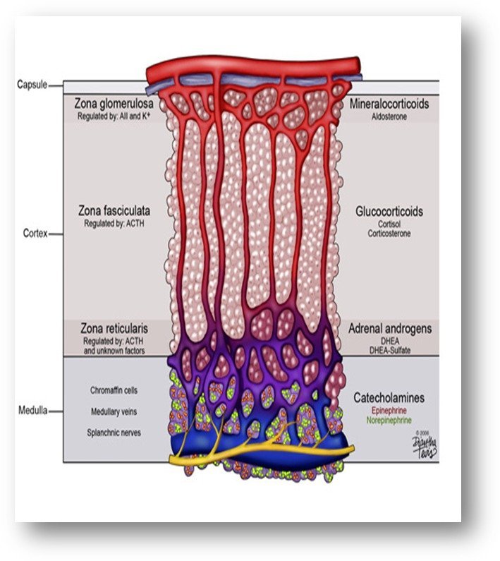
The adrenal gland is composed of two distinct tissues: the outer cortex and the inner medulla. The adrenal cortex tends to be fattier and thus has a more yellow hue. The adrenal medulla is more of a reddish-brown color. A thick capsule consisting of connective tissue surrounds the entire adrenal gland.
The adrenal cortex is much larger than the smaller medulla, which only accounts for approximately 15% of the gland. It is composed of three distinct zones:
Zona Glomerulosa (outer layer)
-
The zona glomerulosa is responsible for the synthesis of mineralocorticoids, of which the most important is aldosterone. This hormone plays an important role in electrolyte balance and regulation of blood pressure.
Zona Fasciculata (middle layer)
-
The zona fasciculata produces glucocorticoids, of which the predominant hormone is cortisol. This hormone plays a role in the regulation of blood sugar via gluconeogenesis. Cortisol also modulates the immune system and modulates the metabolism of fat, protein, and carbohydrates. The secretion of cortisol is under the adrenocorticotropic hormone regulation, which is released from the pituitary gland.
Zona Reticularis (inner zone)
-
The zona reticularis produces androgens and plays a role in the development of secondary sexual characteristics. The primary androgen produced in the zona reticularis is dehydroepiandrosterone (DHEA), which is the most abundant hormone in the body. It serves as a precursor for the synthesis of many other hormones produced by the adrenal gland, such as progesterone, estrogen, cortisol, and testosterone.
The function of these three zones can be remembered by the mnemonic “Salt, Sugar, Sex,” as they correlate to the function of the hormones produced in each layer of the adrenal cortex. The names of these zones can also be recalled by remembering “GFR” for glomerulosa, fasciculata, and reticularis.
The adrenal medulla synthesizes catecholamines. Catecholamines are made from the precursor of dopamine and combined with tyrosine, thus resulting in norepinephrine. Once norepinephrine has been created, it is then methylated via phenylethanolamine N-methyltransferase (PNMT), which is only present in the adrenal medulla.[rx]
Overview of the Adrenal Glands
In mammals, the adrenal glands (also known as the suprarenal glands) are endocrine glands that sit atop the kidneys.
Key Points
- The adrenal glands are are endocrine glands located atop the kidneys.
- They are responsible for releasing three classes of hormones;
mineralocorticoids, glucocorticoids, and androgens along with
catecholamines. - Each adrenal gland is composed to two structures: the adrenal medulla and adrenal cortex.
Key Terms
- adrenal cortex: The outer portion of the adrenal glands that produces
mineralocorticoids, glucocorticoids, and androgens. - Adrenal medulla: The innermost part of the adrenal gland, consisting of cells that secrete adrenaline and noradrenaline.
In mammals, the adrenal glands (also known as the suprarenal glands) are endocrine glands that sit atop the kidneys. They are chiefly responsible for releasing three classes of hormones:
- Mineralocorticoids (aldosterone)
- Glucocorticoids (cortisol)
- Androgens (DHEA)
Along with catecholamines (adrenaline), these hormones control a variety of functions including kidney function, metabolism, fight-or-flight response, and sex hormone levels.
In humans, the adrenal glands are found at the level of the 12th thoracic vertebra sitting above and slightly medial to the kidneys, lying within the renal fascia, and separated from the kidneys by a thin layer of connective tissue. In humans, the right adrenal gland is triangular shaped, while the left adrenal gland is semilunar shaped.
Each adrenal gland has two distinct structures, the outer adrenal cortex and the inner medulla—both produce hormones. The cortex mainly produces mineralcorticoids, glucocorticoids, and androgens, while the medulla chiefly produces adrenaline and nor-adrenaline.
Adrenal Cortex
The adrenal cortex is devoted to the synthesis of corticosteroid and androgen hormones.
Key Points
Specific cortical cells produce particular hormones, including aldosterone, cortisol, and androgens such as androstenedione.
The adrenal cortex comprises three zones, or layers: Zona glomerulosa (outer), Zona fasciculata and Zona reticularis.
The outermost layer, the zona glomerulosa, is the main site for the production of mineralocorticoids, mainly aldosterone.
Zona fasciculata is the layer situated between the glomerulosa and reticularis. This layer is responsible for producing glucocorticoids, such as cortisol.
Zona reticularis is the innermost cortical layer; the zona reticularis produces androgens, mainly DHEA.
Key Terms
adrenal cortex: The outer portion of the adrenal glands that produces hormones essential to homeostasis.
zona glomerulosa: The outermost layer of the adrenal cortex, responsible for producing mineralocorticoids such as aldosterone.
zona fasciculata: The middle layer of the adrenal cortex, responsible for producing glucocorticoids such as cortisol.
zona reticularis: The inner most layer of the adrenal cortex, responsible for producing androgens such as DHEA.
Zones of the Adrenal Cortex
The cortex is regulated by neuroendocrine hormones secreted by the pituitary gland, which are under the control of the hypothalamus, as well as by the renin-angiotensin system. The adrenal cortex has three zones or layers:
- Zona Glomerulosa – The outermost layer, the zona glomerulosa, is the main site for the production of mineralocorticoids, mainly aldosterone, that are largely responsible for the long-term regulation of blood pressure. Aldosterone exerts its effects on the distal convoluted tubule and collecting duct of the kidney, where it causes increased reabsorption of sodium and increased excretion of both potassium (by principal cells) and hydrogen ions (by intercalated cells of the collecting duct). The major stimulus to produce aldosterone is angiotensin II, as ACTH from the pituitary only produces a transient effect. Angiotensin is stimulated by the juxtaglomerular cells when renal blood pressure drops below 90 mmHg.
- Zona fasciculata – Zona fasciculata is the layer situated between the glomerulosa and reticularis. This layer is responsible for producing glucocorticoids, such as 11-deoxycorticosterone, corticosterone, and cortisol in humans. Cortisol enhances the activity of other hormones including glucagon and catecholamines.
- Zona Reticularis – The innermost cortical layer, the zona reticularis, lies directly adjacent to the medulla. It produces androgens, mainly dehydroepiandrosterone (DHEA), DHEA sulfate (DHEA-S), and androstenedione (the precursor to testosterone) in humans.[19] Its small cells form irregular cords and clusters, separated by capillaries and connective tissue. The cells contain relatively small quantities of cytoplasm and lipid droplets and sometimes display brown lipofuscin pigment.[rx]
Hormones of the Adrenal Cortex
The different zones of the adrenal cortex produce different hormones.
Mineralocorticoids
These are produced in the zona glomerulosa. The primary mineralocorticoid is aldosterone. Its secretion is regulated by the oligopeptide angiotensin II. Aldosterone is secreted in response to high extracellular potassium levels, low extracellular sodium levels, and low fluid levels and blood volume. Aldosterone secretion affects metabolism in different ways:
- It increases urinary excretion of potassium ions
- It increases interstitial levels of sodium ions
- It increases water retention and blood volume.
Glucocorticoids
These are produced in the zona fasciculata. The primary glucocorticoid released by the adrenal gland in humans is cortisol. Its secretion is regulated by the hormone ACTH from the anterior pituitary gland. Upon binding to its target, cortisol enhances metabolism in several ways:
- It stimulates the release of amino acids from the body
- It stimulates lipolysis, the breakdown of fat
- It stimulates gluconeogenesis, the production of glucose from newly-released amino acids and lipids
- It increases blood glucose levels in response to stress, by inhibiting glucose uptake into muscle and fat cells
- It strengthens cardiac muscle contractions
- It increases water retention
- It has anti-inflammatory and anti-allergic effects.
Androgens
The zona reticularis produces androgens, the most important of which is
DHEA. In general, these hormones do not have an overall effect in the male body and are converted to more potent androgens such as testosterone and DHT or to estrogens (female sex hormones) in the gonads, acting in this way as a metabolic intermediate.
Adrenal Medulla
The adrenal cortex is devoted to the synthesis of corticosteroid and androgen hormones.
Key Points
The adrenal medulla secretes two water-soluble hormones (norepinephrine and epinephrine) that underly the fight-or-flight response.
To carry out this responsibility, the adrenal medulla receives input from the sympathetic nervous system.
Key Terms
adrenal cortex: The outer portion of the adrenal glands that produces hormones essential to homeostasis.
chromaffin: The cells of the adrenal medulla that secrete adrenaline and noradrenaline in response to nervous stimulation.
The adrenal medulla is the core of the adrenal glands, and is surrounded by the adrenal cortex. The adrenal medulla is responsible for the production of catecholamines, derived from the amino acid tyrosine.
Chromaffin cells are the neuroendocrine cells found in the medulla; they are modified post-synaptic sympathetic neurons that receive sympathetic input. These water-soluble hormones are the major hormones underlying the fight-or-flight response. The adrenal medulla secretes approximately 20% noradrenaline (norepinephrine) and 80% adrenaline (epinephrine). To carry out this responsibility, the adrenal medulla receives input from the sympathetic nervous system through nerve fibers originating in the thoracic spinal cord from T5–T11.
When stimulated, chromaffin cells secrete adrenaline and noradrenaline along with enkephalin and enkephalin-containing peptides into the bloodstream. The secreted adrenaline and noradrenaline play an important role in the fight-or-flight response.
The enkephalins and enkephalin-containing peptides are related to, but also distinct from, the endogenous peptides named endorphins (secreted from the pituitary). All of these peptides bind to opioid receptors and produce analgesic (and other) responses.
Some notable effects of adrenaline and noradrenaline include:
- Increased heart rate and blood pressure.
- Blood vessel constriction in the skin and gastrointestinal tract.
- Smooth muscle dilation.
- Dilating bronchioles and capillaries.
- Increased metabolism.
All of these effects are characteristic of the fight-or-flight response. Receptors for catecholamines are widely distributed throughout the body to allow for a systemic response following secretion.
Blood Supply and Lymphatics
As the adrenal glands produce various systemically important hormones, they require significant blood supply and are extremely well vascularized. The blood supply is tightly controlled by neuroendocrine and paracrine mechanisms, which is one method of regulating the systemic levels of adrenal hormones.[rx]
The three chief sources of blood supply to the adrenal glands include:
-
The superior adrenal arteries, which are small branches coming off the inferior phrenic artery
-
The middle adrenal artery comes directly off the abdominal aorta.
-
The inferior adrenal artery originates from the renal artery bilaterally[rx]
The adrenal glands have one of the greatest blood supply rates per gram of tissue of any organ: up to 60 small arteries may enter each gland.[rx] Three arteries usually supply each adrenal gland:[rx]
- The superior suprarenal artery, a branch of the inferior phrenic artery
- The middle suprarenal artery, a direct branch of the abdominal aorta
- The inferior suprarenal artery, a branch of the renal artery
These blood vessels supply a network of small arteries within the capsule of the adrenal glands. Thin strands of the capsule enter the glands, carrying blood to them.[rx]
Venous blood is drained from the glands by the suprarenal veins, usually one for each gland:[rx]
- The right suprarenal vein drains into the inferior vena cava
- The left suprarenal vein drains into the left renal vein or the left inferior phrenic vein.
The central adrenomedullary vein, in the adrenal medulla, is an unusual type of blood vessel. Its structure is different from the other veins in that the smooth muscle in its tunica media (the middle layer of the vessel) is arranged in conspicuous, longitudinally oriented bundles.[rx]
Variations in adrenal artery origin are very common. The superior adrenal artery can come off the abdominal aorta, celiac axis, or, more rarely, an intercostal artery. The superior adrenal artery is also commonly found as multiple arteries. The middle adrenal artery can come off the inferior phrenic, renal, or superior mesenteric arteries, or the celiac axis. The inferior adrenal arteries can also originate from the abdominal aorta or the inferior phrenic artery.[rx][rx][rx][rx]
The venous drainage from the adrenal glands is dependent on the side of the gland. The left adrenal gland is anatomically further away from the inferior vena cava, and therefore the left adrenal vein drains into the left renal vein. The right adrenal vein is much closer to the inferior vena cava and drains directly into this large vessel. Variations in adrenal venous drainage are common, particularly on the left side. There are reports of venous connections between the left adrenal vein and the left genital vein, and the inferior phrenic vein. Double left adrenal veins are also common.[rx]
Nerves
The function of the adrenal gland is mediated by both synaptic stimulation and hormonal stimulation. Adrenocorticotropic hormone (ACTH) secreted from the anterior pituitary gland activates the adrenal cortex. Subsequently, ACTH activates the respective cortical zones to generate corticosteroids.[rx][rx] However, the adrenal medulla is innervated by preganglionic nerve fibers (type B) arising from the intermediolateral cell column of the spinal cord’s lateral horn from the T5–T8 spinal cord segments. These nerve fibers form the greater splanchnic nerve without entering the paravertebral sympathetic ganglion chain. Some of the nerve fibers from the greater splanchnic nerve synapse at the celiac ganglion. The blood vessels supplying the adrenal glands will then receive their innervation from the celiac ganglion’s postsynaptic fibers.
On the other hand, some fibers of the greater splanchnic nerve circumvent the celiac ganglion and directly enter the adrenal gland to synapse at the chromaffin cells’ membranes. This is the reason why the adrenal medulla acts as a neuroendocrine junction between the two physiological segments. The chromaffin cells release their neurohormones directly into the bloodstream to produce a widespread sympathetic response and apparently act as a special type of postsynaptic neuron.[rx][rx]
Function
Mineralocorticoids
The mineralocorticoids, which include corticosterone, 11-deoxycorticosterone, and more importantly, aldosterone, act on the kidney to increase sodium reabsorption and potassium excretion. Water reabsorption follows increased sodium reabsorption, resulting in an increase in effective circulating volume and therefore increased blood pressure. Specifically, mineralocorticoids achieve this via increased synthesis of epithelial sodium channels (ENaC) and sodium-potassium ATPases on the principal cells of the distal nephron.[rx]
Mineralocorticoids also promote potassium ion secretion at the principal cells because of the gradients produced by the above channels. In high potassium states, aldosterone synthesis is increased to promote potassium excretion. Lastly, mineralocorticoids promote hydrogen ion secretion at the intercalated cells.[rx]
Interestingly, 11-deoxycorticosterone and corticosterone also have mineralocorticoid effects. These are weaker than aldosterone but can produce a strong mineralocorticoid effect when present in excess levels, as in some forms of congenital adrenal hyperplasia (CAH), for example, 11-beta-hydroxylase deficiency resulting in hypertension.[rx]
Glucocorticoids
Cortisol is the major glucocorticoid and increases in response to stress which activates the HPA axis. Therefore, all of its functions can be thought of as allowing the body to function with increased stress. Upon engaging glucocorticoid receptors, cortisol increases the expression of genes that will regulate metabolism, the immune system, cardiovascular function, growth, and reproduction. Cortisol is essential for maintaining blood pressure because it increases the sensitivity of vascular smooth muscle to vasoconstrictors like catecholamines and suppresses the release of vasodilators like nitrous oxide.[rx] Cortisol suppresses the immune system, which is the basis for immunosuppressive drug therapy with glucocorticoids. Regarding metabolism, cortisol increases gluconeogenesis and decreases peripheral glucose uptake. These oppose the actions of insulin, and the net effect is an increase in serum glucose. Cortisol also activates lipolysis and stimulates adipocyte growth, which leads to fat deposition. Generally, growth is inhibited, leading to muscle atrophy, increased bone resorption, and thinning of the skin. Of note, glucocorticoids can act on mineralocorticoid receptors. However, aldosterone effects predominate in the kidney because the renal enzyme, 11-beta-hydroxysteroid dehydrogenase-2 (11-beta-HSD-2) converts cortisol to cortisone.[rx] The 11-beta-HSD-1 converts cortisone into cortisol. Hence, these enzymes add another layer of regulation to cortisol. Licorice toxicity inhibits 11-beta-HSD-2, causing hypertension and hypokalemic alkalosis with normal aldosterone levels. Also, there can be a loss of function mutations in 11-beta-HSD-2, resulting in hypertension with low aldosterone.[rx]
Androgens
The adrenal androgens, primarily DHEA, require peripheral conversion to active sex steroids in the gonads and peripheral tissue. Circulating DHEA-sulfate is the best measure of adrenal androgen excess. Some DHEA is also converted to androstenedione. Ultimately, both are converted to testosterone in peripheral tissues, which is converted to 5-alpha-dihydrotestosterone (DHT), the most potent androgen.[rx] Adrenal androgens do not play a major role in the adult male because the testes are the major source of testosterone. However, adrenal androgens are important in puberty for both males and females and are the main source of circulating testosterone in females. The rise in adrenal gland androgen synthesis is responsible for adrenarche, which precedes gonadarche.[rx]
Catecholamines
Adrenal catecholamines, epinephrine, and norepinephrine are involved in executing the fight-or-flight response of the sympathetic nervous system. They increase blood pressure via alpha-1 receptors on vascular smooth muscle. They help increase serum glucose by activating glycogenolysis and increasing glucagon secretion via beta-2 receptors and decreasing insulin secretion via alpha-2 receptors.[rx]
Hormones of the Adrenal Glands
The role of the adrenal glands in your body is to release certain hormones directly into the bloodstream. Many of these hormones have to do with how the body responds to stress, and some are vital to existence. Both parts of the adrenal glands — the adrenal cortex and the adrenal medulla — perform distinct and separate functions.
Each zone of the adrenal cortex secretes a specific hormone. The key hormones produced by the adrenal cortex include:
Cortisol
Cortisol is a glucocorticoid hormone produced by the zona fasciculata that plays several important roles in the body. It helps control the body’s use of fats, proteins and carbohydrates; suppresses inflammation; regulates blood pressure; increases blood sugar; and can also decrease bone formation.
This hormone also controls the sleep/wake cycle. It is released during times of stress to help your body get an energy boost and better handle an emergency situation.
How Adrenal Glands Work to Produce Cortisol
Adrenal glands produce hormones in response to signals from the pituitary gland in the brain, which reacts to signaling from the hypothalamus, also located in the brain. This is referred to as the hypothalamic pituitary adrenal axis. As an example, for the adrenal gland to produce cortisol, the following occurs:
- The hypothalamus produces corticotropin-releasing hormone (CRH) that stimulates the pituitary gland to secrete adrenocorticotropin hormone (ACTH).
- ACTH then stimulates the adrenal glands to make and release cortisol hormones into the blood.
- Normally, both the hypothalamus and the pituitary gland can sense whether the blood has the appropriate amount of cortisol circulating. If there is too much or too little cortisol, these glands respectively change the amount of CRH and ACTH that gets released. This is referred to as a negative feedback loop.
- Excess cortisol production can occur from nodules in the adrenal gland or excess production of ACTH from a tumor in the pituitary gland or other source.
Aldosterone
This mineralocorticoid hormone produced by the zona glomerulosa plays a central role in regulating blood pressure and certain electrolytes (sodium and potassium). Aldosterone sends signals to the kidneys, resulting in the kidneys absorbing more sodium into the bloodstream and releasing potassium into the urine. This means that aldosterone also helps regulate the blood pH by controlling the levels of electrolytes in the blood.
DHEA and Androgenic Steroids
These hormones produced by the zona reticularis are weak male hormones. They are precursor hormones that are converted in the ovaries into female hormones (estrogens) and in the testes into male hormones (androgens). However, estrogens and androgens are produced in much larger amounts by the ovaries and testes.
Epinephrine (Adrenaline) and Norepinephrine (Noradrenaline)
The adrenal medulla, the inner part of an adrenal gland, controls hormones that initiate the flight or fight response. The main hormones secreted by the adrenal medulla include epinephrine (adrenaline) and norepinephrine (noradrenaline), which have similar functions.
Among other things, these hormones are capable of increasing the heart rate and force of heart contractions, increasing blood flow to the muscles and brain, relaxing airway smooth muscles, and assisting in glucose (sugar) metabolism. They also control the squeezing of the blood vessels (vasoconstriction), helping maintain blood pressure and increasing it in response to stress.
Like several other hormones produced by the adrenal glands, epinephrine and norepinephrine are often activated in physically and emotionally stressful situations when your body needs additional resources and energy to endure unusual strain.
Adrenal Gland Disorders
The two common ways in which adrenal glands cause health issues are by producing too little or too much of certain hormones, which leads to hormonal imbalances. These abnormalities of the adrenal function can be caused by various diseases of the adrenal glands or the pituitary gland.
Adrenal Insufficiency
Adrenal insufficiency is a rare disorder. It may be caused by disease of the adrenal glands (primary adrenal insufficiency, Addison’s disease) or by diseases in the hypothalamus or the pituitary (secondary adrenal insufficiency). It is the opposite of Cushing syndrome and is characterized by low levels of adrenal hormones. The symptoms include weight loss, poor appetite, nausea and vomiting, fatigue, darkening of skin (only in primary adrenal insufficiency), abdominal pain, among other.
The causes of primary adrenal insufficiency may include autoimmune disorders, fungal and other infections, cancer (rarely), and genetic factors.
Although adrenal insufficiency usually develops over time, it can also appear suddenly as an acute adrenal failure (adrenal crisis). It has similar symptoms, but the consequences are more serious, including life-threatening shock, seizures, and coma. These may develop if the condition is left untreated.
Congenital Adrenal Hyperplasia
Adrenal insufficiency can also result from a genetic disorder called congenital adrenal hyperplasia. Children who are born with this disorder are missing an essential enzyme necessary to produce cortisol, aldosterone, or both. At the same time, they often experience an excess of androgen, which may lead to male characteristics in girls and precocious puberty in boys.
Congenital adrenal hyperplasia can remain undiagnosed for years depending on the severity of the enzyme deficiency. In more severe cases, infants may suffer from ambiguous genitalia, dehydration, vomiting, and failure to thrive.
Overactive Adrenal Glands
Sometimes, adrenal glands may develop nodules that produce too many of certain hormones. Nodules 4 centimeters or larger and nodules that show certain features on imaging increase suspicion for malignancy. Both benign and cancerous nodules may produce excessive amounts of certain hormones, which is referred to as a functional nodule. Functional tumors, malignant tumors or nodules greater than 4 centimeters are recommended to be referred for surgical evaluation.
Excess of Cortisol: Cushing Syndrome
Cushing syndrome results from excessive production of cortisol from the adrenal glands. The symptoms may include weight gain and fatty deposits in certain areas of the body, such as the face, below the back of the neck called a buffalo hump and in the abdomen; thinning arms and legs; purple stretch marks on the abdomen; facial hair; fatigue; muscle weakness; easily bruised skin; high blood pressure; diabetes; and other health issues.
Excess cortisol production can also be triggered by overproduction of ACTH by a benign tumor in the pituitary gland or tumor elsewhere in the body. This is known as Cushing’s Disease. Another common cause of Cushing’s syndrome is excessive and prolonged consumption of external steroids, such as prednisone or dexamethasone, which are prescribed to treat many autoimmune or inflammatory diseases (e.g., lupus, rheumatoid arthritis, asthma, inflammatory bowel disease, multiple sclerosis, etc.)
Excess of Aldosterone: Hyperaldosteronism
Hyperaldosteronism results from the overproduction of aldosterone from one or both adrenal glands. This is characterized by an increase in blood pressure that often requires many medications to control. Some people can develop low potassium levels in the blood, which can cause muscle aches, weakness, and spasms. When the cause is adrenal oversecretion, the disease is called Conn syndrome.
Excess of Adrenaline or Noradrenaline: Pheochromocytoma
Pheochromocytoma is a tumor that results in excess production of adrenaline or noradrenaline by the adrenal medulla that often happens in bursts. Occasionally, neural crest tissue, which has similar tissue to the adrenal medulla, may be the cause of the overproduction of these hormones. This is known as a paraganglioma.
Pheochromocytomas may cause persistent or sporadic high blood pressure that may be difficult to control with regular medications. Other symptoms include headaches, sweating, tremors, anxiety, and rapid heartbeat. Some people are genetically predisposed to developing this type of tumor.
Adrenal Cancer
Malignant adrenal tumors (adrenal cancer), such as adrenocortical carcinoma, are rare and often have spread to other organs and tissues by the time they are diagnosed. These tumors tend to grow fairly large and can reach several inches in diameter.
Cancerous adrenal tumors can be functional and release an excess of one or more hormones accompanied by corresponding symptoms, as listed above. Patients may also experience abdominal pain, flank pain or a feeling of abdominal fullness, especially when the adrenal tumor gets very large.
Not all cancers found in adrenal glands originate from the gland itself. The majority of adrenal tumors are metastasis, or cancer spread, from another primary tumor elsewhere in the body.
References


