
Bell’s Palsy – Types, Causes, Symptoms, Diagnosis, Treatment

Patient Tools

Read, save, and share this guide

Use these quick tools to make this medical article easier to read, print, save, or share with a family member.

Medical guide Health (A - Z) Jul 5, 2026 59 reads
Related reading

Patient Mode

Understand this article easily

Switch between simple English and easy Bangla patient notes. This is for education and does not replace a doctor consultation.

Bell's palsy is an idiopathic, acute, unilateral paresis or paralysis of the face in a pattern consistent with peripheral facial nerve dysfunction, and may be partial or complete, occurring with equal frequency on the right and left sides of the face. There is evidence that...

For severe symptoms, danger signs, pregnancy, child illness, or sudden worsening, seek urgent medical care.

বাংলা রোগী নোট এখনো যোগ করা হয়নি। পোস্ট এডিটরে “RX Bangla Patient Mode” বক্স থেকে সহজ বাংলা সারাংশ যোগ করুন।

এই তথ্য শিক্ষা ও সচেতনতার জন্য। এটি ডাক্তারি পরীক্ষা, রোগ নির্ণয় বা প্রেসক্রিপশনের বিকল্প নয়।

Article Summary

Bell's palsy is an idiopathic, acute, unilateral paresis or paralysis of the face in a pattern consistent with peripheral facial nerve dysfunction, and may be partial or complete, occurring with equal frequency on the right and left sides of the face. There is evidence that Bell's palsy is caused by herpes viruses. Additional symptoms of Bell's palsy may include mild pain in or behind the...

Key Takeaways

  • This article explains Types of Bell’s Palsy in simple medical language.
  • This article explains Causes of Bell's palsy in simple medical language.
  • This article explains Symptoms of Bell's palsy in simple medical language.
  • This article explains Diagnosis of Bell's Palsy in simple medical language.
Educational health guideWritten for patient understanding and clinical awareness.
Reviewed content workflowUse writer and reviewer profiles for stronger trust.
Emergency safety firstUrgent warning signs are highlighted below.
Choose your reading view

Patient View highlights a simple learning journey. Clinical View reveals structure, evidence, and editorial completeness.

Seek urgent medical care if you notice

These warning signs are general safety guidance. Local emergency numbers and clinical judgment should always come first.

  • Severe symptoms, breathing difficulty, fainting, confusion, or rapidly worsening illness.
  • New weakness, severe pain, high fever, or symptoms after a serious injury.
  • Any symptom that feels urgent, unusual, or unsafe for the patient.
1

Emergency now

Use emergency care for severe, sudden, rapidly worsening, or life-threatening symptoms.

2

See a doctor

Book a professional medical evaluation if symptoms persist, worsen, recur often, affect daily activities, or occur in a high-risk patient.

3

Learn safely

Use this article to understand possible causes, tests, treatment options, prevention, and questions to ask your clinician.

Before reading

RX Patient Tools

Use these quick guides before reading the article, or return to them when you need help preparing questions for a doctor.

Start here Choose the right pathway for symptoms, reports, medicines, or urgent warning signs. Disease article roadmap Read this topic step by step: meaning, symptoms, warning signs, diagnosis, treatment, prevention, and follow-up. Treatment planner Prepare questions about treatment choices, benefits, risks, side effects, and follow-up. Family & caregiver guide Organize symptoms, reports, medicines, questions, and follow-up safely. Nutrition & diet guide Prepare food, hydration, supplement, and medicine-timing questions safely. Prevention guide Organize risk factors, protective habits, screening, and warning signs. Recovery guide Prepare a safe plan for activity, rehabilitation, warning signs, and follow-up.
Definition

Bell’s palsy is an idiopathic, acute, unilateral paresis or paralysis of the face in a pattern consistent with peripheral facial nerve dysfunction, and may be partial or complete, occurring with equal frequency on the right and left sides of the face. There is evidence that Bell’s palsy is caused by herpes viruses. Additional symptoms of Bell’s palsy may include mild pain in or behind the ear, oropharyngeal or facial numbness, impaired tolerance to ordinary levels of noise, and disturbing taste on the anterior part of the tongue. Severe pain is more suggestive of herpes zoster virus infection (shingles) and possible progression to a Ramsay Hunt syndrome, but another cause should be carefully excluded. Up to 30% of people with acute peripheral facial palsy will not have Bell’s palsy; other causes may include stroke, tumor, trauma, middle ear disease, and Lyme disease.

Bell’s palsy is an unexplained episode of facial muscle weakness or paralysis that begins suddenly and worsens over three to five days. This condition results from damage to the 7th (facial) cranial nerve, and pain and discomfort usually occur on one side of the face or head.

Bell’s palsy is the most common peripheral paralysis of the seventh cranial nerve with an onset that is rapid and unilateral. The diagnosis is one of exclusion and most often made on a physical exam. The facial nerve has both an intracranial, intratemporal, and extratemporal course as its branches. The facial nerve has a motor and parasympathetic function as well as taste to the anterior two-thirds of the tongue. It also controls salivary and lacrimal glands. The motor function of the peripheral facial nerve controls the upper and lower facial muscles. As a result, the diagnosis of Bell’s palsy requires special attention to forehead muscle strength. If forehead strength is preserved, a central cause of weakness should be considered. Although the utility of antivirals has been called into question, treatment is medical with most sources recommending a combination of corticosteroids and antiviral medication.

The Others Names of Bell’s palsy

  • Antoni’s Palsy
  • Facial Nerve Palsy
  • Facial Paralysis
  • Idiopathic Facial Palsy
  • Refrigeration Palsy
  • Idiopathic peripheral facial palsy; Cranial mononeuropathy – Bell palsy

Types of Bell’s Palsy

Bell’s palsy types by severity

Bell’s palsy is often classified by the degree of facial weakness, using the House-Brackmann scale, which ranges from Grade I (normal) to Grade VI (complete paralysis). Clinicians group these into three types:

  1. Mild Bell’s Palsy – House-Brackmann Grades I–II (Mild Dysfunction): Slight weakness noticeable on close inspection, with mostly normal symmetry at rest. Patients can close the eye completely and have good forehead movement Iowa Head and Neck Protocols. This type often resolves quickly and leaves no long-term effects.
  2. Moderate Bell’s Palsy – House-Brackmann Grades III–IV (Moderate Dysfunction): Obvious but not disfiguring weakness, with incomplete eye closure and moderate forehead movement (Grade III) or significant asymmetry and incomplete closure (Grade IV) Medscape. Recovery may take longer and sometimes leaves mild facial asymmetry.
  3. Severe Bell’s Palsy – House-Brackmann Grades V–VI (Severe Dysfunction to Total Paralysis): Barely perceptible motion (Grade V) or no movement at all (Grade VI). Patients cannot close the eye or move the mouth on the affected side. This type carries the highest risk of incomplete recovery without treatment NCBI.

Bell’s Palsy Types by above all conditions

While Bell’s palsy itself refers specifically to idiopathic (unknown-cause) facial nerve paralysis, healthcare professionals often describe subtypes or classifications by severity and presentation:

  • Incomplete vs. Complete Paralysis

    • Incomplete means some facial movement remains; you can still smile a bit or close your eye halfway.

    • Complete means nearly total loss of movement on the affected side.

  • Acute vs. Chronic

    • Acute refers to sudden-onset cases, most typical of Bell’s palsy.

    • Chronic describes facial nerve weakness lasting more than six months, which may hint at other causes.

  • Recurrent Bell’s Palsy

    • A small number of people (about 7%) experience Bell’s palsy more than once. Recurrent episodes may affect the same or opposite side.

  • Bilateral Bell’s Palsy

    • Very rare (fewer than 1% of cases), both sides of the face are affected, often prompting investigation into other conditions.

  • Severity Grading (House-Brackmann Scale)

    • Grade I: Normal function

    • Grade II: Mild dysfunction

    • Grade III: Moderate dysfunction

    • Grade IV: Moderately severe dysfunction

    • Grade V: Severe dysfunction

    • Grade VI: Total paralysis

Each grade helps doctors describe how weak the facial muscles are and track recovery over time.

Causes of Bell’s palsy

Although labeled “idiopathic” (unknown cause), research points to multiple triggers that can inflame or damage the facial nerve. Below are 20 possible causes, each explained in simple terms:

  1. Herpes Simplex Virus Type 1 (HSV-1): This common cold sore virus can hide in nerve cells and reactivate, causing infection, or irritation, often causing pain, swelling, heat, or redness. সহজ বাংলা: শরীরের প্রদাহ; ব্যথা, ফোলা বা লালভাব হতে পারে।" data-rx-term="inflammation" data-rx-definition="Inflammation is the body’s response to injury, infection, or irritation, often causing pain, swelling, heat, or redness. সহজ বাংলা: শরীরের প্রদাহ; ব্যথা, ফোলা বা লালভাব হতে পারে।">inflammation of the facial nerve and leading to weakness Medscape.

  2. Varicella Zoster Virus (VZV): When this chickenpox virus reactivates in the nerve, it causes Ramsay Hunt syndrome, which includes facial paralysis plus painful ear rash Medscape Reference.

  3. Epstein-Barr Virus (EBV): Known for causing mononucleosis, EBV can sometimes trigger Bell’s palsy by inflaming nerve tissue, as seen in rare teen cases Medscape.

  4. Lyme Disease: The bacterium Borrelia burgdorferi, spread by ticks, can infect the facial nerve and cause palsy, especially in areas where Lyme is common Medscape.

  5. Herpes Zoster Oticus (Ramsay Hunt Syndrome): A variant of VZV infection in the ear canal that inflames the facial nerve, causing paralysis, ear pain, and blisters Medscape Reference.

  6. Influenza and Other Respiratory Viruses: Cold and flu viruses can trigger immune reactions that inflame the facial nerve, though this link is less direct Mayo Clinic.

  7. Upper Respiratory Infections: Recent colds or sinus infections may precede Bell’s palsy by causing body-wide infection, or irritation, often causing pain, swelling, heat, or redness. সহজ বাংলা: শরীরের প্রদাহ; ব্যথা, ফোলা বা লালভাব হতে পারে।" data-rx-term="inflammation" data-rx-definition="Inflammation is the body’s response to injury, infection, or irritation, often causing pain, swelling, heat, or redness. সহজ বাংলা: শরীরের প্রদাহ; ব্যথা, ফোলা বা লালভাব হতে পারে।">inflammation that affects the facial nerve Mayo Clinic.

  8. Ear Infections (Otitis Media): Severe middle ear infections can spread inflammation to the facial nerve as it passes near the ear cavity Medscape.

  9. Trauma: Injury to the skull base, temporal bone fractures, or surgical injury near the facial nerve can lead to temporary or permanent palsy Medscape.

  10. Tumors: Growths along the facial nerve pathway—such as acoustic neuromas or parotid gland tumors—can press on the nerve and cause palsy Medscape.

  11. Diabetes Mellitus: High blood sugar can damage small blood vessels and nerves, increasing the risk of Bell’s palsy through diabetic neuropathy Medscape.

  12. Hypertension: Chronic high blood pressure may weaken small blood vessels supplying the facial nerve, leading to ischemia and injury NCBI.

  13. Pregnancy: Hormonal changes and fluid balance shifts—especially in the third trimester—can make nerve swelling more likely, causing palsy NCBI.

  14. Preeclampsia: This pregnancy complication marked by high blood pressure and organ dysfunction can impair blood flow to the facial nerve NCBI.

  15. Sarcoidosis: An autoimmune disease causing small clumps of inflammatory cells can affect the facial nerve, leading to palsy Medscape.

  16. Guillain-Barré Syndrome: A rapid nerve-damaging autoimmune reaction can sometimes present with facial paralysis among its early signs Medscape.

  17. Multiple Sclerosis: Demyelination in the brainstem can rarely affect the facial nerve nucleus, causing central facial palsy that mimics Bell’s palsy Medscape.

  18. Lyme Co-Infection (Babesiosis, Bartonellosis): Tick-borne infections other than Borrelia sometimes accompany Lyme disease and can inflame facial nerves Medscape.

  19. Cold Exposure: Sudden chilling of the face or drafts—sometimes called “cold palsy”—may trigger nerve swelling in susceptible people Mayo Clinic.

  20. Idiopathic (Unknown): In many cases, no clear cause is found, and the palsy resolves on its own; this unexplained form remains true Bell’s palsy Medscape.

What other causes of facial weakness are there?

  • Ramsay-Hunt Syndrome – Viral infection with the chickenpox virus, herpes zoster, which is shingles of the facial nerve. There is usually significant pain and a rash in the ear. Other cranial nerves may be affected leading to hearing loss balance disturbance and numbness.
  • Skull fractures involving the temporal bone – the bone that contains the middle ear. The facial nerve travels through this bone on its way to the facial muscles and so maybe damaged usually with significant trauma to the head.
  • High neck injuries, particularly a stab wound to the upper neck.
  • Tumors anywhere along the course of the facial nerve, which includes the base of the skull, the ear, and the parotid gland just in front of the ear. These will usually present with a gradual loss of weakness to distinguish them from Bell’s palsy
  • Middle ear disease, especially cholesteatoma.
  • Damage to the facial nerve during ear surgery.
  • Loss of power in one side of the face can be a symptom of a stroke but there will be other features present, such as loss of power in a limb, which points to the correct diagnosis.

Symptoms of Bell’s palsy

  • Weakness or paralysis of the upper and lower facial muscles of the affected side
  • Drooping of ipsilateral eyelids
  • Inability to close the eye completely
  • Dry eye due to inability to close eyes completely
  • Excessive tearing of the eye (epiphora)
  • Drooping of the corner of the mouth
  • Ipsilateral impaired/loss of taste sensation
  • Difficulty with eating due to ipsilateral muscle weakness causing food to be trapped on the affected side of the mouth 
  • Dribbling of saliva
  • The altered sensation on the affected side of the face 
  • Pain in or behind the ear
  • Increased sensitivity to sound (hyperacusis) on the affected side if stapedius muscle is involved
  • upper motor neuron lesion – based on innervation, absence of forehead wrinkling is a reliable way of differentiating Bell’s palsy from an upper motor neuron lesion
  • herpes zoster oticus (Ramsay Hunt syndrome)
  • rarer causes including otitis media, HIV infection, sarcoidosis, autoimmune disorders or tumors of the parotid gland.

Bell's Palsy - Types, Causes, Symptoms, Diagnosis, Treatment

Sometimes, you may have a cold shortly before the symptoms of Bell palsy begin. Symptoms most often start suddenly but may take 2 to 3 days to show up. They do not become more severe after that. Many people feel discomfort behind the ear before weakness is noticed. The face feels stiff or pulled to one side, and may look different. Other signs can include:

  1. Twitching
  2. Weakness
  3. Paralysis on one side of the face
  4. Problems smiling or making facial expressions
  5. Difficulty closing your eye
  6. Drooling due to a lack of control over the facial muscles
  7. Difficulty closing one eye
  8. Difficulty eating and drinking; food falls out of one side of the mouth
  9. Drooling due to lack of control over the muscles of the face
  10. Drooping of the face, such as the eyelid or corner of the mouth
  11. Problems smiling, grimacing, or making facial expressions
  12. Twitching or weakness of the muscles in the face

Bell’s palsy symptoms may come on suddenly or develop over a few days. Initial symptoms may include

  1. Pain behind the ear that is followed by weakness and paralysis of the face
  2. Ringing sound in the ear
  3. Slight fever
  4. Slight hearing impairment
  5. A slight increase in sensitivity to sound on the affected side.

Symptoms of full-blown Bell’s palsy may include

  1. Facial weakness or paralysis (look for smooth forehead and problems smiling)—most often on one side
  2. Numbness just before the weakness start
  3. Drooping corner of the mouth
  4. Drooling
  5. Decreased tearing

Inability to close an eye, which can lead to

  1. Dry, red eye
  2. Ulcers forming on the eyes
  3. Infection
  4. Problems with taste
  5. Sound sensitivity in one ear
  6. Earache
  7. Slurred speech

Late symptoms can occur 3-4 months after or second-time onset of Bell’s palsy and can include

  1. Muscle weakness or paralysis
  2. Facial Droop
  3. Impossible or difficult to blink
  4. Difficulty speaking
  5. Difficulty eating and drinking
  6. Nose runs
  7. The nose is constantly stuffed
  8. Difficulty breathing out of nostril on the affected side
  9. Nostril collapse on the affected side
  10. Forehead wrinkles disappear
  11. Sensitivity to sound
  12. Excess or reduced salivation
  13. Facial swelling
  14. Drooling
  15. Diminished or distorted taste
  16. Pain behind ear
  17. Long-lasting tightening of the facial muscles
  18. Tearing from the eye during eating

Diagnosis of Bell’s Palsy

Bell's Palsy - Types, Causes, Symptoms, Diagnosis, Treatment

Diagnosing Bell’s palsy involves ruling out other causes of facial paralysis. Tests fall into five categories:

A. Physical Exam

  1. Cranial Nerve Examination: The doctor checks movement in all facial muscles by asking the patient to smile, raise eyebrows, and close eyes Mayo Clinic.

  2. Ear Inspection: Looking inside the ear canal for vesicles that suggest Ramsay Hunt syndrome Mayo Clinic.

  3. Taste Test: Applying sweet or salty solutions to the front of the tongue to check taste sensation Mayo Clinic.

  4. Corneal Reflex: Gently touching the cornea with a wisp of cotton to see if the eyelid blinks Mayo Clinic.

  5. Hyperacusis Assessment: Placing a stethoscope on the mastoid bone and having the patient listen to a tuning fork to detect increased sound sensitivity Medscape.

  6. Salivary Flow Test: Collecting saliva from each side of the mouth after sour stimulation to compare gland function Medscape.

  7. Stapedial Reflex Test: Measuring the muscle reflex in the middle ear that usually dampens loud sounds Medscape.

  8. Nasal Airflow Test: Checking for symmetry in nasal breathing to assess facial muscle control Mayo Clinic.

  • Facial Symmetry Inspection
    Observe resting face for droop of brow, eyelid, and mouth.

  • Voluntary Movement Testing
    Ask patient to raise eyebrows, close eyes tightly, smile, and puff cheeks to assess muscle function.

  • Bell’s Phenomenon Observation
    Notice upward rotation of the eyeball when attempting eye closure.

  • House-Brackmann Grading
    Rate facial nerve function on a scale from I (normal) to VI (total paralysis).

  • General Neurologic Exam
    Check other cranial nerves, motor strength, and sensation to exclude stroke or central lesions.

Manual Tests

  1. Schirmer’s Test
    A small strip of paper under the lower eyelid measures tear production to detect dry eye risk.

  2. Tinel’s Sign
    Tapping over the stylomastoid foramen (where the facial nerve exits the skull) can reproduce tingling if the nerve is irritated.

  3. Muscle Strength Grading
    The doctor applies light resistance as you try to close your eye or smile to grade muscle power.

  4. Sensory Testing
    Although rare in Bell’s palsy, gentle touch to facial skin checks for numbness or altered sensation.

  5. Corneal Reflex Test
    Touch cornea with a wisp of cotton to assess trigeminal (V) sensory and facial (VII) motor response.

  6. Gustatory Taste Test
    Apply salt or sweet solutions to the tongue’s anterior two-thirds to evaluate taste sensation.

  7. Stapedius Muscle Reflex (Acoustic Reflex)
    Use tuning fork or reflex hammer to assess stapedius contraction in response to loud sound.

  8. Resistance Testing
    Manually resist eyebrow lifting or mouth elevation to grade muscle strength.

C. Laboratory and Pathological Tests

  1. Complete Blood Count (CBC): To look for signs of infection or inflammation Medscape.

  2. Erythrocyte Sedimentation Rate (ESR): A general marker of inflammation that can be elevated in autoimmune or infectious causes Medscape.

  3. Blood Glucose and HbA1c: To screen for diabetes as a risk factor Medscape.

  4. Lyme Serology: Antibody tests for Borrelia burgdorferi if the patient is from an area where Lyme is common Medscape.

  5. HIV and Syphilis Testing: To exclude these infections as causes of facial palsy Medscape.

D. Electrodiagnostic Tests

  1. Electroneuronography (ENoG): Measures the speed and strength of nerve signals along the facial nerve to assess damage Wikipedia.

  2. Electromyography (EMG): Records electrical activity in facial muscles to pinpoint the location and severity of nerve injury Wikipedia.

  3. Maximal and Minimal Stimulation Test (MST): Evaluates the lowest and highest electrical currents needed to produce a muscle response, giving prognostic information Medscape.

  4. Electroneurography (ENoG)
    Records muscle response to a standard electrical stimulus of the facial nerve to quantify damage.

  5. Blink Reflex Study
    Electrical stimulation near the ear triggers a blink; delayed or absent response indicates nerve dysfunction.

E. Imaging Tests

  1. Magnetic Resonance Imaging (MRI) with Gadolinium: Visualizes the facial nerve along its entire course to detect tumors, inflammation, or demyelination .

  2. Computed Tomography (CT) Scan: Helps rule out fractures, bone lesions, or structural causes along the nerve pathway Wikipedia. CT scan —a type of x-ray that uses a computer to make pictures of structures inside the head to see if there is an infection, tumor, bone fracture , or other problem in the area of the facial nerve

  3. The nerve excitability test – determines the excitation threshold by recording the minimum electrical stimulus required to produce visible muscle contraction. A difference greater than 3.5 mA between affected and unaffected sides is considered to be significant in terms of poorer outcomes. Measuring the peak-to-peak amplitude of the evoked compound action potential of the involved side compared to the normal side has prognostic importance. If there is a 90% or greater reduction in the amplitude of the affected side, the prognosis is poor.[]

  4. Currently the trigeminal blink reflex – is the only test to measure the intracranial pathway of the facial nerve and also a useful test to study various post paralysis sequelae such as synkinesis and hemifacial spasms. With the recovery of facial function, the ipsilateral R1 latency becomes less prolonged and the amount of initial prolongation of this response correlates with greater loss of facial motor function.

  5. Gadolinium contrast magnetic resonance (MRI) – study reveals enhancement of internal acoustic meatal segment on the affected side; however, this is a non-specific finding. MRI should not be done routinely and should be the investigation to look for other possible causes for acute facial paralysis especially if there is little or no recovery of function.

Treatment of Bell’s palsy

Non-Pharmacological Treatments

Each of these therapies aims to reduce inflammation, maintain muscle tone, or retrain facial muscles to speed recovery and prevent permanent muscle contractures.

  1. Acupuncture
    Involves inserting fine needles at specific points on the face and body. It may modulate nerve function and reduce pain, though high-quality studies are lacking Frontiers.

  2. Biofeedback Training
    Uses visual or auditory feedback to help patients relearn controlled muscle movements by monitoring muscle activity and guiding relaxation techniques Mayo Clinic.

  3. Botulinum Toxin Injections
    Though a drug, it’s delivered as a procedure: small doses of Botox are injected into unaffected muscles to reduce synkinesis (unwanted muscle contractions) and improve symmetry Mayo Clinic.

  4. Facial Exercise Therapy
    Structured exercises (e.g., smiling, raising eyebrows) maintain muscle tone and stimulate the nerve. Systematic reviews show improved facial function with early exercise AAFP.

  5. Mime Therapy
    A specialized exercise program combining gentle stretch and coordination exercises to improve facial symmetry and emotional expression ScienceDirect.

  6. Massage
    Gentle manual massage of facial muscles and soft tissues reduces stiffness, improves circulation, and can alleviate discomfort Frontiers.

  7. Thermotherapy (Warm Compresses)
    Applying moist heat to the affected side relaxes muscles and eases pain by improving blood flow Wikipedia.

  8. Electrostimulation
    Low-level electrical currents applied to paralyzed muscles may promote nerve conduction and prevent atrophy; evidence is preliminary Frontiers.

  9. Kabat Rehabilitation (PNF Techniques)
    Proprioceptive neuromuscular facilitation techniques use specific patterns of movement and resistance to retrain facial muscle coordination Frontiers.

  10. Hyperbaric Oxygen Therapy
    Breathing pure oxygen in a pressurized chamber may reduce nerve swelling and support healing in severe cases; evidence remains very tentative Wikipedia.

  11. Ultrasound Therapy
    Pulsed ultrasound waves applied to the face enhance circulation and may stimulate nerve repair; studies show benefit when combined with exercise and massage Nose Creek Physiotherapy.

  12. Transcutaneous Electrical Nerve Stimulation (TENS)
    Surface electrodes deliver mild electrical currents to reduce pain and improve muscle activation Nose Creek Physiotherapy.

  13. Eye Protection Measures
    Frequent use of tear‐like eye drops, gel, or ointment during the day and wearing an eye patch or tape at night protects the cornea when blinking is impaired Wikipedia.

  14. Kinesio Taping
    Sports taping techniques applied to facial muscles can reduce synkinesis and guide correct muscle movement MedStar Health.

  15. Breathing and Meditation Practices
    Guided breathwork and mindfulness calm the nervous system, reducing stress-related exacerbations and supporting recovery Facial Palsy UK.

  16. Peer Support and Psychological Interventions
    Group or one-on-one support reduces distress, normalizes the experience, and encourages adherence to therapy Facial Palsy UK.

  17. Yoga
    Gentle yoga poses and stretches improve overall circulation and help manage stress, indirectly supporting nerve healing Number Analytics.

  18. Cognitive-Behavioral Therapy (CBT)
    Addresses anxiety or depression that may accompany Bell’s palsy, improving mental well-being and treatment engagement Number Analytics.

  19. Deep Breathing Exercises
    Simple diaphragmatic breathing sessions reduce muscle tension and stress, promoting a healing environment audreylemd.com.

  20. Low-Impact Motion Exercises (Walking, Stretching)
    General physical activity boosts circulation, which may enhance nutrient delivery to injured nerves Nose Creek Physiotherapy.

  21. Physical Therapy – In Bell’s palsy various physical therapies, such as exercise, biofeedback, laser, electrotherapy, massage, and thermotherapy are used to hasten recovery. However, the evidence for the efficacy of any of these therapies is lacking. Cochrane systemic review of the efficacy of physical therapies, electrostimulation, and exercises, on the outcome of Bell’s palsy, concluded that there was no significant benefit or harm from any of these physical therapies for Bell’s palsy. There was limited evidence that improvement began earlier in the exercise group.[] Another systematic review examined the effects of facial exercises associated either with mirror or electromyogram biofeedback with respect to complications of delayed recovery in Bell’s palsy and concluded that because of the small number of randomized controlled trials, it was not possible to analyze if the exercises, were effective.[] However, the possibility that facial exercise reduces time to recover and sequelae need confirming with good-quality randomized controlled trials.[]

Drug Treatments

These medications target inflammation, viral replication, pain, and prevent corneal damage.

  1. Acyclovir (Antiviral)
    Dosage: 400 mg five times daily for 7 days
    Purpose: Inhibit herpes simplex viral replication
    Mechanism: Guanosine analog terminates viral DNA synthesis
    Side Effects: Headache, nausea Wikipedia

  2. Valacyclovir (Antiviral)
    Dosage: 1 g three times daily for 7 days
    Purpose: Prodrug of acyclovir with better bioavailability
    Mechanism: Converted to acyclovir, then inhibits viral DNA polymerase
    Side Effects: Fatigue, headache Wikipedia

  3. Ibuprofen (NSAID)
    Dosage: 400 mg every 4–6 hours as needed
    Purpose: Relieve pain and mild inflammation
    Mechanism: Inhibits cyclooxygenase (COX) enzymes
    Side Effects: GI upset, renal impairment Mayo Clinic

  4. Artificial Tears (Lubricant Eye Drops)
    Dosage: Instill one drop every 2 hours while awake
    Purpose: Prevent corneal drying and irritation
    Mechanism: Provides tear film supplementation
    Side Effects: Mild eye irritation Wikipedia

  5. Ocular Lubricating Ointment
    Dosage: Apply thin strip to lower eyelid at bedtime
    Purpose: Protect cornea during sleep
    Mechanism: Creates a protective lipid barrier
    Side Effects: Blurred vision upon application Wikipedia

  6. Botulinum Toxin A
    Dosage: 1–10 units injected into overactive facial muscles
    Purpose: Reduce synkinesis and unwanted contractions
    Mechanism: Blocks acetylcholine release at neuromuscular junction
    Side Effects: Injection site pain, temporary weakness Mayo Clinic

  7. Gabapentin (Neuropathic Pain)
    Dosage: Start 300 mg at bedtime, titrate to 900–1,200 mg/day
    Purpose: Alleviate neuropathic pain around the ear
    Mechanism: Modulates α₂δ subunit of voltage-gated calcium channels
    Side Effects: Dizziness, fatigue Dr.Oracle

  8. Topical Lidocaine Gel
    Dosage: Apply to area of pain 3–4 times/day
    Purpose: Local analgesia for ear or facial pain
    Mechanism: Blocks sodium channels in nerve membranes
    Side Effects: Local numbness Wikipedia

  9. Acetaminophen
    Dosage: 500–1,000 mg every 4–6 hours as needed
    Purpose: Pain relief when NSAIDs are contraindicated
    Mechanism: Central COX inhibition and modulation of serotonergic pathways
    Side Effects: Hepatotoxicity in overdose Mayo Clinic

  1. Gabapentin (Anticonvulsant)

    • Class: Calcium channel modulator

    • Dosage & Time: 300 mg three times daily, adjusted for pain control

    • Purpose: Alleviate nerve-related facial pain

    • Mechanism: Modulates α₂δ subunit of voltage-gated calcium channels, reducing excitatory neurotransmitter release

    • Side Effects: Drowsiness, dizziness Wikipedia

  2. Carbamazepine – (Anticonvulsant)200 mg twice daily, max 800 mg/day for severe neuralgic pain; mechanism: blocks high-frequency discharges. S/E: mild liver enzyme rise, sleepiness.

    • Class: Sodium channel blocker

    • Dosage & Time: 100 mg twice daily, titrating up to 200 mg three times daily

    • Purpose: Treat synkinesis and neuropathic pain

    • Mechanism: Stabilizes hyperexcited neural membranes by blocking sodium channels

    • Side Effects: Drowsiness, nausea, risk of blood dyscrasias Wikipedia

  3. Botulinum Toxin Type B

    • Class: Neurotoxin

    • Dosage & Time: 2.5–5 units injected into targeted muscles every 3–4 months

    • Purpose: Reduce unwanted muscle contractions (synkinesis)

    • Mechanism: Blocks acetylcholine release at the neuromuscular junction, relaxing overactive muscles

    • Side Effects: Temporary weakness in nearby muscles, injection site pain Wikipedia

  4. Artificial Tears (Carboxymethylcellulose)

  • Class: Ophthalmic lubricant

  • Dosage & Time: 1–2 drops every 2–4 hours while awake

  • Purpose: Protect cornea when eyelid closure is incomplete

  • Mechanism: Mimics natural tears, maintaining moisture on the ocular surface

  • Side Effects: Mild irritation or blurred vision Wikipedia

  1. Methylcobalamin (active vitamin B12 injection) – 500–1000 µg IM three times a week for 4–8 weeks in people with low B-vitamin levels; nourishes myelin and speeds nerve remyelination. S/E: rare acne-like rash.AAFPPMC
  2. Botulinum toxin type A (chemodenervation) – 2.5-4 units injected into over-active muscles every 3-4 months after the acute phase to treat synkinesis and asymmetry; blocks acetylcholine release. S/E: transient local weakness.PMCPubMed

  3. Artificial-tear ointment with hyaluronic acid (ocular lubricant) – ribbon in lower lid at bedtime; purpose: physical barrier preventing night-time corneal drying; minimal systemic effects.AAO-HNS

  4. Botulinum Toxin (Botox) Injections – These injections give you the option to treat the unaffected or the affected side of your face. These injections can help your facial muscles to relax to reduce unwanted muscle movements. They can also reduce muscle activity in the unaffected side of your face to maintain a balance. Botox injections may also help treat tears when eating and to keep you from winking an eye when smiling, eating, or laughing.
  5. Vitamin therapy – Vitamins B-12, B-6, and zinc may help nerve growth.
  6. Prednisolone – The rationale for the use of corticosteroids in the acute phase of Bell’s palsy is that inflammation and edema of the facial nerve are implicated in causing Bell’s palsy and corticosteroids have a potent anti-inflammatory action which should minimize nerve damage and thereby improve the outcome. Randomized, double-blind, placebo-controlled trials have provided compelling evidence that treatment with prednisolone improves outcomes in patients with Bell’s palsy and shortens the time to complete recovery.[]
  7. Methylprednisolone – should be used in all patients with facial palsy of less than 72 h duration who do not have contraindications to steroid therapy. The prednisolone dose used was 60 mg per day for 5 days then reduced by 10 mg per day (for a total treatment time of 10 days)[] and 50 mg per day (in two divided doses) for 10 days.[] The reported adverse rates were low. Treatment with prednisolone is likely to be cost-effective.[]
  8. Antiviral Agents – The rationale for the use of antiviral agents is the evidence that the inflammation of the facial nerve in Bell’s palsy might be related to the herpes simplex virus (HSV). In an autopsy study, latent HSV type-1 has been isolated from the majority of the geniculate ganglia samples.[] HSV-1 genome was detected in 79% of facial nerve endoneurial fluid in patients with Bell’s palsy, but not in the controls.[] However, the benefit of acyclovir or valacyclovir, either as single agents or in combination with prednisolone in Bell’s palsy has not been definitively established.[,, ] Thus with the available evidence acyclovir or valacyclovir should not be routine and treatment with acyclovir is highly unlikely to be considered cost-effective.[]
  9. Combination therapy – A randomized controlled trial found that at nine months of diagnosis, the facial function had recovered in 94.4% of patients who took prednisolone alone, 85.4% of those who took aciclovir alone, and 92.7% of those who received both. There were no serious adverse effects in any group. The study concluded that early treatment with prednisolone alone increases the likelihood of complete recovery and there was no additional benefit of treatment with aciclovir alone or combining with prednisolone.However, a systematic review also found that treatment with prednisolone reduced the chances of incomplete recovery but using an antiviral drug had an additional benefit.

Dietary Molecular Supplements

These nutrients support nerve health, reduce inflammation, and may inhibit viral triggers.

  1. Vitamin B₁₂ (Methylcobalamin) – 1,000 µg/day oral
    Supports myelin formation and nerve regeneration Life Extension

  2. Acetyl-L-Carnitine – 500 mg twice daily
    Enhances nerve metabolism and repair Life Extension

  3. Curcumin – 500 mg twice daily with meals
    Potent anti-inflammatory and antioxidant action Life Extension

  4. Omega-3 Fatty Acids – 1,000 mg EPA/DHA daily
    Modulates inflammatory cytokines, supports neuron membrane fluidity Life Extension

  5. Ginkgo Biloba – 120 mg/day in divided doses
    Increases microcirculation and may protect nerve cells Life Extension

  6. L-Lysine – 1,000 mg/day
    May inhibit herpes virus replication Life Extension

  7. Zinc – 25 mg/day
    Essential for immune function and antioxidant enzyme activity Life Extension

  8. Reishi Mushroom Extract – 1,000 mg/day
    Immunomodulatory and antiviral properties Life Extension

  9. Vitamin C – 500 mg twice daily
    Antioxidant that supports collagen and nerve repair PMC

  10. Magnesium – 300 mg/day
    Supports nerve conduction and reduces muscle excitability Life Extension


Regenerative and Stem Cell Therapies

These emerging treatments aim to harness cell-based repair mechanisms for facial nerve recovery.

  1. Uncultured Umbilical Cord-Derived MSC Transplantation
    Eight injections of allogeneic MSCs directly into the affected facial region over 32 months led to complete recovery in a 7-year chronic case by promoting regeneration and reducing inflammation PubMed.

  2. Adipose-Derived Stem Cell (ADSC) Therapy
    Single sentinel case used autologous fat grafting enriched with ADSCs, showing improved facial movement by delivering trophic growth factors to injured nerves BioMed Central.

  3. Mesenchymal Stem Cell (MSC) Immunomodulation
    IV or local MSCs modulate macrophage polarization toward anti-inflammatory M2 phenotype via AKT–FoxO1 pathway, enhancing remyelination and nerve repair in animal models jmsgr.tamhsc.edu.

  4. Extracellular Vesicles (EVs) from Stem Cells
    EVs harvested from ADSCs deliver microRNAs and proteins that stimulate nerve regeneration without requiring live cell transplantation BioMed Central.

  5. Peripheral Blood Mononuclear Cells (PBMCs) with Platelet-Rich Plasma
    Combined PBMC and PRP injections provided both cellular and growth-factor support, resulting in improved muscle function and nerve recovery in a small series PMC.

  6. Platelet-Rich Plasma (PRP) Therapy
    Single or serial PRP injections concentrate neurotrophic factors (e.g., NT-3, GDNF) at the injury site, promoting axonal sprouting and functional recovery in preclinical models PMC.


Surgical Procedures

Reserved for cases with poor spontaneous recovery or severe degeneration.

  1. Middle Fossa Decompression
    Drilling away bone over the facial canal at the skull base to relieve nerve pressure; studies show mixed outcomes and it is not routinely recommended AAFP.

  2. Transmastoid Decompression
    Accesses the facial canal via the mastoid bone to release constriction; clinical benefit remains uncertain AAFP.

  3. Cross-Facial Nerve Grafting
    Harvests sural nerve grafts to connect functional facial nerve branches on the healthy side to paralyzed muscles, enabling reinnervation Mayo Clinic.

  4. Free Muscle Transfer (e.g., Gracilis Transplantation)
    Transplants a segment of thigh muscle with its nerve and blood supply to restore dynamic smile function; requires microsurgical technique Mayo Clinic.

  5. Nerve Transfer Surgery (Masseteric to Facial Nerve)
    Uses a branch of the trigeminal nerve (masseteric) to reinnervate facial muscles, offering improved symmetry and voluntary control Mayo Clinic.

  6. Nerve repair or nerve grafts – Facial nerve regeneration occurs at a rate of one millimeter per day. If a nerve has been cut or removed, the direct microscopic repair is the best option.
  7. Nerve transposition – Often the tongue nerve (hypoglossal nerve) or the other facial nerve can be connected to the existing facial nerve. For example, the patient can then train themselves to move their face by moving their tongue.
  8. Muscle transposition or sling procedures – The temporalis muscle or masseter muscle (some of the only muscles on the face not supplied by the facial nerve), can be moved down and connected to the corner of the mouth to allow movement of the face.
  9. Muscle transfers –  Free muscles from the leg (gracilis) can be used to provide both muscle bulk

Often, no treatment is needed. Symptoms often begin to improve right away. But, it may take weeks or even months for the muscles to get stronger. Your provider may give you lubricating eye drops or eye ointments to keep the surface of the eye moist if you can’t close it completely. You may need to wear an eye patch while you sleep. Sometimes, medicines may be used, but it isn’t known how much they help. If medicines are used, they are started right away.

Physiotherapy

  • Relaxation techniques – Relaxing by using techniques such as meditation and yoga may relieve muscle tension and chronic pain.
  • Acupuncture – Placing thin needles into a specific point in your skin helps stimulate nerves and muscles, which may offer some relief.
  • Biofeedback training – By teaching you to use your thoughts to control your body, you may help gain better control over your facial muscles.
  • Hyperbaric oxygen –  may improve the time to recovery and the proportion of people who make a full recovery compared with corticosteroids; however, the evidence for this is weak. We don’t know whether facial nerve decompression surgery is beneficial in Bell’s palsy.
  • Facial retraining –  may improve recovery of facial motor function scores including stiffness and lip mobility, and may reduce the risk of motor synkinesis in Bell’s palsy, but the evidence is too weak to draw conclusions.
  • The efficacy of acupuncture – remains unknown because the available studies are of low quality (poor primary study design or inadequate reporting practices).[Rx] There is very tentative evidence for hyperbaric oxygen therapy in severe disease.[Rx]
  • During the earliest stages of Bells palsy, when muscles are completely flaccid, it is advisable to limit therapy to:
  • Moist heat (to ease soreness and reduce swelling)
  • Massage (also to ease soreness, plus to provide a degree of motion & stimulation to the muscles and increase circulation)
  • Mental exercises (to retain the “memory” of facial motions)

A special form of physiotherapy called facial retraining can also help minimize the asymmetrical appearance of the face that occurs when one side is weakened. Examples of facial retraining exercises can include but are not limited to

  • Sniffle. Wrinkle’s nose. Flared nostrils.
  • Curl upper lip up and raise and protruding upper lip.
  • Compress lips together. Pucker lips & attempt to whistle.
  • Smile without showing teeth; then smile showing teeth.
  • Try moving your lips into a small smile slowly. Then gently pucker slowly using equal strength from both sides.
  • Draw the angle of the mouth upward so as to deepen the furrow from the side of the nose to the side of the mouth.
  • Harden (wrinkle) the chin; “stick out” the chin (like a boxer).
  • Using your index finger and thumb pull the corners of your lips toward the center. Slowly and smoothly push out and up into a smile. Continue the movement up to the cheekbone. Use firm pressure.
  • Placing 4 fingertips on the eyebrow rub using a firm stroke up to the hairline. Return downward to the eyebrow. Do the same type of massage in a circular motion on your cheeks and chin, and outward to your ear.
  • Try to close the eye slowly and gently, without letting your mouth pull up or your eyebrow moves downward.
  • Raise eyebrows and hold for 10 -15 seconds (watch out for synkinesis – hold the brow at a point before the corner of your mouth starts to move or your cheek tries to help). Wrinkle forehead.
  • Frown and draw eyebrows downward.
  • Gently wink with one eye and then the other to the best of your ability.
  • Open eyes widely, but without involving your eyebrow. Stop if you see any inappropriate muscle actions.

Bell’s palsy develops rapidly, with symptoms ‘peaking’ within 48 hours of their onset in many patients, and maximal by 5 days overall. Patients often mistakenly believe they have had a stroke, because of the facial paralysis. In the majority of cases (60-80%), the condition gradually disappears completely within weeks to months. In some, there may be slight residual paralysis that is barely noticeable to others. Unfortunately, some people are left with complications of the condition, which include:

  • Obvious residual paralysis (e.g. asymmetrical smile, drooling of liquids, inability to close eye, abnormal blinking, tear pooling)
  • Dry eye
  • Affected taste
  • Distorted nerve functions (e.g. tear production with eating, blinking when opening mouth).

In 7-10% of people, Bell’s palsy will recur.

Hyperacusis (distorted hearing) and associated diseases such as diabetes are associated with a poor outcome. Factors that increase the likelihood of better outcome include:

  • Young age
  • Incomplete paralysis at the outset
  • Recovery of taste within 1 week
  • Early recovery (within 10-21 days).
  • The use of facial massage, neuromuscular retraining (e.g. using a mirror to practice the use of facial muscles), acupressure, or acupuncture may help enhance the recovery process.

Prevention Strategies

While idiopathic, risk may be reduced by bolstering overall health and avoiding triggers.

  1. Stress Management (Mindfulness, Therapy)
    Chronic stress can trigger viral reactivation; practices like meditation help keep stress in check drpanossian.com.

  2. Regular Exercise
    Improves circulation and immune resilience, potentially lowering infection risk drpanossian.com.

  3. Adequate Sleep
    Supports immune function and nerve repair processes drpanossian.com.

  4. Balanced Diet with Fruits & Vegetables
    Provides antioxidants and nutrients essential for nerve health drpanossian.com.

  5. Avoid Excessive Alcohol & Caffeine
    Both can impair immunity and hydration, increasing susceptibility to viral triggers drpanossian.com.

  6. Blood Sugar Control (for Diabetics)
    High glucose impairs nerve healing; tight glycemic control reduces risk Wikipedia.

  7. Good Oral Hygiene
    Prevents dental infections that can spread to facial nerves MedStar Health.

  8. Boost Immune Function (Diet, Supplements)
    Adequate micronutrients and lifestyle support may deter viral reactivation Dr. Axe.

  9. Protect from Cold Drafts
    Keep the face warm in cold weather to prevent nerve irritation (common clinical advice).

  10. Influenza Vaccination
    Reduces risk of respiratory infections linked to Bell’s palsy onset (general preventive recommendation).


When to See a Doctor

  • Onset of facial weakness (especially if within the first 72 hours)

  • Severe ear or facial pain

  • Inability to close the eye fully

  • Worsening after 2 weeks without improvement

  • Bilateral facial paralysis (rare, may indicate serious pathology)

  • Associated stroke-like symptoms (arm/leg weakness, speech changes)

  • Rash on ear or mouth (suggests Ramsay Hunt syndrome)

  • Persistent corneal discomfort despite lubrication

  • Fluctuating or jerky facial movements (synkinesis)

  • No sign of recovery after 3 months (consider specialist referral) Mayo Clinic


Dietary Do’s and Don’ts

Do Eat:

  1. Whole grains (brown rice, oats)

  2. Fresh fruits (berries, citrus)

  3. Vegetables (leafy greens)

  4. Lean proteins (poultry, fish)

  5. Omega-3 sources (salmon, chia seeds)

  6. Nuts and seeds (walnuts, flaxseed)

  7. Low-fat dairy (yogurt, cottage cheese)

  8. Legumes (beans, lentils)

  9. Healthy fats (olive oil, avocado)

  10. Hydrating fluids (water, herbal teas)

Don’t Eat:

  1. Refined carbs (white bread, pastries)

  2. Sugary drinks (sodas, energy drinks)

  3. Excessive salt (processed snacks)

  4. Fried foods (French fries, fried chicken)

  5. Trans fats (margarine, baked goods)

  6. High-sugar sweets (candy, doughnuts)

  7. Caffeinated beverages (strong coffee, energy shots) drpanossian.com

  8. Alcoholic drinks

  9. Artificial sweeteners

  10. Excessive red meat

How can you care for yourself at home?

Facial exercises – As the nerve in your face begins to work again, doing simple exercises—such as tightening and relaxing your facial muscles—may make those muscles stronger and help you recover more quickly. Massaging your forehead, cheeks, and lips with oil or cream may also help.

Eye care – If you can’t blink or close your eye fully, your eye may become dry. A dry eye can lead to sores and serious vision problems. To help protect the eye and keep it moist:

  • Use your finger to close and open your eyelid often throughout the day.
  • Use eye drops (“artificial tears”) or ointment. Those that contain methylcellulose are a good choice and don’t require a prescription. You may want to use drops during the day and ointment at night while you sleep. Ask your doctor how often to use the drops.
  • Wear an eye patch while you sleep, and wear glasses or goggles the rest of the time.

Mouth care – If you have no feeling and little saliva on one side of your tongue, food may get stuck there, leading to gum disease or tooth decay. Brush and floss your teeth often and well to help prevent these problems. To prevent swallowing problems, eat slowly, and chew your food well. Eating soft, smooth foods, such as yogurt, may also help.

Home Remedies for Bell’s Palsy 

You can improve your chances of recovering faster by taking medications. It is also important to protect your eye if you cannot close your eyelids. Here are some treatment options for quick recovery:
Bell's Palsy - Types, Causes, Symptoms, Diagnosis, Treatment
www .rxharun.com

Eye Protection

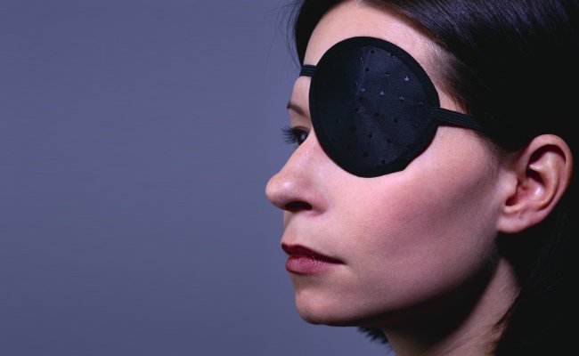
You can always damage your eye if your eyelids do not close properly. It may also lead to dryness. Your doctor will give you goggles or an eye pad for protection along with eye drops to keep your eye moist. There is also an option of taping the lower and upper lid together to keep your eye closed while you are sleeping.

2.

Bell's Palsy - Types, Causes, Symptoms, Diagnosis, Treatment
rx

Try Heat Compress

Apply soaked warm washcloth on face at least 3 times a day to find some comfort. The heat will help to relax facial muscles and prevent atrophy.

3.       Massage with Olive Oil

Bell's Palsy - Types, Causes, Symptoms, Diagnosis, Treatment
www.rxharun.com

Regular massage with olive oil can increase blood circulation, which will help to improve your paralysis by stimulating damaged nerve. Start with your lower face. Place your thumb and index finger on each corner of your mouth, then push your lips forward and repeat for about 15 times. Next, massage your forehead and around your eyebrows. After that, move to your cheeks. Remember to apply some pressure to massage in a circular motion. Each part of the face shall be massaged for at least 2-3 minutes.

4.        Try Castor Oil

Bell's Palsy - Types, Causes, Symptoms, Diagnosis, Treatment
www.rxharun.com

Castor oil helps because it’s effective in dealing with swelling, repairing damaged nerve and strengthening facial muscles. To do that, apply a cotton cloth that has been soaked in heated castor oil on the face for at least 3 times a day. For better result, you can use something such as flannel to retain the heat.

5. Eat Some Celery

Bell's Palsy - Types, Causes, Symptoms, Diagnosis, Treatment
www.rxharun.com

The vitamins and minerals in celery are beneficial for nerve health and can help to repair damaged nerve. In addition, celery can also boost your immune system. Eat celery, leaves and stick, raw several times a day.

6.

Bell's Palsy - Types, Causes, Symptoms, Diagnosis, Treatment
www.rxharun.com

Have Some Ginger and Turmeric

The anti-inflammatory properties in ginger and turmeric are very effective in relieving Bell’s palsy symptoms. You can either add them to your food while cooking or take their supplements.

7.        Take Vitamin Supplements
Bell's Palsy - Types, Causes, Symptoms, Diagnosis, Treatment
www.rxharun.com

Vitamins such as vitamin B-12, B-6, and zinc can greatly speed up Bell’s palsy recovery. Vitamin B-12 is helpful for neurological conditions while vitamin B-6 helps nerve growth. Vitamin B-1 also helps with its thiamine in treating never inflammation. However, consult your doctor for proper doses before taking them.

Ayurvedic Treatment for Bell’s Palsy

Brahmi Capsules

Brahmi capsules of Planet Ayurveda are packed with pure extract of the best quality Brahmi. Brahmi is known as Bacopa monnieri scientifically.

Pack Size: 1 Bottle 60 Capsules

Dosage: 1 capsule twice daily with plain water after meals

Ashwagandha Capsules

Ashwagandha capsules of Planet Ayurveda are packed with pure extract of the best quality Ashwagandha. Ashwagandha is Withania somnifera scientifically.

Pack Size: 1 Bottle 60 Capsules

Dosage: 1 capsule twice daily with plain water after meals

Curcumin Capsules

Curcumin capsules of Planet Ayurveda are packed with a standardized extract of Curcumin, an active compound of turmeric.

Pack Size: 1 Bottle 60 Capsules

Dosage: 1 capsule twice daily with plain water after meals

Chandraprabha Vati

Chandraprabha Vati is a classical natural formulation, a blend of many ingredients that are explained in ayurvedic texts. Some of them are Vacha (Acorus calamus), Mustak (Ciprus rotundus), Pippali (Piper longum), Maricha (Piper nigrum), Haridra (Curcuma longa), Vidanga (Emblica ribes), etc.

Overall Chandraprabha Vati maintains healthy body functions.

Pack Size: 1 Bottle of 120 Tablets

Dosage: 2 tablets twice daily with plain water after meals

Nirgundi Oil

Nirgundi oil contains Nirgundi (Vitex Segundo) with other ingredients like Haridra (Curcuma longa), Manjista (Rubia cordifolia), Kalahari (Dichrostachys cinerea).

Pack Size: 1 Bottle 100ml

Dosage: for external use. Apply on the affected area once to twice daily and massage gently.

Bell’s Palsy (Homeopathy)

Aconitum napellus

When one side of a person’s face becomes paralyzed, especially after being exposed to wind or cold air, this remedy may be helpful. A feeling of fear and agitation and a sudden onset of symptoms are strong indications for Aconitum napellus.

Agaricus

This remedy may be indicated in Bell’s palsy when the facial muscles on one side are stiff, and grimacing or twitching occurs in other parts of the face. People who need this remedy are often excitable, with senses that are over acute. Many people who need this remedy have deep anxiety about their health.

Cadmium sulphur atom

Facial paralysis (usually left-sided) that starts after exposure to wind, and is accompanied by chilliness or overwhelming weakness, suggests a need for this remedy. The person’s mouth may look distorted, and completely closing one of the eyes often is impossible.

Causticum

This remedy can be helpful when facial paralysis has developed gradually (most often on the right side). Opening and closing the mouth can be difficult, and the person may accidentally bite the tongue or the inside of the cheek. The person may be weak but restless and tends to feel best when keeping warm.

Cocculus

One-sided facial paralysis, with pain or tension, felt in the other cheek, especially when opening the mouth, suggests a need for this remedy. Weakness, dizziness, or numbness are other indications. The person may feel worse from lack of sleep or from being emotionally upset.

Nux vomica

One-sided facial paralysis (more often on the left) in a person who is irritable, impatient, and hypersensitive to odors, sounds, and light may indicate a need for this remedy. Cramping and constricting feelings may be felt, and problems may be worse from the cold.

Platina (also called Platinum)

This remedy may be indicated for painless paralysis of the face, with facial distortion that raises one eyebrow or creates a “haughty” look. The person may also experience numbness in the lips and cheeks, or other body parts

Complications

Due to the complexity of our facial muscles and their function, a number of problems can arise following the prolonged experience of having Bell’s palsy

  • Contracture – Shortening of the facial muscles over time may make the affected side of the face appear to be slightly ‘lifted’ in comparison to the unaffected side, and the affected eye may appear smaller than the unaffected eye. The fold between the outer edge of the nostril and the corner of the mouth may seem deeper due to the increased contraction of cheek muscles on that side.
  • Crocodile tears – This means that the affected eye waters involuntarily, particularly whilst eating. This is due to faulty ‘re-wiring’ of the nerves during the recovery phase.
  • Lagophthalmos – This is an inability to close the affected eye, which if prolonged may result in eye dryness and/or corneal ulceration. (This complication can be assisted/prevented by the use of artificial tears and taping the eye down at night.) In rare cases, the vision may be permanently damaged if care is not taken.
  • Synkinesis – This means that when intentionally trying to move one part of the face, another part automatically moves. For example, on smiling the eye on the affected side automatically closes. Similarly, on raising the eyebrows or closing the eyes, involuntary contraction of the cheek or neck muscles occurs. Find out more about synkinesis.
  • People with persistent symptoms of Bell’s palsy may experience psychological problems including stress, anxiety, depression and low self-esteem
  • altered sense of taste
  • facial pain, pain in or behind the ear (in fewer than 50% of cases)
  • no tears in one eye
  • sensitivity to noise on the affected side

There’s none of the confusion, language difficulty, or crushing headache that’s commonly seen in stroke victims. You may have some trouble talking, but it’s purely muscular, whereas stroke victims have difficulty finding the words they need.

Usually, the symptoms appear at once. Occasionally, they worsen over a few days. Steady, progressive paralysis over several weeks is not a sign of Bell’s palsy.

The most serious complication seen in Bell’s palsy is permanent mild facial paralysis. This is found in a minority of cases. Overall, about 80% to 90% recover completely over weeks to months, and most of the rest improve.

Another complication is increased risk of stroke. Stroke prevention and follow-up is recommended.

Incomplete recovery is more likely in people who

  • are over age 55
  • feel pain other than ear pain
  • have weakness/paralysis on both sides of the face – this occurs in 1% of cases
  • show a lack of tears

People who don’t recover completely may be left with one or more of the following symptoms

  • abnormal blink
  • asymmetrical smile
  • buccinator paralysis (food caught in cheek of paralyzed side)
  • corneal damage
  • drooling of liquids from corner of paralyzed mouth
  • dry eye
  • excessive perspiration
  • frozen muscle in the nostril area
  • hyperacusis (perceiving sounds as unusually loud)
  • impaired taste
  • impaired speech
  • synkinesis

Bell's Palsy - Types, Causes, Symptoms, Diagnosis, Treatment

RX Medical Knowledge Graph

Explore this medical topic

Continue through verified related conditions, investigations, medicines, and patient guides. These links are educational and do not replace professional medical advice.

RX Clinical Pathway Engine

Continue through a complete learning pathway

Move from understanding the topic to symptoms, tests, treatment, medicines, monitoring, and prevention.

Search the complete library
  1. Understand the condition Begin with the essential facts and a clear explanation of the topic.
  2. Recognize symptoms Learn common symptoms, signs, and patterns of presentation.
  3. Know when to seek help Review urgent warning signs and when professional assessment may be needed.
  4. Understand causes and risks Explore causes, risk factors, mechanisms, and contributing conditions.
  5. Explore tests and diagnosis Learn how clinicians assess the condition and which investigations may be discussed.
  6. Learn treatment approaches Review general treatment categories and management principles.
  7. Understand medicines safely Continue to medicine education, uses, precautions, and monitoring.
  8. Plan monitoring and follow-up Understand monitoring, complications, rehabilitation, and follow-up learning.
  9. Review prevention and self-care Explore prevention, healthy routines, and questions to discuss with a clinician.

Conditions & Diseases

Background, symptoms, causes, diagnosis, and care.

Explore this library

Tests & Investigations

Laboratory, imaging, screening, and diagnostic education.

Explore this library

Medicines

Uses, safety, monitoring, and related medicine knowledge.

Explore this library

Cancer Knowledge

Cancer types, screening, oncology, and treatment education.

Explore this library
Doctor visit helper

Prepare before seeing a doctor

A simple rural-patient checklist to help you explain symptoms clearly, ask better questions, and avoid unsafe self-treatment.

Safety note: This is not a prescription or diagnosis. For severe symptoms, pregnancy danger signs, children with serious illness, chest pain, breathing difficulty, stroke-like weakness, or major injury, seek urgent care.

Which doctor may help?

Start with a registered doctor or the nearest qualified health center.

What to tell the doctor

  • Write when the problem started and how it changed.
  • Bring old prescriptions, investigation reports, and current medicines.
  • Write allergies, pregnancy status, diabetes, kidney/liver disease, and major past illnesses.
  • Bring one family member if the patient is weak, elderly, confused, or a child.

Questions to ask

  • What is the most likely cause of my symptoms?
  • Which danger signs mean I should go to hospital quickly?
  • Which tests are necessary now, and which can wait?
  • How should I take medicines safely and what side effects should I watch for?
  • When should I come for follow-up?

Tests to discuss

  • Vital signs: temperature, pulse, blood pressure, oxygen saturation
  • Basic physical examination by a clinician
  • CBC, urine test, blood sugar, or imaging only when clinically needed

Avoid these mistakes

  • Do not use antibiotics, steroid tablets/injections, or strong painkillers without proper medical advice.
  • Do not hide pregnancy, kidney disease, ulcer, allergy, or blood thinner use.
  • Do not delay emergency care when danger signs are present.

Medicine safety and first-aid guide

This section is for patient education only. It does not replace a doctor, pharmacist, or emergency care.

Safe first steps

  • Avoid heavy lifting, sudden bending, and prolonged bed rest.
  • Use comfortable posture and gentle movement as tolerated.
  • Discuss physiotherapy, X-ray, or MRI only when clinically needed.

OTC medicine safety

  • For mild back pain, pain-relief medicine may be discussed with a doctor or pharmacist.
  • Avoid repeated painkiller use if you have kidney disease, stomach ulcer, uncontrolled blood pressure, or are taking blood thinners.

Avoid these mistakes

  • Do not start antibiotics without a proper medical decision.
  • Do not use steroid tablets or injections casually for quick relief.
  • Do not delay emergency care because of home remedies.

Get urgent help if

  • Back pain with leg weakness, numbness around private area, loss of urine/stool control, fever, cancer history, or major injury needs urgent care.
Medicine names, dose, and timing must be decided by a qualified clinician or pharmacist after checking age, pregnancy, allergy, other diseases, and current medicines.

For rural patients and family caregivers

Patient health record and symptom diary

Write your symptoms, medicines already taken, test results, and questions before visiting a doctor. This note stays on your device unless you print or copy it.

Doctor to discuss: Medicine doctor / pediatrician for children / qualified clinician
Tests to discuss with doctor
  • Temperature chart and hydration assessment
  • CBC with platelet count if fever persists or dengue/other infection is possible
  • Urine test, malaria/dengue tests, chest evaluation, or blood culture only when clinically indicated
Questions to ask
  • What is the most likely cause of my symptoms?
  • Which warning signs mean I should go to emergency care?
  • Which tests are really needed now?
  • Which medicines are safe for my age, pregnancy status, allergy, kidney/liver/stomach condition, and current medicines?
  • Do I need antibiotics, or is this more likely viral?

Emergency warning signs such as chest pain, severe breathing difficulty, sudden weakness, confusion, severe dehydration, major injury, or loss of bladder/bowel control need urgent medical care. Do not wait for online information.

Safe pathway to proper treatment

Care roadmap for: Bell’s Palsy – Types, Causes, Symptoms, Diagnosis, Treatment

Use this simple roadmap to understand the next safe steps. It is educational and does not replace examination by a doctor.

Go to emergency care if you notice:
  • Severe or rapidly worsening symptoms
  • Breathing difficulty, chest pain, fainting, confusion, severe weakness, major injury, or severe dehydration
Doctor / service to discuss: Qualified healthcare provider; specialist depends on symptoms and examination.
  1. Step 1

    Check danger signs first

    If danger signs are present, seek emergency care and do not wait for online information.

  2. Step 2

    Record the symptom story

    Write when symptoms started, severity, medicines already taken, allergies, pregnancy status, and test results.

  3. Step 3

    Visit a qualified clinician

    A doctor, nurse, or qualified healthcare provider can examine you and decide which tests or treatment are needed.

  4. Step 4

    Do only useful tests

    Do tests after clinical assessment. Avoid unnecessary tests, random antibiotics, or repeated medicines without diagnosis.

  5. Step 5

    Follow up and return early if worse

    If symptoms worsen, new warning signs appear, or treatment is not helping, return for review quickly.

Rural patient practical tips
  • Take a written symptom diary and all previous prescriptions/test reports.
  • Do not hide medicines already taken, even herbal or over-the-counter medicines.
  • Ask which warning signs mean urgent referral to hospital.

This roadmap is for education. A real diagnosis and treatment plan requires history, examination, and clinical judgment.

RX Patient Help

Ask a health question safely

Write your symptom story. A health professional or site editor can review it before any answer is prepared. This box is not for emergency care.

Emergency first: Severe chest pain, breathing trouble, unconsciousness, stroke signs, severe injury, heavy bleeding, or rapidly worsening symptoms need urgent local medical care now.

Frequently Asked Questions

Types of Bell’s Palsy Bell’s palsy types by severity Bell’s palsy is often classified by the degree of facial weakness, using the House-Brackmann scale, which ranges from Grade I (normal) to Grade VI (complete paralysis). Clinicians group these into three types: Mild Bell’s Palsy - House-Brackmann Grades I–II (Mild Dysfunction): Slight weakness noticeable on close inspection, with mostly normal symmetry at rest. Patients can close the eye completely and have good forehead movement Iowa Head and Neck Protocols. This type often resolves quickly and leaves no long-term effects. Moderate Bell’s Palsy - House-Brackmann Grades III–IV (Moderate Dysfunction): Obvious but not disfiguring weakness, with incomplete eye closure and moderate forehead movement (Grade III) or significant asymmetry and incomplete closure (Grade IV) Medscape. Recovery may take longer and sometimes leaves mild facial asymmetry. Severe Bell’s Palsy - House-Brackmann Grades V–VI (Severe Dysfunction to Total Paralysis): Barely perceptible motion (Grade V) or no movement at all (Grade VI). Patients cannot close the eye or move the mouth on the affected side. This type carries the highest risk of incomplete recovery without treatment NCBI. Bell’s Palsy Types by above all conditions While Bell’s palsy itself refers specifically to idiopathic (unknown-cause) facial nerve paralysis, healthcare professionals often describe subtypes or classifications by severity and presentation: Incomplete vs. Complete Paralysis Incomplete means some facial movement remains; you can still smile a bit or close your eye halfway. Complete means nearly total loss of movement on the affected side. Acute vs. Chronic Acute refers to sudden-onset cases, most typical of Bell’s palsy. Chronic describes facial nerve weakness lasting more than six months, which may hint at other causes. Recurrent Bell’s Palsy A small number of people (about 7%) experience Bell’s palsy more than once. Recurrent episodes may affect the same or opposite side. Bilateral Bell’s Palsy Very rare (fewer than 1% of cases), both sides of the face are affected, often prompting investigation into other conditions. Severity Grading (House-Brackmann Scale) Grade I: Normal function Grade II: Mild dysfunction Grade III: Moderate dysfunction Grade IV: Moderately severe dysfunction Grade V: Severe dysfunction Grade VI: Total paralysis Each grade helps doctors describe how weak the facial muscles are and track recovery over time. Causes of Bell's palsy Although labeled “idiopathic” (unknown cause), research points to multiple triggers that can inflame or damage the facial nerve. Below are 20 possible causes, each explained in simple terms: Herpes Simplex Virus Type 1 (HSV-1): This common cold sore virus can hide in nerve cells and reactivate, causing inflammation of the facial nerve and leading to weakness Medscape. Varicella Zoster Virus (VZV): When this chickenpox virus reactivates in the nerve, it causes Ramsay Hunt syndrome, which includes facial paralysis plus painful ear rash Medscape Reference. Epstein-Barr Virus (EBV): Known for causing mononucleosis, EBV can sometimes trigger Bell’s palsy by inflaming nerve tissue, as seen in rare teen cases Medscape. Lyme Disease: The bacterium Borrelia burgdorferi, spread by ticks, can infect the facial nerve and cause palsy, especially in areas where Lyme is common Medscape. Herpes Zoster Oticus (Ramsay Hunt Syndrome): A variant of VZV infection in the ear canal that inflames the facial nerve, causing paralysis, ear pain, and blisters Medscape Reference. Influenza and Other Respiratory Viruses: Cold and flu viruses can trigger immune reactions that inflame the facial nerve, though this link is less direct Mayo Clinic. Upper Respiratory Infections: Recent colds or sinus infections may precede Bell’s palsy by causing body-wide inflammation that affects the facial nerve Mayo Clinic. Ear Infections (Otitis Media): Severe middle ear infections can spread inflammation to the facial nerve as it passes near the ear cavity Medscape. Trauma: Injury to the skull base, temporal bone fractures, or surgical injury near the facial nerve can lead to temporary or permanent palsy Medscape. Tumors: Growths along the facial nerve pathway—such as acoustic neuromas or parotid gland tumors—can press on the nerve and cause palsy Medscape. Diabetes Mellitus: High blood sugar can damage small blood vessels and nerves, increasing the risk of Bell’s palsy through diabetic neuropathy Medscape. Hypertension: Chronic high blood pressure may weaken small blood vessels supplying the facial nerve, leading to ischemia and injury NCBI. Pregnancy: Hormonal changes and fluid balance shifts—especially in the third trimester—can make nerve swelling more likely, causing palsy NCBI. Preeclampsia: This pregnancy complication marked by high blood pressure and organ dysfunction can impair blood flow to the facial nerve NCBI. Sarcoidosis: An autoimmune disease causing small clumps of inflammatory cells can affect the facial nerve, leading to palsy Medscape. Guillain-Barré Syndrome: A rapid nerve-damaging autoimmune reaction can sometimes present with facial paralysis among its early signs Medscape. Multiple Sclerosis: Demyelination in the brainstem can rarely affect the facial nerve nucleus, causing central facial palsy that mimics Bell’s palsy Medscape. Lyme Co-Infection (Babesiosis, Bartonellosis): Tick-borne infections other than Borrelia sometimes accompany Lyme disease and can inflame facial nerves Medscape. Cold Exposure: Sudden chilling of the face or drafts—sometimes called “cold palsy”—may trigger nerve swelling in susceptible people Mayo Clinic. Idiopathic (Unknown): In many cases, no clear cause is found, and the palsy resolves on its own; this unexplained form remains true Bell’s palsy Medscape. What other causes of facial weakness are there?

Ramsay-Hunt Syndrome - Viral infection with the chickenpox virus, herpes zoster, which is shingles of the facial nerve. There is usually significant pain and a rash in the ear. Other cranial nerves may be affected leading to hearing loss balance disturbance and numbness. Skull fractures involving the temporal bone - the bone that contains the middle ear. The facial nerve travels through this bone on its way to the facial muscles and so maybe damaged usually with significant trauma to…

Symptoms of Bell's palsy Weakness or paralysis of the upper and lower facial muscles of the affected side Drooping of ipsilateral eyelids Inability to close the eye completely Dry eye due to inability to close eyes completely Excessive tearing of the eye (epiphora) Drooping of the corner of the mouth Ipsilateral impaired/loss of taste sensation Difficulty with eating due to ipsilateral muscle weakness causing food to be trapped on the affected side of the mouth  Dribbling of saliva The altered sensation on the affected side of the face  Pain in or behind the ear Increased sensitivity to sound (hyperacusis) on the affected side if stapedius muscle is involved upper motor neuron lesion – based on innervation, absence of forehead wrinkling is a reliable way of differentiating Bell’s palsy from an upper motor neuron lesion herpes zoster oticus (Ramsay Hunt syndrome) rarer causes including otitis media, HIV infection, sarcoidosis, autoimmune disorders or tumors of the parotid gland. Sometimes, you may have a cold shortly before the symptoms of Bell palsy begin. Symptoms most often start suddenly but may take 2 to 3 days to show up. They do not become more severe after that. Many people feel discomfort behind the ear before weakness is noticed. The face feels stiff or pulled to one side, and may look different. Other signs can include: Twitching Weakness Paralysis on one side of the face Problems smiling or making facial expressions Difficulty closing your eye Drooling due to a lack of control over the facial muscles Difficulty closing one eye Difficulty eating and drinking; food falls out of one side of the mouth Drooling due to lack of control over the muscles of the face Drooping of the face, such as the eyelid or corner of the mouth Problems smiling, grimacing, or making facial expressions Twitching or weakness of the muscles in the face Bell's palsy symptoms may come on suddenly or develop over a few days. Initial symptoms may include Pain behind the ear that is followed by weakness and paralysis of the face Ringing sound in the ear Slight fever Slight hearing impairment A slight increase in sensitivity to sound on the affected side. Symptoms of full-blown Bell's palsy may include Facial weakness or paralysis (look for smooth forehead and problems smiling)—most often on one side Numbness just before the weakness start Drooping corner of the mouth Drooling Decreased tearing Inability to close an eye, which can lead to Dry, red eye Ulcers forming on the eyes Infection Problems with taste Sound sensitivity in one ear Earache Slurred speech Late symptoms can occur 3-4 months after or second-time onset of Bell's palsy and can include Muscle weakness or paralysis Facial Droop Impossible or difficult to blink Difficulty speaking Difficulty eating and drinking Nose runs The nose is constantly stuffed Difficulty breathing out of nostril on the affected side Nostril collapse on the affected side Forehead wrinkles disappear Sensitivity to sound Excess or reduced salivation Facial swelling Drooling Diminished or distorted taste Pain behind ear Long-lasting tightening of the facial muscles Tearing from the eye during eating Diagnosis of Bell's Palsy Diagnosing Bell’s palsy involves ruling out other causes of facial paralysis. Tests fall into five categories: A. Physical Exam Cranial Nerve Examination: The doctor checks movement in all facial muscles by asking the patient to smile, raise eyebrows, and close eyes Mayo Clinic. Ear Inspection: Looking inside the ear canal for vesicles that suggest Ramsay Hunt syndrome Mayo Clinic. Taste Test: Applying sweet or salty solutions to the front of the tongue to check taste sensation Mayo Clinic. Corneal Reflex: Gently touching the cornea with a wisp of cotton to see if the eyelid blinks Mayo Clinic. Hyperacusis Assessment: Placing a stethoscope on the mastoid bone and having the patient listen to a tuning fork to detect increased sound sensitivity Medscape. Salivary Flow Test: Collecting saliva from each side of the mouth after sour stimulation to compare gland function Medscape. Stapedial Reflex Test: Measuring the muscle reflex in the middle ear that usually dampens loud sounds Medscape. Nasal Airflow Test: Checking for symmetry in nasal breathing to assess facial muscle control Mayo Clinic. Facial Symmetry InspectionObserve resting face for droop of brow, eyelid, and mouth. Voluntary Movement TestingAsk patient to raise eyebrows, close eyes tightly, smile, and puff cheeks to assess muscle function. Bell’s Phenomenon ObservationNotice upward rotation of the eyeball when attempting eye closure. House-Brackmann GradingRate facial nerve function on a scale from I (normal) to VI (total paralysis). General Neurologic ExamCheck other cranial nerves, motor strength, and sensation to exclude stroke or central lesions. Manual Tests Schirmer’s TestA small strip of paper under the lower eyelid measures tear production to detect dry eye risk. Tinel’s SignTapping over the stylomastoid foramen (where the facial nerve exits the skull) can reproduce tingling if the nerve is irritated. Muscle Strength GradingThe doctor applies light resistance as you try to close your eye or smile to grade muscle power. Sensory TestingAlthough rare in Bell’s palsy, gentle touch to facial skin checks for numbness or altered sensation. Corneal Reflex TestTouch cornea with a wisp of cotton to assess trigeminal (V) sensory and facial (VII) motor response. Gustatory Taste TestApply salt or sweet solutions to the tongue’s anterior two-thirds to evaluate taste sensation. Stapedius Muscle Reflex (Acoustic Reflex)Use tuning fork or reflex hammer to assess stapedius contraction in response to loud sound. Resistance TestingManually resist eyebrow lifting or mouth elevation to grade muscle strength. C. Laboratory and Pathological Tests Complete Blood Count (CBC): To look for signs of infection or inflammation Medscape. Erythrocyte Sedimentation Rate (ESR): A general marker of inflammation that can be elevated in autoimmune or infectious causes Medscape. Blood Glucose and HbA1c: To screen for diabetes as a risk factor Medscape. Lyme Serology: Antibody tests for Borrelia burgdorferi if the patient is from an area where Lyme is common Medscape. HIV and Syphilis Testing: To exclude these infections as causes of facial palsy Medscape. D. Electrodiagnostic Tests Electroneuronography (ENoG): Measures the speed and strength of nerve signals along the facial nerve to assess damage Wikipedia. Electromyography (EMG): Records electrical activity in facial muscles to pinpoint the location and severity of nerve injury Wikipedia. Maximal and Minimal Stimulation Test (MST): Evaluates the lowest and highest electrical currents needed to produce a muscle response, giving prognostic information Medscape. Electroneurography (ENoG)Records muscle response to a standard electrical stimulus of the facial nerve to quantify damage. Blink Reflex StudyElectrical stimulation near the ear triggers a blink; delayed or absent response indicates nerve dysfunction. E. Imaging Tests Magnetic Resonance Imaging (MRI) with Gadolinium: Visualizes the facial nerve along its entire course to detect tumors, inflammation, or demyelination . Computed Tomography (CT) Scan: Helps rule out fractures, bone lesions, or structural causes along the nerve pathway Wikipedia. CT scan —a type of x-ray that uses a computer to make pictures of structures inside the head to see if there is an infection, tumor, bone fracture , or other problem in the area of the facial nerve The nerve excitability test - determines the excitation threshold by recording the minimum electrical stimulus required to produce visible muscle contraction. A difference greater than 3.5 mA between affected and unaffected sides is considered to be significant in terms of poorer outcomes. Measuring the peak-to-peak amplitude of the evoked compound action potential of the involved side compared to the normal side has prognostic importance. If there is a 90% or greater reduction in the amplitude of the affected side, the prognosis is poor.[rx] Currently the trigeminal blink reflex - is the only test to measure the intracranial pathway of the facial nerve and also a useful test to study various post paralysis sequelae such as synkinesis and hemifacial spasms. With the recovery of facial function, the ipsilateral R1 latency becomes less prolonged and the amount of initial prolongation of this response correlates with greater loss of facial motor function. Gadolinium contrast magnetic resonance (MRI) - study reveals enhancement of internal acoustic meatal segment on the affected side; however, this is a non-specific finding. MRI should not be done routinely and should be the investigation to look for other possible causes for acute facial paralysis especially if there is little or no recovery of function. Treatment of Bell's palsy Non-Pharmacological Treatments Each of these therapies aims to reduce inflammation, maintain muscle tone, or retrain facial muscles to speed recovery and prevent permanent muscle contractures. AcupunctureInvolves inserting fine needles at specific points on the face and body. It may modulate nerve function and reduce pain, though high-quality studies are lacking Frontiers. Biofeedback TrainingUses visual or auditory feedback to help patients relearn controlled muscle movements by monitoring muscle activity and guiding relaxation techniques Mayo Clinic. Botulinum Toxin InjectionsThough a drug, it’s delivered as a procedure: small doses of Botox are injected into unaffected muscles to reduce synkinesis (unwanted muscle contractions) and improve symmetry Mayo Clinic. Facial Exercise TherapyStructured exercises (e.g., smiling, raising eyebrows) maintain muscle tone and stimulate the nerve. Systematic reviews show improved facial function with early exercise AAFP. Mime TherapyA specialized exercise program combining gentle stretch and coordination exercises to improve facial symmetry and emotional expression ScienceDirect. MassageGentle manual massage of facial muscles and soft tissues reduces stiffness, improves circulation, and can alleviate discomfort Frontiers. Thermotherapy (Warm Compresses)Applying moist heat to the affected side relaxes muscles and eases pain by improving blood flow Wikipedia. ElectrostimulationLow-level electrical currents applied to paralyzed muscles may promote nerve conduction and prevent atrophy; evidence is preliminary Frontiers. Kabat Rehabilitation (PNF Techniques)Proprioceptive neuromuscular facilitation techniques use specific patterns of movement and resistance to retrain facial muscle coordination Frontiers. Hyperbaric Oxygen TherapyBreathing pure oxygen in a pressurized chamber may reduce nerve swelling and support healing in severe cases; evidence remains very tentative Wikipedia. Ultrasound TherapyPulsed ultrasound waves applied to the face enhance circulation and may stimulate nerve repair; studies show benefit when combined with exercise and massage Nose Creek Physiotherapy. Transcutaneous Electrical Nerve Stimulation (TENS)Surface electrodes deliver mild electrical currents to reduce pain and improve muscle activation Nose Creek Physiotherapy. Eye Protection MeasuresFrequent use of tear‐like eye drops, gel, or ointment during the day and wearing an eye patch or tape at night protects the cornea when blinking is impaired Wikipedia. Kinesio TapingSports taping techniques applied to facial muscles can reduce synkinesis and guide correct muscle movement MedStar Health. Breathing and Meditation PracticesGuided breathwork and mindfulness calm the nervous system, reducing stress-related exacerbations and supporting recovery Facial Palsy UK. Peer Support and Psychological InterventionsGroup or one-on-one support reduces distress, normalizes the experience, and encourages adherence to therapy Facial Palsy UK. YogaGentle yoga poses and stretches improve overall circulation and help manage stress, indirectly supporting nerve healing Number Analytics. Cognitive-Behavioral Therapy (CBT)Addresses anxiety or depression that may accompany Bell’s palsy, improving mental well-being and treatment engagement Number Analytics. Deep Breathing ExercisesSimple diaphragmatic breathing sessions reduce muscle tension and stress, promoting a healing environment audreylemd.com. Low-Impact Motion Exercises (Walking, Stretching)General physical activity boosts circulation, which may enhance nutrient delivery to injured nerves Nose Creek Physiotherapy. Physical Therapy - In Bell's palsy various physical therapies, such as exercise, biofeedback, laser, electrotherapy, massage, and thermotherapy are used to hasten recovery. However, the evidence for the efficacy of any of these therapies is lacking. Cochrane systemic review of the efficacy of physical therapies, electrostimulation, and exercises, on the outcome of Bell's palsy, concluded that there was no significant benefit or harm from any of these physical therapies for Bell's palsy. There was limited evidence that improvement began earlier in the exercise group.[Rx] Another systematic review examined the effects of facial exercises associated either with mirror or electromyogram biofeedback with respect to complications of delayed recovery in Bell's palsy and concluded that because of the small number of randomized controlled trials, it was not possible to analyze if the exercises, were effective.[Rx] However, the possibility that facial exercise reduces time to recover and sequelae need confirming with good-quality randomized controlled trials.[Rx] Drug Treatments These medications target inflammation, viral replication, pain, and prevent corneal damage. Acyclovir (Antiviral)Dosage: 400 mg five times daily for 7 daysPurpose: Inhibit herpes simplex viral replicationMechanism: Guanosine analog terminates viral DNA synthesisSide Effects: Headache, nausea Wikipedia Valacyclovir (Antiviral)Dosage: 1 g three times daily for 7 daysPurpose: Prodrug of acyclovir with better bioavailabilityMechanism: Converted to acyclovir, then inhibits viral DNA polymeraseSide Effects: Fatigue, headache Wikipedia Ibuprofen (NSAID)Dosage: 400 mg every 4–6 hours as neededPurpose: Relieve pain and mild inflammationMechanism: Inhibits cyclooxygenase (COX) enzymesSide Effects: GI upset, renal impairment Mayo Clinic Artificial Tears (Lubricant Eye Drops)Dosage: Instill one drop every 2 hours while awakePurpose: Prevent corneal drying and irritationMechanism: Provides tear film supplementationSide Effects: Mild eye irritation Wikipedia Ocular Lubricating OintmentDosage: Apply thin strip to lower eyelid at bedtimePurpose: Protect cornea during sleepMechanism: Creates a protective lipid barrierSide Effects: Blurred vision upon application Wikipedia Botulinum Toxin ADosage: 1–10 units injected into overactive facial musclesPurpose: Reduce synkinesis and unwanted contractionsMechanism: Blocks acetylcholine release at neuromuscular junctionSide Effects: Injection site pain, temporary weakness Mayo Clinic Gabapentin (Neuropathic Pain)Dosage: Start 300 mg at bedtime, titrate to 900–1,200 mg/dayPurpose: Alleviate neuropathic pain around the earMechanism: Modulates α₂δ subunit of voltage-gated calcium channelsSide Effects: Dizziness, fatigue Dr.Oracle Topical Lidocaine GelDosage: Apply to area of pain 3–4 times/dayPurpose: Local analgesia for ear or facial painMechanism: Blocks sodium channels in nerve membranesSide Effects: Local numbness Wikipedia AcetaminophenDosage: 500–1,000 mg every 4–6 hours as neededPurpose: Pain relief when NSAIDs are contraindicatedMechanism: Central COX inhibition and modulation of serotonergic pathwaysSide Effects: Hepatotoxicity in overdose Mayo Clinic Gabapentin (Anticonvulsant) Class: Calcium channel modulator Dosage & Time: 300 mg three times daily, adjusted for pain control Purpose: Alleviate nerve-related facial pain Mechanism: Modulates α₂δ subunit of voltage-gated calcium channels, reducing excitatory neurotransmitter release Side Effects: Drowsiness, dizziness Wikipedia Carbamazepine - (Anticonvulsant)200 mg twice daily, max 800 mg/day for severe neuralgic pain; mechanism: blocks high-frequency discharges. S/E: mild liver enzyme rise, sleepiness. Class: Sodium channel blocker Dosage & Time: 100 mg twice daily, titrating up to 200 mg three times daily Purpose: Treat synkinesis and neuropathic pain Mechanism: Stabilizes hyperexcited neural membranes by blocking sodium channels Side Effects: Drowsiness, nausea, risk of blood dyscrasias Wikipedia Botulinum Toxin Type B Class: Neurotoxin Dosage & Time: 2.5–5 units injected into targeted muscles every 3–4 months Purpose: Reduce unwanted muscle contractions (synkinesis) Mechanism: Blocks acetylcholine release at the neuromuscular junction, relaxing overactive muscles Side Effects: Temporary weakness in nearby muscles, injection site pain Wikipedia Artificial Tears (Carboxymethylcellulose) Class: Ophthalmic lubricant Dosage & Time: 1–2 drops every 2–4 hours while awake Purpose: Protect cornea when eyelid closure is incomplete Mechanism: Mimics natural tears, maintaining moisture on the ocular surface Side Effects: Mild irritation or blurred vision Wikipedia Methylcobalamin (active vitamin B12 injection) – 500–1000 µg IM three times a week for 4–8 weeks in people with low B-vitamin levels; nourishes myelin and speeds nerve remyelination. S/E: rare acne-like rash.AAFPPMC Botulinum toxin type A (chemodenervation) – 2.5-4 units injected into over-active muscles every 3-4 months after the acute phase to treat synkinesis and asymmetry; blocks acetylcholine release. S/E: transient local weakness.PMCPubMed Artificial-tear ointment with hyaluronic acid (ocular lubricant) – ribbon in lower lid at bedtime; purpose: physical barrier preventing night-time corneal drying; minimal systemic effects.AAO-HNS Botulinum Toxin (Botox) Injections - These injections give you the option to treat the unaffected or the affected side of your face. These injections can help your facial muscles to relax to reduce unwanted muscle movements. They can also reduce muscle activity in the unaffected side of your face to maintain a balance. Botox injections may also help treat tears when eating and to keep you from winking an eye when smiling, eating, or laughing. Vitamin therapy - Vitamins B-12, B-6, and zinc may help nerve growth. Prednisolone - The rationale for the use of corticosteroids in the acute phase of Bell's palsy is that inflammation and edema of the facial nerve are implicated in causing Bell's palsy and corticosteroids have a potent anti-inflammatory action which should minimize nerve damage and thereby improve the outcome. Randomized, double-blind, placebo-controlled trials have provided compelling evidence that treatment with prednisolone improves outcomes in patients with Bell's palsy and shortens the time to complete recovery.[Rx–Rx] Methylprednisolone - should be used in all patients with facial palsy of less than 72 h duration who do not have contraindications to steroid therapy. The prednisolone dose used was 60 mg per day for 5 days then reduced by 10 mg per day (for a total treatment time of 10 days)[Rx] and 50 mg per day (in two divided doses) for 10 days.[Rx] The reported adverse rates were low. Treatment with prednisolone is likely to be cost-effective.[Rx] Antiviral Agents - The rationale for the use of antiviral agents is the evidence that the inflammation of the facial nerve in Bell's palsy might be related to the herpes simplex virus (HSV). In an autopsy study, latent HSV type-1 has been isolated from the majority of the geniculate ganglia samples.[Rx] HSV-1 genome was detected in 79% of facial nerve endoneurial fluid in patients with Bell's palsy, but not in the controls.[Rx] However, the benefit of acyclovir or valacyclovir, either as single agents or in combination with prednisolone in Bell's palsy has not been definitively established.[Rx–Rx,Rx, Rx] Thus with the available evidence acyclovir or valacyclovir should not be routine and treatment with acyclovir is highly unlikely to be considered cost-effective.[Rx] Combination therapy - A randomized controlled trial found that at nine months of diagnosis, the facial function had recovered in 94.4% of patients who took prednisolone alone, 85.4% of those who took aciclovir alone, and 92.7% of those who received both. There were no serious adverse effects in any group. The study concluded that early treatment with prednisolone alone increases the likelihood of complete recovery and there was no additional benefit of treatment with aciclovir alone or combining with prednisolone.7However, a systematic review also found that treatment with prednisolone reduced the chances of incomplete recovery but using an antiviral drug had an additional benefit.14 Dietary Molecular Supplements These nutrients support nerve health, reduce inflammation, and may inhibit viral triggers. Vitamin B₁₂ (Methylcobalamin) – 1,000 µg/day oralSupports myelin formation and nerve regeneration Life Extension Acetyl-L-Carnitine – 500 mg twice dailyEnhances nerve metabolism and repair Life Extension Curcumin – 500 mg twice daily with mealsPotent anti-inflammatory and antioxidant action Life Extension Omega-3 Fatty Acids – 1,000 mg EPA/DHA dailyModulates inflammatory cytokines, supports neuron membrane fluidity Life Extension Ginkgo Biloba – 120 mg/day in divided dosesIncreases microcirculation and may protect nerve cells Life Extension L-Lysine – 1,000 mg/dayMay inhibit herpes virus replication Life Extension Zinc – 25 mg/dayEssential for immune function and antioxidant enzyme activity Life Extension Reishi Mushroom Extract – 1,000 mg/dayImmunomodulatory and antiviral properties Life Extension Vitamin C – 500 mg twice dailyAntioxidant that supports collagen and nerve repair PMC Magnesium – 300 mg/daySupports nerve conduction and reduces muscle excitability Life Extension Regenerative and Stem Cell Therapies These emerging treatments aim to harness cell-based repair mechanisms for facial nerve recovery. Uncultured Umbilical Cord-Derived MSC TransplantationEight injections of allogeneic MSCs directly into the affected facial region over 32 months led to complete recovery in a 7-year chronic case by promoting regeneration and reducing inflammation PubMed. Adipose-Derived Stem Cell (ADSC) TherapySingle sentinel case used autologous fat grafting enriched with ADSCs, showing improved facial movement by delivering trophic growth factors to injured nerves BioMed Central. Mesenchymal Stem Cell (MSC) ImmunomodulationIV or local MSCs modulate macrophage polarization toward anti-inflammatory M2 phenotype via AKT–FoxO1 pathway, enhancing remyelination and nerve repair in animal models jmsgr.tamhsc.edu. Extracellular Vesicles (EVs) from Stem CellsEVs harvested from ADSCs deliver microRNAs and proteins that stimulate nerve regeneration without requiring live cell transplantation BioMed Central. Peripheral Blood Mononuclear Cells (PBMCs) with Platelet-Rich PlasmaCombined PBMC and PRP injections provided both cellular and growth-factor support, resulting in improved muscle function and nerve recovery in a small series PMC. Platelet-Rich Plasma (PRP) TherapySingle or serial PRP injections concentrate neurotrophic factors (e.g., NT-3, GDNF) at the injury site, promoting axonal sprouting and functional recovery in preclinical models PMC. Surgical Procedures Reserved for cases with poor spontaneous recovery or severe degeneration. Middle Fossa DecompressionDrilling away bone over the facial canal at the skull base to relieve nerve pressure; studies show mixed outcomes and it is not routinely recommended AAFP. Transmastoid DecompressionAccesses the facial canal via the mastoid bone to release constriction; clinical benefit remains uncertain AAFP. Cross-Facial Nerve GraftingHarvests sural nerve grafts to connect functional facial nerve branches on the healthy side to paralyzed muscles, enabling reinnervation Mayo Clinic. Free Muscle Transfer (e.g., Gracilis Transplantation)Transplants a segment of thigh muscle with its nerve and blood supply to restore dynamic smile function; requires microsurgical technique Mayo Clinic. Nerve Transfer Surgery (Masseteric to Facial Nerve)Uses a branch of the trigeminal nerve (masseteric) to reinnervate facial muscles, offering improved symmetry and voluntary control Mayo Clinic. Nerve repair or nerve grafts - Facial nerve regeneration occurs at a rate of one millimeter per day. If a nerve has been cut or removed, the direct microscopic repair is the best option. Nerve transposition - Often the tongue nerve (hypoglossal nerve) or the other facial nerve can be connected to the existing facial nerve. For example, the patient can then train themselves to move their face by moving their tongue. Muscle transposition or sling procedures - The temporalis muscle or masseter muscle (some of the only muscles on the face not supplied by the facial nerve), can be moved down and connected to the corner of the mouth to allow movement of the face. Muscle transfers -  Free muscles from the leg (gracilis) can be used to provide both muscle bulk Often, no treatment is needed. Symptoms often begin to improve right away. But, it may take weeks or even months for the muscles to get stronger. Your provider may give you lubricating eye drops or eye ointments to keep the surface of the eye moist if you can't close it completely. You may need to wear an eye patch while you sleep. Sometimes, medicines may be used, but it isn't known how much they help. If medicines are used, they are started right away. Physiotherapy Relaxation techniques - Relaxing by using techniques such as meditation and yoga may relieve muscle tension and chronic pain. Acupuncture - Placing thin needles into a specific point in your skin helps stimulate nerves and muscles, which may offer some relief. Biofeedback training - By teaching you to use your thoughts to control your body, you may help gain better control over your facial muscles. Hyperbaric oxygen -  may improve the time to recovery and the proportion of people who make a full recovery compared with corticosteroids; however, the evidence for this is weak. We don't know whether facial nerve decompression surgery is beneficial in Bell's palsy. Facial retraining -  may improve recovery of facial motor function scores including stiffness and lip mobility, and may reduce the risk of motor synkinesis in Bell's palsy, but the evidence is too weak to draw conclusions. The efficacy of acupuncture - remains unknown because the available studies are of low quality (poor primary study design or inadequate reporting practices).[Rx] There is very tentative evidence for hyperbaric oxygen therapy in severe disease.[Rx] During the earliest stages of Bells palsy, when muscles are completely flaccid, it is advisable to limit therapy to: Moist heat (to ease soreness and reduce swelling) Massage (also to ease soreness, plus to provide a degree of motion & stimulation to the muscles and increase circulation) Mental exercises (to retain the "memory" of facial motions) A special form of physiotherapy called facial retraining can also help minimize the asymmetrical appearance of the face that occurs when one side is weakened. Examples of facial retraining exercises can include but are not limited to Sniffle. Wrinkle's nose. Flared nostrils. Curl upper lip up and raise and protruding upper lip. Compress lips together. Pucker lips & attempt to whistle. Smile without showing teeth; then smile showing teeth. Try moving your lips into a small smile slowly. Then gently pucker slowly using equal strength from both sides. Draw the angle of the mouth upward so as to deepen the furrow from the side of the nose to the side of the mouth. Harden (wrinkle) the chin; "stick out" the chin (like a boxer). Using your index finger and thumb pull the corners of your lips toward the center. Slowly and smoothly push out and up into a smile. Continue the movement up to the cheekbone. Use firm pressure. Placing 4 fingertips on the eyebrow rub using a firm stroke up to the hairline. Return downward to the eyebrow. Do the same type of massage in a circular motion on your cheeks and chin, and outward to your ear. Try to close the eye slowly and gently, without letting your mouth pull up or your eyebrow moves downward. Raise eyebrows and hold for 10 -15 seconds (watch out for synkinesis - hold the brow at a point before the corner of your mouth starts to move or your cheek tries to help). Wrinkle forehead. Frown and draw eyebrows downward. Gently wink with one eye and then the other to the best of your ability. Open eyes widely, but without involving your eyebrow. Stop if you see any inappropriate muscle actions. Bell's palsy develops rapidly, with symptoms 'peaking' within 48 hours of their onset in many patients, and maximal by 5 days overall. Patients often mistakenly believe they have had a stroke, because of the facial paralysis. In the majority of cases (60-80%), the condition gradually disappears completely within weeks to months. In some, there may be slight residual paralysis that is barely noticeable to others. Unfortunately, some people are left with complications of the condition, which include: Obvious residual paralysis (e.g. asymmetrical smile, drooling of liquids, inability to close eye, abnormal blinking, tear pooling) Dry eye Affected taste Distorted nerve functions (e.g. tear production with eating, blinking when opening mouth). In 7-10% of people, Bell's palsy will recur. Hyperacusis (distorted hearing) and associated diseases such as diabetes are associated with a poor outcome. Factors that increase the likelihood of better outcome include: Young age Incomplete paralysis at the outset Recovery of taste within 1 week Early recovery (within 10-21 days). The use of facial massage, neuromuscular retraining (e.g. using a mirror to practice the use of facial muscles), acupressure, or acupuncture may help enhance the recovery process. Prevention Strategies While idiopathic, risk may be reduced by bolstering overall health and avoiding triggers. Stress Management (Mindfulness, Therapy)Chronic stress can trigger viral reactivation; practices like meditation help keep stress in check drpanossian.com. Regular ExerciseImproves circulation and immune resilience, potentially lowering infection risk drpanossian.com. Adequate SleepSupports immune function and nerve repair processes drpanossian.com. Balanced Diet with Fruits & VegetablesProvides antioxidants and nutrients essential for nerve health drpanossian.com. Avoid Excessive Alcohol & CaffeineBoth can impair immunity and hydration, increasing susceptibility to viral triggers drpanossian.com. Blood Sugar Control (for Diabetics)High glucose impairs nerve healing; tight glycemic control reduces risk Wikipedia. Good Oral HygienePrevents dental infections that can spread to facial nerves MedStar Health. Boost Immune Function (Diet, Supplements)Adequate micronutrients and lifestyle support may deter viral reactivation Dr. Axe. Protect from Cold DraftsKeep the face warm in cold weather to prevent nerve irritation (common clinical advice). Influenza VaccinationReduces risk of respiratory infections linked to Bell’s palsy onset (general preventive recommendation). When to See a Doctor Onset of facial weakness (especially if within the first 72 hours) Severe ear or facial pain Inability to close the eye fully Worsening after 2 weeks without improvement Bilateral facial paralysis (rare, may indicate serious pathology) Associated stroke-like symptoms (arm/leg weakness, speech changes) Rash on ear or mouth (suggests Ramsay Hunt syndrome) Persistent corneal discomfort despite lubrication Fluctuating or jerky facial movements (synkinesis) No sign of recovery after 3 months (consider specialist referral) Mayo Clinic Dietary Do’s and Don’ts Do Eat: Whole grains (brown rice, oats) Fresh fruits (berries, citrus) Vegetables (leafy greens) Lean proteins (poultry, fish) Omega-3 sources (salmon, chia seeds) Nuts and seeds (walnuts, flaxseed) Low-fat dairy (yogurt, cottage cheese) Legumes (beans, lentils) Healthy fats (olive oil, avocado) Hydrating fluids (water, herbal teas) Don’t Eat: Refined carbs (white bread, pastries) Sugary drinks (sodas, energy drinks) Excessive salt (processed snacks) Fried foods (French fries, fried chicken) Trans fats (margarine, baked goods) High-sugar sweets (candy, doughnuts) Caffeinated beverages (strong coffee, energy shots) drpanossian.com Alcoholic drinks Artificial sweeteners Excessive red meat How can you care for yourself at home?

Facial exercises - As the nerve in your face begins to work again, doing simple exercises—such as tightening and relaxing your facial muscles—may make those muscles stronger and help you recover more quickly. Massaging your forehead, cheeks, and lips with oil or cream may also help. Eye care - If you can't blink or close your eye fully, your eye may become dry. A dry eye can lead to sores and serious vision problems. To help protect the eye and keep it…

A global war against illness

Help this medical guide reach someone who may need it

Share reliable health information with a patient, family member, caregiver, or colleague. Reading and awareness can help people ask better questions and seek appropriate care.

Continue exploring

Explore this topic across the RX Medical Library

Open a focused A–Z pathway or continue with closely related indexed articles. These links are educational and do not replace personal medical care.

Search this topic
Diseases A–Z Drugs A–Z Lab Tests A–Z Cancer A–Z
