Kidneys – Anatomy, Nerve Supply, Function

Patient Tools

Read, save, and share this guide

Use these quick tools to make this medical article easier to read, print, save, or share with a family member.

On this page3 sections

Article Summary

The kidneys are bean-shaped organs, with medial concavity and lateral convexity, weighing anywhere from 150 to 200 g in males and about 120 to 135 g in females. The dimensions are usually a length of 10 to 12 cm, a width of 5 to 7 cm, and a thickness of 3 to 5 cm. Each kidney is about the size of a closed fist. They...

Key Takeaways

  • This article explains Structure and Function of Kidneys in simple medical language.
  • This article explains Blood Supply of Kidneys in simple medical language.
  • This article explains Nerves of Kidneys in simple medical language.
Before reading

RX Patient Tools

Use these quick guides before reading the article, or return to them when you need help preparing questions for a doctor.

Start here Choose the right pathway for symptoms, reports, medicines, or urgent warning signs. Disease article roadmap Read this topic step by step: meaning, symptoms, warning signs, diagnosis, treatment, prevention, and follow-up. Treatment planner Prepare questions about treatment choices, benefits, risks, side effects, and follow-up. Family & caregiver guide Organize symptoms, reports, medicines, questions, and follow-up safely. Nutrition & diet guide Prepare food, hydration, supplement, and medicine-timing questions safely. Prevention guide Organize risk factors, protective habits, screening, and warning signs. Recovery guide Prepare a safe plan for activity, rehabilitation, warning signs, and follow-up.
Educational health guideWritten for patient understanding and clinical awareness.
Reviewed content workflowUse writer and reviewer profiles for stronger trust.
Emergency safety firstUrgent warning signs are highlighted below.
Definition

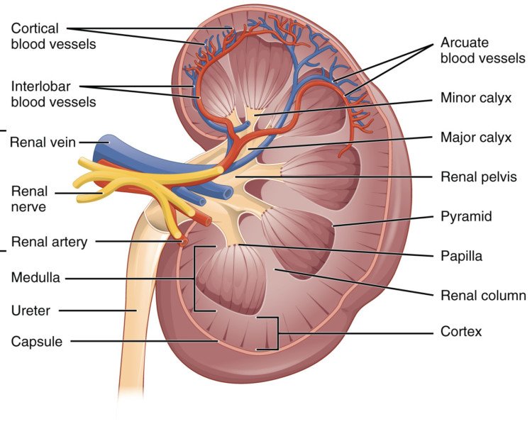
The are bean-shaped organs, with medial concavity and lateral convexity, weighing anywhere from 150 to 200 g in males and about 120 to 135 g in females. The dimensions are usually a length of 10 to 12 cm, a width of 5 to 7 cm, and a thickness of 3 to 5 cm. Each is about the size of a closed fist. They are located retroperitoneally on the posterior abdominal wall and are found between the transverse processes of T12 and L3. Both of the upper poles are usually oriented slightly medially and posteriorly relative to the lower poles. If the upper poles are oriented laterally, this could suggest a horseshoe kidney or a superior pole renal mass.  Further, the right kidney is usually slightly more inferior in position than the left kidney, likely because of the .

Kidneys - Anatomy, Nerve Supply, Function

The following are the kidneys relative to surrounding organs

  • Superiorly – on top of each kidney and separated by renal , are the suprarenal glands (), the right pyramidal suprarenal gland oriented apically on the right kidney and the left crescentic suprarenal gland oriented more medially on the left kidney
  • The right kidney is posterior to the ascending , the second part of the medially, and the liver, separated by the hepatorenal recess
  • The left kidney is posterior to the descending colon, its renal hilum lateral to the tail of the , superomedial aspect adjacent to the greater curvature of the stomach, and left upper pole adjacent to the and connected by splenorenal
  • Posteriorly the rests over the upper third of each kidney with the 12th rib passing posteriorly over the upper pole. The kidneys usually sit located over the medial aspect of the psoas muscle and the lateral aspect of the quadratus lumborum. The proximal will typically pass over the psoas muscle on their way to the bony .
  • At the medial margin of each kidney lies the renal hilum, where the renal enters, and the and leave the renal . The renal vein is found anterior to the renal artery, which is anterior to the renal pelvis. The renal pelvis is the flattened, superior end of the . It receives 2 or 3 major calyces, each of which receives 2 or 3 minor calyces. The minor calyces are indented by the renal papillae, which are the apices of the renal pyramids. A pyramid and its cortical tissue comprise a lobe.
  • Each kidney is covered by a two-layered capsule and is surrounded by perinephric fat, Gerota’s fascia, Zuckerkandl fascia, and perinephric fat. The entire area immediately involving the kidneys is considered the retroperitoneum.

Structure and Function of Kidneys

The kidney is composed of two regions: the cortex and medulla.

  • The cortex is composed of renal corpuscles,
  • convoluted tubules,
  • straight tubules,
  • collecting tubules,
  • collecting ducts, and vasculature.
  • Medullary rays, comprised of straight tubules and collecting ducts, extend into the cortex from the medulla.
  • The medulla also contains the vasa recta, a network of integral to the countercurrent exchange system.
  • Pyramids are conical structures formed by the collecting of tubules in the medulla, oriented with the base towards the cortex and apices towards the hilum.
  • The papillae at the apices of the pyramids extend into minor calyces and drain via the collecting ducts at their tips, the area cribrosa. A collecting duct and the group of that it drains is referred to as a lobule.

Blood Supply of Kidneys

About 20% of the total cardiac output goes to the kidneys.

  • These highly vascular organs are supplied via the renal , which branch off of the inferior to the superior mesenteric artery and enter the hilum of the kidney at L2.
  • The longer right renal artery passes posterior to the inferior vena cava (IVC).
  • Both of the renal arteries divide close to the renal hilum giving off five segmental arteries. The first branch is the posterior segmental artery, and it supplies the posterior segment of the kidney.

The remaining 4 main segmental arteries all arise from the anterior branch of the renal artery and are named according to the segment of the kidney they supply

  • The superior segmental artery,
  • The anterosuperior segmental artery,
  • The anteroinferior segmental artery, and
  • The inferior segmental artery.
  • Accessory renal arteries, leftover embryologically in approximately 25% of people due to failure of vessel degeneration, may arise from the aorta or renal artery and usually enter the poles.

Nerves of Kidneys

Innervation to the kidneys and a portion of the proximal ureters and suprarenal glands, is communicated via the

  • Renal nerve plexus consisting of sympathetic and parasympathetic fibers.
  • Splanchnic nerves.
  • Sympathetic efferents nerve
  • The sensory renal afferent nerves are situated primarily in the renal pelvis, following along the renal artery or proximal ureter to the pelvic wall, and serve an important role in sympathetic outflow and blood pressure regulation. Some travel along the ureter or pelvis in a parallel fashion, while others are oriented circumferentially. They innervate the pelvic wall, renal artery, and renal vein. No sensory fibers are found in the medulla, and few can be observed in the cortex.

References

Doctor visit helper

Prepare before seeing a doctor

A simple rural-patient checklist to help you explain symptoms clearly, ask better questions, and avoid unsafe self-treatment.

Safety note: This is not a prescription or diagnosis. For severe symptoms, pregnancy danger signs, children with serious illness, chest pain, breathing difficulty, stroke-like weakness, or major injury, seek urgent care.

Which doctor may help?

General physician, urologist, nephrologist, or gynecologist depending on symptoms.

What to tell the doctor

  • Write burning, frequency, fever, flank pain, blood in urine, pregnancy, diabetes, and previous UTI history.

Questions to ask

  • Is this UTI, stone, prostate problem, diabetes-related, or another cause?
  • Do I need urine culture before antibiotics?

Tests to discuss

  • Urine routine/microscopy
  • Urine culture for recurrent/severe infection or treatment failure
  • Blood sugar and kidney function when indicated
  • Ultrasound if stone/obstruction/recurrent symptoms

Avoid these mistakes

  • Avoid self-starting antibiotics; wrong antibiotic can cause resistance.
  • Seek urgent care for fever with flank pain, pregnancy, vomiting, confusion, or inability to pass urine.

Medicine safety and first-aid guide

This section is for patient education only. It does not replace a doctor, pharmacist, or emergency care.

Safe first steps

  • Stop activity and seek urgent medical evaluation.
  • Chest pain should not be managed only with home medicine.
  • Discuss ECG and cardiac blood tests with emergency care when appropriate.

OTC medicine safety

  • Do not take random painkillers to hide chest pain before medical evaluation.

Avoid these mistakes

  • Do not start antibiotics without a proper medical decision.
  • Do not use steroid tablets or injections casually for quick relief.
  • Do not delay emergency care because of home remedies.

Get urgent help if

  • Chest pressure, sweating, breathlessness, fainting, pain spreading to arm/jaw/back, or known heart disease needs emergency care.
Medicine names, dose, and timing must be decided by a qualified clinician or pharmacist after checking age, pregnancy, allergy, other diseases, and current medicines.

For rural patients and family caregivers

Patient health record and symptom diary

Write your symptoms, medicines already taken, test results, and questions before visiting a doctor. This note stays on your device unless you print or copy it.

Doctor to discuss: Doctor / qualified healthcare provider
Tests to discuss with doctor
  • Basic vital signs: temperature, pulse, blood pressure, oxygen level if needed
  • Relevant blood, urine, imaging, or specialist tests only after clinical assessment
Questions to ask
  • What is the most likely cause of my symptoms?
  • Which warning signs mean I should go to emergency care?
  • Which tests are really needed now?
  • Which medicines are safe for my age, pregnancy status, allergy, kidney/liver/stomach condition, and current medicines?

Emergency warning signs such as chest pain, severe breathing difficulty, sudden weakness, confusion, severe dehydration, major injury, or loss of bladder/bowel control need urgent medical care. Do not wait for online information.

Safe pathway to proper treatment

Care roadmap for: Kidneys – Anatomy, Nerve Supply, Function

Use this simple roadmap to understand the next safe steps. It is educational and does not replace examination by a doctor.

Go to emergency care if you notice:
  • Severe or rapidly worsening symptoms
  • Breathing difficulty, chest pain, fainting, confusion, severe weakness, major injury, or severe dehydration
Doctor / service to discuss: Qualified healthcare provider; specialist depends on symptoms and examination.
  1. Step 1

    Check danger signs first

    If danger signs are present, seek emergency care and do not wait for online information.

  2. Step 2

    Record the symptom story

    Write when symptoms started, severity, medicines already taken, allergies, pregnancy status, and test results.

  3. Step 3

    Visit a qualified clinician

    A doctor, nurse, or qualified healthcare provider can examine you and decide which tests or treatment are needed.

  4. Step 4

    Do only useful tests

    Do tests after clinical assessment. Avoid unnecessary tests, random antibiotics, or repeated medicines without diagnosis.

  5. Step 5

    Follow up and return early if worse

    If symptoms worsen, new warning signs appear, or treatment is not helping, return for review quickly.

Rural patient practical tips
  • Take a written symptom diary and all previous prescriptions/test reports.
  • Do not hide medicines already taken, even herbal or over-the-counter medicines.
  • Ask which warning signs mean urgent referral to hospital.

This roadmap is for education. A real diagnosis and treatment plan requires history, examination, and clinical judgment.