MSCT Scan

Patient Tools

Read, save, and share this guide

Use these quick tools to make this medical article easier to read, print, save, or share with a family member.

Medical guide Rx Lab Test (A - Z) Jul 6, 2026 35 reads
Related reading

Patient Mode

Understand this article easily

Switch between simple English and easy Bangla patient notes. This is for education and does not replace a doctor consultation.

Multi-Slice Computed Tomography (MSCT) alternative to diagnostic coronary angiography is emerging as a non-invasive clinically reliable, fast-growing less invasive imaging technique alternative to conventional angiography, multislice CT angiography has been recognized as the method of choice for detecting and diagnosing head,  neck, and heart vascular...

For severe symptoms, danger signs, pregnancy, child illness, or sudden worsening, seek urgent medical care.

বাংলা রোগী নোট এখনো যোগ করা হয়নি। পোস্ট এডিটরে “RX Bangla Patient Mode” বক্স থেকে সহজ বাংলা সারাংশ যোগ করুন।

এই তথ্য শিক্ষা ও সচেতনতার জন্য। এটি ডাক্তারি পরীক্ষা, রোগ নির্ণয় বা প্রেসক্রিপশনের বিকল্প নয়।

Article Summary

Multi-Slice Computed Tomography (MSCT) alternative to diagnostic coronary angiography is emerging as a non-invasive clinically reliable, fast-growing less invasive imaging technique alternative to conventional angiography, multislice CT angiography has been recognized as the method of choice for detecting and diagnosing head,  neck, and heart vascular disease, abdominal aortic aneurysm, aortic dissection, pulmonary embolism, severe coronary calcifications causing overestimation of severity of stenosis, arrhythmias precluding the...

Key Takeaways

  • This article explains Indications in simple medical language.
  • This article explains Side Effects in simple medical language.
Educational health guideWritten for patient understanding and clinical awareness.
Reviewed content workflowUse writer and reviewer profiles for stronger trust.
Emergency safety firstUrgent warning signs are highlighted below.
Choose your reading view

Patient View highlights a simple learning journey. Clinical View reveals structure, evidence, and editorial completeness.

Seek urgent medical care if you notice

These warning signs are general safety guidance. Local emergency numbers and clinical judgment should always come first.

  • Severe symptoms, breathing difficulty, fainting, confusion, or rapidly worsening illness.
  • New weakness, severe pain, high fever, or symptoms after a serious injury.
  • Any symptom that feels urgent, unusual, or unsafe for the patient.
1

Emergency now

Use emergency care for severe, sudden, rapidly worsening, or life-threatening symptoms.

2

See a doctor

Book a professional medical evaluation if symptoms persist, worsen, recur often, affect daily activities, or occur in a high-risk patient.

3

Learn safely

Use this article to understand possible causes, tests, treatment options, prevention, and questions to ask your clinician.

Before reading

RX Patient Tools

Use these quick guides before reading the article, or return to them when you need help preparing questions for a doctor.

Start here Choose the right pathway for symptoms, reports, medicines, or urgent warning signs. Disease article roadmap Read this topic step by step: meaning, symptoms, warning signs, diagnosis, treatment, prevention, and follow-up. Treatment planner Prepare questions about treatment choices, benefits, risks, side effects, and follow-up. Family & caregiver guide Organize symptoms, reports, medicines, questions, and follow-up safely. Nutrition & diet guide Prepare food, hydration, supplement, and medicine-timing questions safely. Prevention guide Organize risk factors, protective habits, screening, and warning signs. Recovery guide Prepare a safe plan for activity, rehabilitation, warning signs, and follow-up.
Definition

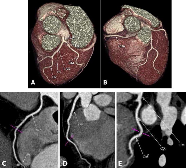
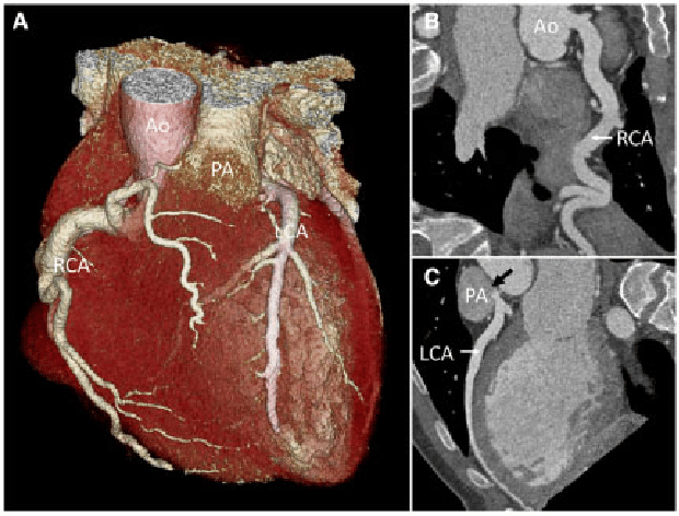
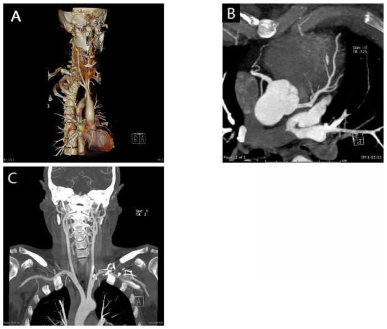
Multi-Slice Computed Tomography (MSCT) alternative to diagnostic coronary angiography is emerging as a non-invasive clinically reliable, fast-growing less invasive imaging technique alternative to conventional angiography, multislice CT angiography has been recognized as the method of choice for detecting and diagnosing head,  neck, and heart vascular disease, abdominal aortic aneurysm, aortic dissection, pulmonary embolism, severe coronary calcifications causing overestimation of severity of stenosis, arrhythmias precluding the use of MSCT, and the relatively high radiation exposure. The diagnostic accuracy of MSCT is high to detect significantly (luminal diameter > 50%) coronary stenosis with a rapid and accurate diagnosis while avoiding unnecessary invasive coronary angiography procedures.[]

Rapid improvements in multislice CT scanners over the last decade have allowed this technique to become a potentially effective alternative to invasive coronary angiography in patients with suspected coronary artery disease. High diagnostic value has been achieved with multislice CT angiography with the use of 64- and more slice CT scanners. In addition, multislice CT angiography shows accurate detection and analysis of coronary calcium, characterization of coronary plaques, as well as prediction of the disease progression and rapid technical evolution from the early generation of 4-slice CT scanners to the latest models such as 64-slice, 256-slice, and 320-slice CT scanners. [].

MSCT Scan MSCT Scan

Indications

Multi-Slice Computed Tomography (MSCT) image analysis included defining the three-dimensional anatomical location of the tumor, and cancerous cell, evaluating the maximum axial diameter, margin fracture, and density of the bony lesions, and assessing associate anatomical relationships of feeding arteries, vessel, capillary, and vertebral artery on the pre-contrast images and contrast-enhanced CT images. It can also help in dissecting vertebra overlapping superior and inferior borders of the spinal tumor, complete, incomplete vertebral fracture, stroke, vascular perforation in the brain, separating and protecting peripheral vessels around the tumor, as well as a resecting tumor for revealing intratumoral vessels to achieve maximal resection 3D image of the tumor and minimal damage to the spinal cord.

Thoraco‐abdominal aorta

  • Diagnosis of congenital and degenerative aortic diseases
  • Assessment of acute aortic injuries and dissections
  • Evaluation of visceral arteries (coeliac, superior mesenteric, and renal arteries)
  • Preoperative planning and follow up
  • Tumor staging and surgical planning

Renal arteries

  • Assessment of anatomy for donor transplants
  • Diagnosis of renal artery stenosis in hypertensives or deteriorating renal function
  • Assessment of renal arteries post‐intervention (renal artery stenting)

Peripheral arterial system

  • Assessment of peripheral vascular disease
  • Assessment of bypass grafts

Carotid/intracranial circulation

  • Characterization of the atherosclerotic disease
  • Assessment of aortic arch vessels
  • Verification of internal carotid artery stenosis
  • Preoperative planning of endovascular and surgical treatment of intracranial aneurysms and vascular malformations

Cardiac imaging

  • Atypical chest pain
  • Patients with intermediate-risk
  • Young patients with a high risk for coronary disease
  • Coronary artery anomalies
  • Non‐invasive follow‐up following percutaneous transluminal angioplasty and stenting
  • Assessment of myocardial scars, aneurysms, tumors, and thrombi
  • Assessment of coronary artery bypass grafts
  • Assessment of the pulmonary veins before and following radiofrequency ablation
  • the diagnostic accuracy of coronary artery disease; the prognostic value of coronary artery disease with regard to the prediction of major cardiac events; detection and quantification of coronary calcium and characterization of coronary plaques.

Bone and spine

  • Intraspinal or intervertebral foramen cervical dumbbell tumors,
  • tumors beyond the intervertebral foramen
  • Spinous process, supraspinal and interspinous ligament,
  • The contralateral paravertebral muscle thus
  • Investigate stability and flexibility problems of cervical vertebrae and
  • alleviates cervical stiffness.
  • Reveals the contour of the tumor as well as compression upon the spinal cord, the extent of spinal edema as well as mutual relationships among the tumor, dura mater, and nerve root [,].
  • At the same time, VRT is conducive to analyzing tumors at different levels for intratumoral vascular route and evaluating infliction and erosion extent of vessels thus assessing neurosurgical difficulties and risks.
  • Owing to its high resolution based on the reconstruction thickness at the sub-millimeter level,
  • VRT formulates images from different perspectives for locating vertebral artery, confirming vascular route and compression degree as well as visualizing peripheral vessels around the tumor.
  • sparing nerve roots: nerve roots above the tumor or enwrapped by the tumor could be dissected and protected, while nerve roots coursing through the tumor should be resected due to their involvement in the tumor original.

Analysis of coronary artery lesions

Systematic analyses of a coronary artery MDCT study took into consideration the following steps:

  • (1) Analysis of images reconstructed from different phases of the cardiac cycle, in order to choose those where the coronary arterial tree is best filled with contrast and where movement artifacts are the least.
  • (2) A complete review of axial images that constitute the cardiac volume, paying attention to cardiac anatomy, degree of opacification of chamber and walls of the heart, and aspect of extracardiac structures.
  • (3) Optimization of images aimed to improve the visualization of coronary arteries, by using specific post-processing protocols.
  • (4) Analysis of the coronary artery tree, for which is fundamental the following systematization:
  • □ Examination of the anatomical distribution of coronary arteries aimed to identify normal variants and congenital abnormalities of the origin of vessels.
    □ Detection and localization of coronary artery lesions, carefully avoiding sections and angulations or interposed structures with potential image artifacts.
  • □ Evaluation of composition and morphology of the ulcer. সহজ বাংলা: শরীরের অস্বাভাবিক দাগ, ক্ষত বা ফোলা অংশ।" data-rx-term="lesion" data-rx-definition="A lesion is an abnormal area of tissue such as a spot, wound, patch, lump, or ulcer. সহজ বাংলা: শরীরের অস্বাভাবিক দাগ, ক্ষত বা ফোলা অংশ।">lesion. In regard to the composition of the plaque, a distinction was made between calcified and non-calcified plaques. Plaques with a mean attenuation of 130 HU or greater were graded as calcified, whereas plaques with a mean attenuation of less than 130 HU were graded as non-calcified. Calcified plaques were identified on nonenhanced scans, and non-calcified plaques were identified on contrast-enhanced scans.
  • □ Qualitative and quantitative assessment of obstruction of the vessel caused by the ulcer. সহজ বাংলা: শরীরের অস্বাভাবিক দাগ, ক্ষত বা ফোলা অংশ।" data-rx-term="lesion" data-rx-definition="A lesion is an abnormal area of tissue such as a spot, wound, patch, lump, or ulcer. সহজ বাংলা: শরীরের অস্বাভাবিক দাগ, ক্ষত বা ফোলা অংশ।">lesion.

A classification of atherosclerotic coronary artery lesions is possible by applying this systematic analysis of MDCT. This classification can be made according to the following aspects:

  • The number of vessels involved.

□ The location: proximal, middle, or distal portions of the vessel.

□ The extension of the ulcer. সহজ বাংলা: শরীরের অস্বাভাবিক দাগ, ক্ষত বা ফোলা অংশ।" data-rx-term="lesion" data-rx-definition="A lesion is an abnormal area of tissue such as a spot, wound, patch, lump, or ulcer. সহজ বাংলা: শরীরের অস্বাভাবিক দাগ, ক্ষত বা ফোলা অংশ।">lesion: focal or diffuse.

□ The degree of obstruction.

a. Non-significant stenosis (less than 60 % of the vessel lumen, including mild and moderate degrees of obstruction).

b. Significant stenosis (equal or more than 60 %, including critical subocclusive and occlusive lesions.

□ The components of the ulcer. সহজ বাংলা: শরীরের অস্বাভাবিক দাগ, ক্ষত বা ফোলা অংশ।" data-rx-term="lesion" data-rx-definition="A lesion is an abnormal area of tissue such as a spot, wound, patch, lump, or ulcer. সহজ বাংলা: শরীরের অস্বাভাবিক দাগ, ক্ষত বা ফোলা অংশ।">lesion:

a. Non-calcified, mixed, or “soft” lesions.

b. Calcified lesions: The calcium component of the lesion can be focal, diffuse, eccentric or concentric.

Currently, MSCTA has been applied for late-phase image reconstruction in the following ways [] multiplanar reconstruction (MPR), shaded surface display (SSD), bone scintigraphy, maximum intensity projection (MIP), and recently volume rendering technique (VRT) which surpass its predecessors as a supreme technique in reconstructing three-dimensional spatial relationships.

Side Effects

Potential adverse effects of contrast medium injection and radiation exposure side effects were explained to all the patients by a radiologist, and written informed consent was obtained before the procedure.

MSCT Scan MSCT Scan MSCT Scan MSCT Scan MSCT Scan

References
RX Medical Knowledge Graph

Explore this medical topic

Continue through verified related conditions, investigations, medicines, and patient guides. These links are educational and do not replace professional medical advice.


RX Clinical Pathway Engine

Continue through a complete learning pathway

Move from understanding the topic to symptoms, tests, treatment, medicines, monitoring, and prevention.

Search the complete library
  1. Understand the condition Begin with the essential facts and a clear explanation of the topic.
  2. Recognize symptoms Learn common symptoms, signs, and patterns of presentation.
  3. Know when to seek help Review urgent warning signs and when professional assessment may be needed.
  4. Understand causes and risks Explore causes, risk factors, mechanisms, and contributing conditions.
  5. Explore tests and diagnosis Learn how clinicians assess the condition and which investigations may be discussed.
  6. Learn treatment approaches Review general treatment categories and management principles.
  7. Understand medicines safely Continue to medicine education, uses, precautions, and monitoring.
  8. Plan monitoring and follow-up Understand monitoring, complications, rehabilitation, and follow-up learning.
  9. Review prevention and self-care Explore prevention, healthy routines, and questions to discuss with a clinician.

Conditions & Diseases

Background, symptoms, causes, diagnosis, and care.

Explore this library

Tests & Investigations

Laboratory, imaging, screening, and diagnostic education.

Explore this library

Medicines

Uses, safety, monitoring, and related medicine knowledge.

Explore this library

Cancer Knowledge

Cancer types, screening, oncology, and treatment education.

No strong indexed relationship is available yet.

Explore this library
Doctor visit helper

Prepare before seeing a doctor

A simple rural-patient checklist to help you explain symptoms clearly, ask better questions, and avoid unsafe self-treatment.

Safety note: This is not a prescription or diagnosis. For severe symptoms, pregnancy danger signs, children with serious illness, chest pain, breathing difficulty, stroke-like weakness, or major injury, seek urgent care.

Which doctor may help?

Start with a registered doctor or the nearest qualified health center.

What to tell the doctor

  • Write when the problem started and how it changed.
  • Bring old prescriptions, investigation reports, and current medicines.
  • Write allergies, pregnancy status, diabetes, kidney/liver disease, and major past illnesses.
  • Bring one family member if the patient is weak, elderly, confused, or a child.

Questions to ask

  • What is the most likely cause of my symptoms?
  • Which danger signs mean I should go to hospital quickly?
  • Which tests are necessary now, and which can wait?
  • How should I take medicines safely and what side effects should I watch for?
  • When should I come for follow-up?

Tests to discuss

  • Vital signs: temperature, pulse, blood pressure, oxygen saturation
  • Basic physical examination by a clinician
  • CBC, urine test, blood sugar, or imaging only when clinically needed

Avoid these mistakes

  • Do not use antibiotics, steroid tablets/injections, or strong painkillers without proper medical advice.
  • Do not hide pregnancy, kidney disease, ulcer, allergy, or blood thinner use.
  • Do not delay emergency care when danger signs are present.

Medicine safety and first-aid guide

This section is for patient education only. It does not replace a doctor, pharmacist, or emergency care.

Safe first steps

  • Avoid heavy lifting, sudden bending, and prolonged bed rest.
  • Use comfortable posture and gentle movement as tolerated.
  • Discuss physiotherapy, X-ray, or MRI only when clinically needed.

OTC medicine safety

  • For mild back pain, pain-relief medicine may be discussed with a doctor or pharmacist.
  • Avoid repeated painkiller use if you have kidney disease, stomach ulcer, uncontrolled blood pressure, or are taking blood thinners.

Avoid these mistakes

  • Do not start antibiotics without a proper medical decision.
  • Do not use steroid tablets or injections casually for quick relief.
  • Do not delay emergency care because of home remedies.

Get urgent help if

  • Back pain with leg weakness, numbness around private area, loss of urine/stool control, fever, cancer history, or major injury needs urgent care.
Medicine names, dose, and timing must be decided by a qualified clinician or pharmacist after checking age, pregnancy, allergy, other diseases, and current medicines.

For rural patients and family caregivers

Patient health record and symptom diary

Write your symptoms, medicines already taken, test results, and questions before visiting a doctor. This note stays on your device unless you print or copy it.

Doctor to discuss: Emergency care / cardiology / medicine doctor
Tests to discuss with doctor
  • ECG as early as possible when chest pain suggests heart risk
  • Troponin or cardiac blood tests if doctor suspects heart attack
  • Blood pressure, oxygen level, chest examination, and other tests as advised urgently
Questions to ask
  • What is the most likely cause of my symptoms?
  • Which warning signs mean I should go to emergency care?
  • Which tests are really needed now?
  • Which medicines are safe for my age, pregnancy status, allergy, kidney/liver/stomach condition, and current medicines?
  • Is this heart-related, and do I need emergency observation?

Emergency warning signs such as chest pain, severe breathing difficulty, sudden weakness, confusion, severe dehydration, major injury, or loss of bladder/bowel control need urgent medical care. Do not wait for online information.

Safe pathway to proper treatment

Care roadmap for: MSCT Scan

Use this simple roadmap to understand the next safe steps. It is educational and does not replace examination by a doctor.

Go to emergency care if you notice:
  • Severe or rapidly worsening symptoms
  • Breathing difficulty, chest pain, fainting, confusion, severe weakness, major injury, or severe dehydration
Doctor / service to discuss: Qualified healthcare provider; specialist depends on symptoms and examination.
  1. Step 1

    Check danger signs first

    If danger signs are present, seek emergency care and do not wait for online information.

  2. Step 2

    Record the symptom story

    Write when symptoms started, severity, medicines already taken, allergies, pregnancy status, and test results.

  3. Step 3

    Visit a qualified clinician

    A doctor, nurse, or qualified healthcare provider can examine you and decide which tests or treatment are needed.

  4. Step 4

    Do only useful tests

    Do tests after clinical assessment. Avoid unnecessary tests, random antibiotics, or repeated medicines without diagnosis.

  5. Step 5

    Follow up and return early if worse

    If symptoms worsen, new warning signs appear, or treatment is not helping, return for review quickly.

Rural patient practical tips
  • Take a written symptom diary and all previous prescriptions/test reports.
  • Do not hide medicines already taken, even herbal or over-the-counter medicines.
  • Ask which warning signs mean urgent referral to hospital.

This roadmap is for education. A real diagnosis and treatment plan requires history, examination, and clinical judgment.

RX Patient Help

Ask a health question safely

Write your symptom story. A health professional or site editor can review it before any answer is prepared. This box is not for emergency care.

Emergency first: Severe chest pain, breathing trouble, unconsciousness, stroke signs, severe injury, heavy bleeding, or rapidly worsening symptoms need urgent local medical care now.

Frequently Asked Questions

Is this article a replacement for a doctor?

No. It is educational content only. Patients should consult a qualified clinician for diagnosis and treatment.

When should I seek urgent care?

Seek urgent care for severe symptoms, rapidly worsening condition, breathing difficulty, severe pain, neurological changes, or any emergency warning sign.

A global war against illness

Help this medical guide reach someone who may need it

Share reliable health information with a patient, family member, caregiver, or colleague. Reading and awareness can help people ask better questions and seek appropriate care.

Continue exploring

Explore this topic across the RX Medical Library

Open a focused A–Z pathway or continue with closely related indexed articles. These links are educational and do not replace personal medical care.

Search this topic
Diseases A–Z Drugs A–Z Lab Tests A–Z Cancer A–Z
Diseases A–Z

45 Best Meditation Books

Below you will likely see some familiar titles, but you will also discover a few new…