RX Patient Tools
Use these quick guides before reading the article, or return to them when you need help preparing questions for a doctor.
Cervical spondylosis is a generalized disease process affecting all levels of the cervical spine. Cervical spondylosis encompasses a sequence of degenerative changes in the intervertebral discs, osteophytosis of the vertebral bodies, hypertrophy of the facets and laminal arches, and ligamentous and segmental instability. The natural history of cervical spondylosis is associated with the aging process. Senescent and pathologic processes are thus morphologically indistinguishable. Clinical manifestations of cervical spondylosis may arise when morphologic sequelae are superimposed on a developmentally narrow spinal canal. The two clinical syndromes of spondylotic pain, numbness, tingling, or weakness. সহজ বাংলা: নার্ভ রুট চাপা/জ্বালায় ব্যথা বা অবশভাব।" data-rx-term="radiculopathy" data-rx-definition="Radiculopathy means nerve-root irritation or compression causing pain, numbness, tingling, or weakness. সহজ বাংলা: নার্ভ রুট চাপা/জ্বালায় ব্যথা বা অবশভাব।">radiculopathy and myelopathy are distinct, yet they may overlap.[rx]
Cervical spondylosis is a term that encompasses a wide range of progressive degenerative changes that affect all the components of the cervical spine (i.e., intervertebral discs, facet joints, joints of Luschka, ligaments Flava, and laminae). It is a natural process of aging and presents in the majority of people after the fifth decade of life.[rx]
Causes of Cervical Spondylosis
A clinical syndrome caused by compression on the spinal cord that is characterized by
- clumsiness in hands
- gait imbalance
- degenerative cervical spondylosis (CSM)
- compression usually caused by anterior degenerative changes (osteophytes, disc osteophyte complex)
- degenerative spondylolisthesis and hypertrophy of ligamentum flavum may contribute
- the most common cause of cervical weakness, numbness, balance trouble, or coordination problems. সহজ বাংলা: স্পাইনাল কর্ডের সমস্যা।" data-rx-term="myelopathy" data-rx-definition="Myelopathy means spinal cord dysfunction, often causing weakness, numbness, balance trouble, or coordination problems. সহজ বাংলা: স্পাইনাল কর্ডের সমস্যা।">myelopathy
Congenital stenosis
Symptoms usually begin when congenital narrowing combined with spondylotic degenerative changes in older patients
Neurologic injury
- mechanism of injury can be
- direct cord compression
- ischemic injury secondary to compression of the anterior spinal artery
Associated conditions
- lumbar spinal stenosis
- tandem stenosis occurs in the lumbar and cervical spine in ~20% of patients
- tends to be slowly progressive and rarely improves with nonoperative modalities
- progression characterized by steplike deterioration with periods of stable symptoms
- early recognition and treatment prior to spinal cord damage is critical for good clinical outcomes
Risk Factors for Cervical Spondylosis
The lists below are the factors that you will have a higher risk of getting neck pain and cervical spondylosis:
- Genetics – if your family has a history of neck pain
- Smoking – clearly linked to increased neck pain
- Occupation – jobs with lots of neck motion and overhead work
- Mental health issues – depression/anxiety
- Injuries/trauma – car wreck or on-the-job injury
“Red flag” features and the conditions they may suggest
Malignancy, infection, or inflammation
-
Fever, night sweats
-
Unexpected weight loss
-
History of inflammatory arthritis, malignancy, infection, tuberculosis, HIV infection, drug dependency, or immunosuppression
-
Excruciating pain
-
Intractable night pain
-
Cervical lymphadenopathy
-
Exquisite tenderness over a vertebral body
Myelopathy
-
Gait disturbance or clumsy hands, or both
-
Objective neurological deficit—upper motor neuron signs in the legs and lower motor neuron signs in the arms
-
Sudden onset in a young patient suggests disc prolapse
Other
-
History of severe osteoporosis
-
History of neck surgery
-
Drop attacks, especially when moving the neck, suggest vascular disease
-
Intractable or increasing pain
Symptoms of Cervical spondylosis
The pain can be from minor to major and it becomes worse when looking up or down for a long period of time, such example is reading a book or driving. To improve the pain, most people tend to takes rest or even lay down. The pain usually becomes worse in the morning and at the end of the day.
Symptoms of cervical spondylosis include
- Neck stiffness and pain
- Numbness and weakness in the upper limbs
- Difficulty in walking, losing balance, or weakness in limbs
- Difficulty in turning the head fully or bending the neck, which may hinder drive
- Muscle spasms in neck and shoulders
- Headaches
- Grinding and popping feeling in the neck when rotating the head
- Loss of bladder and bowel control.
- Axial neck pain (oftentimes absent)
- Occipital headache common
- Extremity paresthesias
- Diffuse non-dermatomal numbness and tingling
- Weakness and clumsiness
- Weakness and decreased manual dexterity (dropping object, difficulty manipulating fine objects)
- Gait instability patient feels “unstable” on feet
- Weakness walking up and downstairs
- Gait changes are the most important clinical predictor
- Urinary retention rare and only appear late in disease progression, not very useful in diagnosis due to the high prevalence of urinary conditions in this patient population
- Cervical pain aggravated by movement
- Referred pain (occiput, between the shoulder blades, upper limbs)
- Retro-orbital or temporal pain (from C1 to C2)
- Cervical stiffness—reversible or irreversible
- Vague numbness, tingling, or weakness in upper limbs
- Dizziness or vertigo
- Poor balance
- Rarely, syncope triggers a migraine pseudo-angina
- Poorly localized tenderness
- Limited range of movement (forward flexion, backward extension, lateral flexion, and rotation to both sides)
- Minor neurological changes like inverted supinator jerks (unless complicated by myelopathy or radiculopathy)
Diagnosis of Cervical Spondylosis
Classically, symptomatic cervical spondylosis presents as one or more of the following three primary clinical syndromes
Axial neck pain
-
Commonly complain of stiffness and pain in the cervical spine that is most severe in the upright position and relieved with bed rest when removing the load from the neck
-
Neck motion, especially in hyperextension and side-bending, typically increases the pain
-
In upper and lower cervical spine disease, patients may report radiating pain into the back of the ear or occiput versus radiating pain into the superior trapezius or periscapular musculature, respectively
-
Occasionally, patients can present with atypical symptoms of cervical angina such as jaw pain or chest pain.
Cervical radiculopathy
-
Radicular symptoms usually follow a myotomal distribution depending on the nerve root(s) involved and can present as unilateral or bilateral neck pain, arm pain, scapular pain, paresthesia, and arm or hand weakness
-
Pain is exacerbated by head tilt toward the affected side or by hyperextension and side-bending toward the affected side.
Cervical myelopathy
-
Typically has an insidious onset with or without neck pain (frequently absent)
-
Can initially present with hand weakness and clumsiness, resulting in the inability to complete tasks requiring fine motor coordination (e.g., buttoning a shirt, tying shoelaces, picking up small objects)
-
Frequent reports of gait instability and unexplained falls
-
Urinary symptoms (i.e., incontinence) are rare and typically appear late in disease progression
Physical exam of Cervical spondylosis
-
Other non-specific neck pain lesions—acute neck strain, postural neck ache, or whiplash
-
Fibromyalgia – and psychogenic neck pain
-
Mechanical lesions – disc prolapse or diffuse idiopathic skeletal hyperostosis
-
Inflammatory disease – rheumatoid arthritis, ankylosing spondylitis, or polymyalgia rheumatica
-
Metabolic diseases – Paget’s disease, osteoporosis, gout, or pseudo-gout
-
Infections – osteomyelitis or tuberculosis
-
Malignancy – primary tumors, secondary deposits, or myeloma
Motor signs
-
Weakness in triceps and hand intrinsics
-
Atrophy of intrinsic hand muscles
-
Clumsiness with fine motor skills
-
The proximal weakness of the lower extremities
- weakness usually difficult to detect on physical exam
- lower extremity weakness is more concerning finding
- finger escape sign when the patient holds fingers extended and adducted, the small finger spontaneously abducts due to the weakness of intrinsic muscle grip and release test normally a patient can make a fist and release 20 times in 10 seconds. myelopathic patients may struggle to do this
Upper motor neuron signs
-
Hoffman’s sign (quick flexion of both the thumb and index finger when the middle fingernail is snapped)
-
Inverted radial reflex (flexion of the fingers in response to the brachioradialis reflex)
-
Pathological clonus
-
Babinski sign
Sensory dysfunction
-
Glove-like sensory loss in hands
-
Proprioceptive dysfunction
Proprioception dysfunction
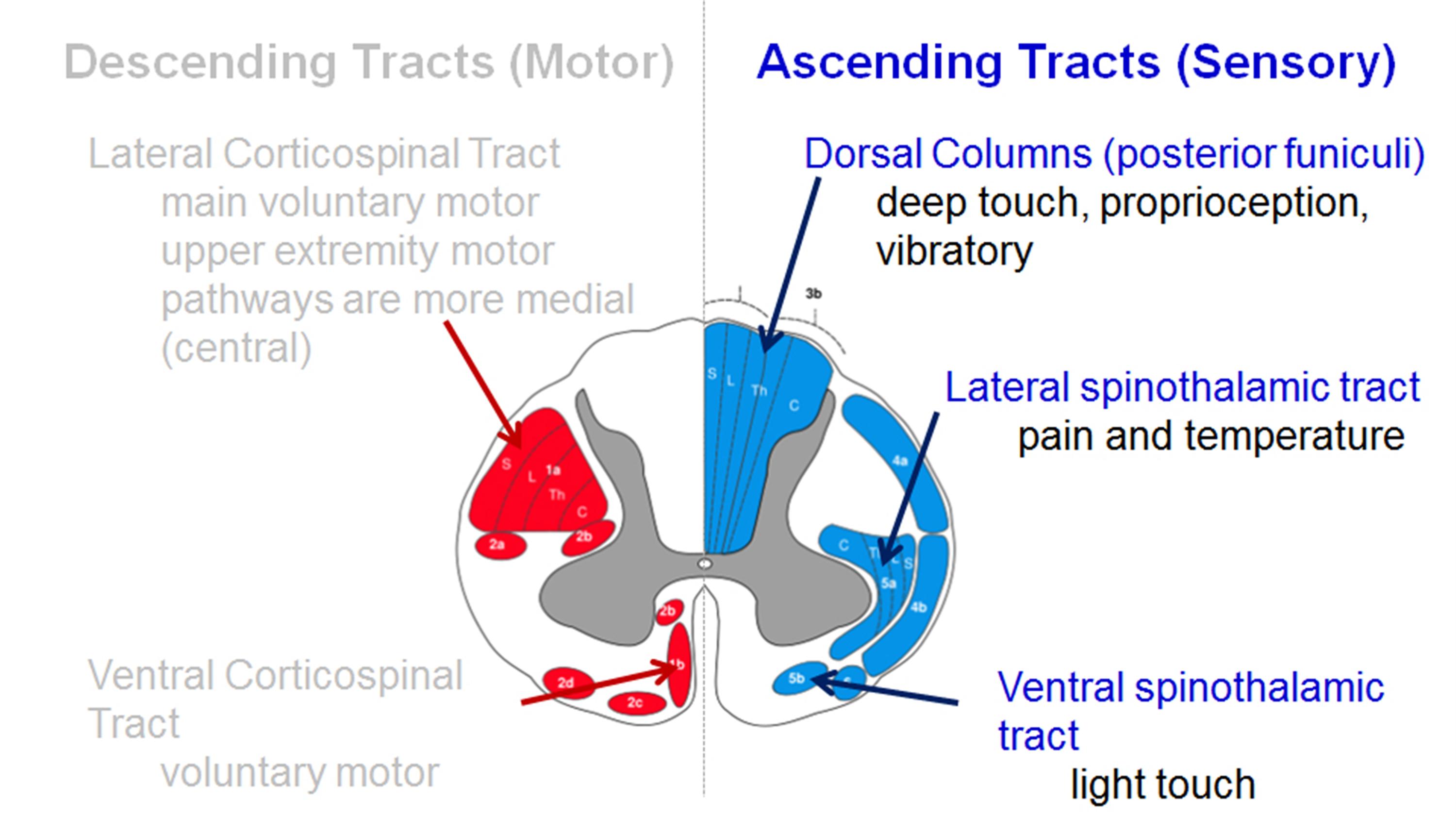
Decreased pain sensation
- pinprick testing should be done to look for a global decrease in sensation or dermatomal changes
- due to the involvement of lateral spinothalamic tract
- vibratory changes are usually only found in the severe case of long-standing myelopathy
Assessment tools
-
Lhermitte sign
-
Romberg test
-
9-Hole peg test
-
Grip and release test (observe a decreasing number of cycles)
-
Timed gait, 30-m walking test
-
Tandem gait
Upper motor neuron signs (spasticity)
- Hyperreflexia – may be absent when there is concomitant peripheral nerve disease (cervical or lumbar nerve root compression, spinal stenosis, diabetes)
- Inverted radial reflex – tapping distal brachioradialis tendon produces ipsilateral finger flexion
- Hoffmann’s sign – snapping patients distal phalanx of the middle finger leads to spontaneous flexion of other fingers
Sustained clonus
- > three beats defined as sustained clonus
- sustained clonus has poor sensitivity (~13%) but high specificity (~100%) for cervical myelopathy
Babinski test
- considered positive with the extension of the great toe
Gait and balance
- toe-to-heel walk patient has difficulty performing
- Romberg test patient stands with arms held forward and eyes closed loss of balance consistent with posterior column dysfunction
- provocative tests: Lhermitte Sign >test is positive when extreme cervical flexion leads to electric shock-like sensations that radiate down the spine and into the extremities
| Motor dysfunction | |
| Upper extremities | |
| 0 | Unable to move hands |
| 1 | Unable to eat with a spoon but able to move hands |
| 2 | Unable to button shirt but able to eat with a spoon |
| 3 | Able to button shirt with great difficulty |
| 4 | Able to button shirt with slight difficulty |
| Lower extremities | |
| 0 | Complete loss of motor & sensory function |
| 1 | Sensory preservation without the ability to move legs |
| 2 | Able to move legs but unable to walk |
| 3 | Able to walk on a flat floor with a walking aid (cane or crutch) |
| 4 | Able to walk up- &/or downstairs w/aid of a handrail |
| 5 | Moderate-to-significant lack of stability but able to walk up &/or downstairs without a handrail |
| 6 | Mild lack of stability but able to walk unaided with smooth reciprocation |
| 7 | No dysfunction |
| Sensory dysfunction | |
| Upper extremities | |
| 0 | Complete loss of hand sensation |
| 1 | Severe sensory loss or pain |
| 2 | Mild sensory loss |
| 3 | No sensory loss |
| Sphincter dysfunction | |
| 0 | Unable to micturate voluntarily |
| 1 | Marked difficulty in micturition |
| 2 | Mild-to-moderate difficulty in micturition |
| 3 | Normal micturition |
[Rx]
Radiographs
- recommended views cervical AP, lateral, oblique, flexion, and extension views
- general findings degenerative changes of uncovertebral and facet joints
- osteophyte formation
- disc space narrowing
- decreased sagittal diameter
- cord compression occurs with canal diameter is < 13mm
- lateral radiograph important to look for the diameter of the spinal canal
- a Pavlov ratio of less than 0.8 suggests a congenitally narrow spinal canal predisposing to stenosis and cord compression
Sagittal alignment
- C2 to C7 alignment determined by tangential lines on the posterior edge of the C2 and C7 body on lateral radiographs in neutral position
- Local kyphosis angle the angle between the lines drawn at the posterior margin of most cranial and caudal vertebral bodies forming the maximum local kyphosis
- Oblique radiograph important to look for foraminal stenosis which often caused by uncovertebral joint arthrosis
- Flexion and extension views important to look for angular or translational instability look for compensatory subluxation above or below the spondylotic/stiff segment
- Sensitivity/specificity changes often do not correlate with symptoms 70% of patients by 70 yrs of age will have degenerative changes seen on plain x -rays
X-ray
- Plain radiographs are an appropriate initial imaging study for neck and upper extremity pain in the absence of “red flag” symptoms. However, degenerative changes seen on imaging often poorly correlate with the presence of neck pain.[rx]
- Common radiographic findings include osteophyte formation, disc space narrowing, endplate sclerosis, degenerative changes of uncovertebral and facet joints, and calcified/ossified soft tissues. AP, lateral, and oblique views of the spine are adequate to evaluate for foraminal stenosis, sagittal alignment, and size of the spinal canal.
- The Torg-Pavlov ratio is obtainable by comparing the sagittal diameter of the spinal canal to the sagittal diameter of the vertebral body. The normal value is 1.0, with a ratio of <0.8 indicating cervical stenosis. Flexion and extension views also merit consideration if there is a concern for ligamentous instability.
MRI
- MRI is a study of choice to evaluate the degree of the spinal cord and nerve root compression effacement of CSF indicates functional stenosis
- Signal changes on T1-weighted images correlate with a poorer prognosis following surgical decompression spinal cord signal changes
- Seen as bright signal on T2 images (myelomalacia) compression ratio of < 0.4 carries poor prognosis CR = smallest AP diameter of cord / largest transverse diameter of the cord
- Sensitivity/specificity has a high rate of false-positive (28% greater than 40 will have findings of HNP or foraminal stenosis)
- CT without contrast can provide complementary information with an MRI and is more useful to evaluate OPLL and osteophytes
- CT myelography more invasive than an MRI but gives excellent information regarding degrees of spinal cord compression
- Useful in patients that cannot have an MRI (pacemaker), or have artifact (local hardware)
- Contrast is given via C1-C2 puncture and allowed to diffuse caudally, or given via a lumbar puncture and allowed to diffuse proximally by putting the patient in Trendelenburg position.
Computed tomography (CT)
- CT provides a good definition of bony structures and is more sensitive than plain radiographs in assessing intervertebral foraminal stenosis in the setting of uncovertebral or facet hypertrophy. However, it is less sensitive than MRI for the evaluation of soft tissues and nerve root compression.
CT myelogram
- CT is most useful when combined with the injection of intrathecal contrast (myelography) to better evaluate the location and amount of neural compression. It is more invasive than an MRI but can be a consideration in patients who have a contraindication to MRI (e.g., pacemaker) or have an artifact from the hardware.
Discogram
- Provocative discography is rarely necessary for cervical spondylosis. It is useful for the evaluation of patients who are experiencing cervical discogenic pain or have multiple herniations in which surgery is a strong possibility. However, the diagnostic procedure remains controversial as it may accelerate the degeneration of normal discs.[rx]
Electromyogram (EMG)
- EMG can be useful in supplementing neuroimaging findings in the diagnosis of cervical radiculopathy. It is especially valuable in differentiating nerve root compression from other possible concomitant neurologic conditions, including peripheral neuropathies, entrapment neuropathies, brachial plexopathies, myopathies, and motor neuron diseases.
Nerve conduction studies high false-negative rate may be useful to distinguish peripheral from the central process (ALS)
- Normal aging mild symptoms of myelopathy often confused with a “normal aging” process
- Stroke
- Movement disorders
- Vitamin B12 deficiency
- Amyotrophic lateral sclerosis (ALS)
- Multiple sclerosis
Treatment of Cervical spondylosis
Nonoperative
Observation, NSAIDs, therapy, and lifestyle modifications
- indications of mild disease with no functional impairment
- the function is a more important determinant for surgery than physical exam finding
- patients who are poor candidates for surgery
- modalities medications (NSAIDS, gabapentin)
- immobilization (hard collar in slight flexion)
- physical therapy for neck strengthening, balance, and gait training
- traction and chiropractic modalities are not likely to benefit and do have some risks
- be sure to watch patients carefully for progression
- outcomes improved nonoperative outcomes associated with patients with larger transverse area of the spinal cord (>70mm2)
- some studies have shown improvement with immobilization in patients with very mild symptoms
Operative
surgical decompression, restoration of lordosis, stabilization
- indications significant functional impairment AND 1-2 level disease lordotic, neutral or kyphotic alignment
- techniques appropriate procedure depends on cervical alignment number of stenotic levels
- medical conditions (e.g., goiter)
- location of compression
- anterior cervical diskectomy/corpectomy and fusion
- posterior laminectomy and fusion
- posterior laminoplasty
- combined anterior and posterior procedure
- cervical disk arthroplasty
- outcomes prospective studies show improvement in overall pain, function, and neurologic symptoms with operative treatment early recognition and treatment prior to spinal cord damage is critical for good clinical outcomes
Goals
optimal surgical treatment depends on the individual. Things to consider include
- number of stenotic levels
- sagittal alignment of the spine
- the degree of existing motion and desire to maintain
- medical comorbidities (eg, dysphasia)
- simplified treatment algorithm
Anterior Decompression and Fusion (ACDF) alone
Indications
- the mainstay of treatment in most patients with single or two level disease
- fixed cervical kyphosis of > 10 degrees anterior procedure can correct kyphosis
- compression arising from 2 or fewer disc segments
- pathology is anterior (OPLL, soft discs, disc osteophyte complexes)
- uses Smith-Robinson anterior approach
Decompression of corpectomy and strut graft may be required for multilevel spondylosis two level corpectomies tend to be biomechanically vulnerable (preferable to combine single-level corpectomy with adjacent level diskectomy)
- 7% to 20% rates of graft dislodgement with cervical corpectomy with associated severe complications, including death, reported.
- fixation anterior plating functions to increase fusion rates and preserve the position of the interbody cage or strut graft
- pros & cons advantages compared to posterior approach lower infection rate blood loss less postoperative pain disadvantages avoid in patients with poor swallowing function
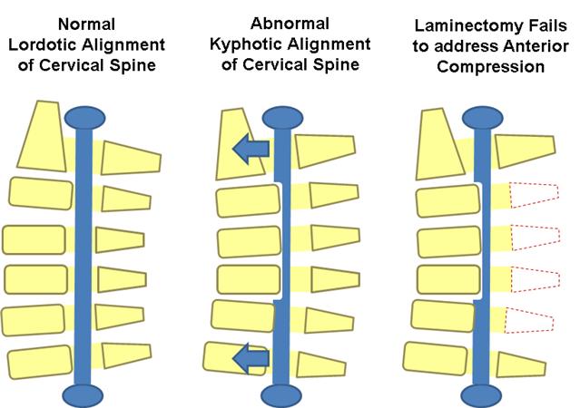
Laminectomy with posterior fusion
- multilevel compression with kyphosis of < 10 degrees,> 13 degrees of fixed kyphosis is a contraindication for a posterior procedure
- in the flexible kyphotic spine, posterior decompression and fusion may be indicated if kyphotic deformity can be corrected prior to instrumentation
Contraindications
- fixed kyphosis of > 10 degrees is a contraindication to posterior decompression
- will not adequately decompress spinal cord as it is “bowstringing” anterior
Pros & cons
- fusion may improve neck pain associated with degenerative facets
- not effective in patients with > 10 degrees fixed kyphosis
Laminoplasty
- gaining in popularity
- useful when maintaining motion is desired
- avoids complications of fusion so may be indicated in patients at high risk of pseudoarthrosis
- cervical kyphosis > 13 degrees is a contraindication to posterior decompression
will not adequately decompress spinal cord as it is “bowstringing” anterior - severe axial neck pain is a relative contraindication and these patients should be fused
Technique
- the volume of the canal is expanded by hinged-door laminoplasty followed by fusion
usually performed from C3 to C7 - open door technique hinges created unilaterally at the junction of lateral mass and lamina and opening on opposite side opening held open by bone, suture anchors, or special plates
- French door technique hinge created bilaterally and the opening created midline
Pros & cons advantages
- lower complication rate than multilevel anterior decompression especially in patients with OPLL a motion-preserving technique pseudoarthrosis not a concern in patients with poor healing potential (diabetes, chronic steroid users) can be combined with a subsequent anterior procedure
- allows for decompression of multilevel stenotic myelopathy without compromising stability and motion (avoids postlaminectomy kyphosis)
Disadvantages
- higher average blood loss than anterior procedures
- postoperative neck pain
- still associated with loss of motion outcomes equivalent to multilevel anterior decompression and fusion
- Combined anterior and posterior surgery multilevel stenosis in the rigid kyphotic spine
- multi-level anterior cervical corpectomies
- postlaminectomy kyphosis
Laminectomy alone
- indications rarely indicated due to risk of post laminectomy kyphosis
- pros & cons progressive kyphosis 11 to 47% incidence if laminectomy performed alone without fusion
Surgical Infection
- higher rate of surgical infection with posterior approach than anterior approach
- Pseudoarthrosis incidence 12% for single level fusions, 30% for multilevel fusions treatment
- treat with either posterior wiring or plating or repeat anterior decompression and plating if patient has symptoms of radiculopathy
- reported to occur in ~ 4.6% of patients after surgery for cervical compression myelopathy
- no significant differences between patients undergoing anterior decompression and fusion and posterior laminoplasty
- occurs immediately postop to weeks following surgery
Mechanism
- mechanism is controversial
- in laminectomy patients, it is thought to be caused by tethering of nerve root with dorsal migration of spinal cord following removal of posterior elements
Prognosis
- patients with postoperative C5 palsy generally have a good prognosis for functional recovery, but recovery takes time
- Recurrent laryngeal nerve injury approach in the past it has been postulated that the RLN is more vulnerable to injury on the right due to a more aberrant pathway recent studies have shown there is not an increased injury rate with a right sided approach
- treatment if you have a postoperative RLN palsy, watch over time
- if not improved over 6 weeks, then ENT consult to scope patient and inject Teflon
- if you are performing revision anterior cervical surgery, and there is any suspicion of an RLN from the first operation, obtain ENT consult to establish prior injury
- if a patient has prior RLN nerve injury, perform revision surgery on the same as the prior injury/approach to prevent a bilateral RLN injury
- Hardware failure and migration 7-20% with two-level anterior corpectomies two-level corpectomies should be stabilized from behind
- Postlaminectomy kyphosis treat with anterior/posterior procedure
- Postoperative axial neck pain
- Vertebral artery injury
- Esophageal Injury
- Dysphagia & alteration in speech
Keywords: degenerative disc disease, cervical spondylosis, cervical spondylotic myelopathy, cervical spine stenosis, anterior cervical discectomy and fusion, cervical laminoplasty, cervical disk replacement
Complications
In a 2019 cohort study by El-Yahochouchi et al., the overall incidence of immediate and delayed adverse events following an epidural steroid injection was 2.4% and 4.9%, respectively.[rx] Complications include:
-
Neurologic injury
-
Epidural abscess
-
Epidural hematoma
-
Increased pain
-
Vasovagal reactions
-
Central steroid response (e.g., facial flushing, nonpositional headaches)
-
Endocrinologic effects (e.g., hyperglycemia, hypothalamic-pituitary axis suppression, decreased bone density)
Complications from anterior and posterior cervical spine surgery include [18][24]:
-
Injury to spinal cord and nerve roots
-
Infection
-
Dural tear and CSF leak
-
Recurrent laryngeal, superior laryngeal, and hypoglossal nerve injuries
-
Esophageal injury and dysphagia
-
Vertebral and carotid artery injuries
-
Tracheal injury
-
Adjacent segment degeneration
-
Pseudoarthrosis
-
Post-laminectomy kyphosis
References
- https://www.ncbi.nlm.nih.gov/pmc/articles/PMC1819511/
- https://www.ncbi.nlm.nih.gov/pmc/articles/PMC5582708/
- https://www.ncbi.nlm.nih.gov/pubmed/17204889
- https://www.ncbi.nlm.nih.gov/pubmed/2536306
- https://www.ncbi.nlm.nih.gov/books/NBK551557/
- https://en.wikipedia.org/wiki/Cervical%20Spondylosis?redirect=no
- https://www.sciencedirect.com/cervical-spondylosis-and-other-disorders-of-the-cervical-spine
- https://www.sciencedirect.com/science/article/pii/B9780750613613500136
- https://www.sciencedirect.com/topics/medicine-and-dentistry/spondylosis
- https://www.sciencedaily.com/releases/2011/04/110411164007.htm
- https://books.google.com/books/about/Manage_and_Cure_Neck_Pain_Cervical_Spond.html?
- https://books.google.com/books/about/Cervical_Spondylosis.html?id=Z6izAAAAIAAJ
- https://books.google.com/books/about/Cervical_Arthrosis.html?id=vhxsAAAAMAAJ
- https://link.springer.com/chapter/10.1007/978-1-59745-106-2_6
- http://link.springer.com/chapter/10.1007/978-3-642-67605-5_21
- https://www.science.gov/topicpages/s/single-level+cervical+spondylosis.html
- https://www.science.gov/topicpages/c/cervical+spondylotic+neuropathy
- https://worldwidescience.org/topicpages/o/onset+cervical+radiculopathy.html


