
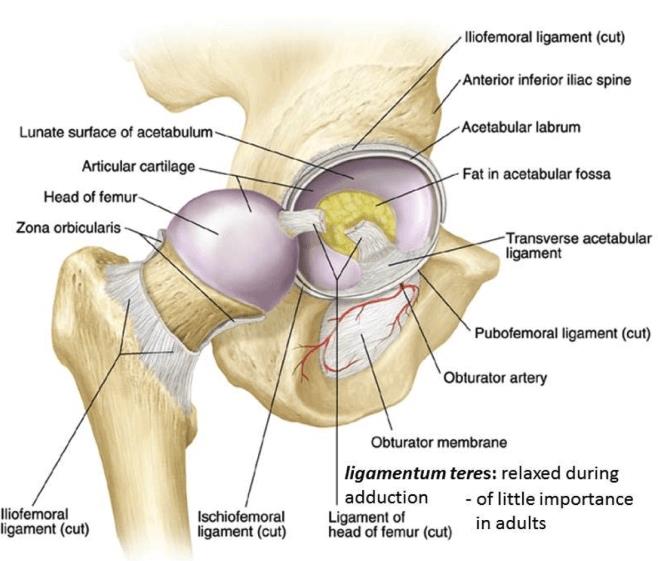
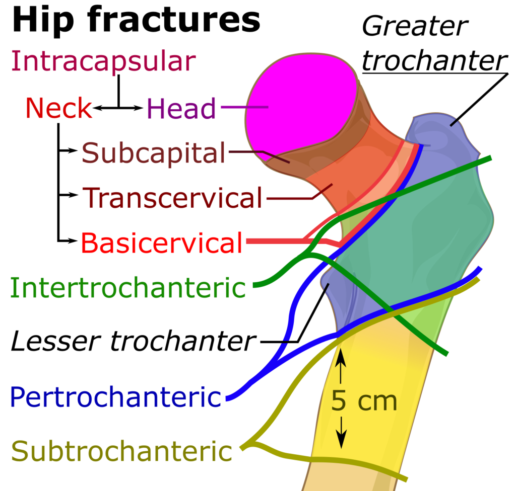
Transcervical femoral neck fracture may be defined as intracapsular fracture, two-parts oblique transcervical fractures go through the base of the femoral neck at its junction between the sub-capital and basicervical femoral neck region with the intertrochanteric region in which the break crosses into the surface of a joint and are limited muscle attachment and maximum cartilaginous part of the femur head. In this type, femur neck fractures are more complicated and may have the greatest chance of osteoporosis, avascular necrosis formation due to limited blood supply.
Causes of Transcervical Femoral Neck Fracture
Causes of Transcervical Femoral Neck Fracture are following
- The repetitive impact – to the lower limb bone with weight-bearing exercises occupational work cause microfractures, which consolidate to stress and avulsion fractures. [rx]
- Heavy influence – The force of a jump or fall from height can result in a broken knee. It can happen in foot bone fractures even if you jump from a high altitude, lift accident in a big shopping mall, or multilevel apartment.
- Missteps – You can cause a fracture of the ankle if you put your foot, and knee down awkwardly abnormally. Your ankle might twist, Suddenly change direction, sprinting, kicking, leaping, or roll your foot joint to the side as you put weight on it. It can also happen in stare up or stare down unconscious.
- Sports – High-impact sports such as football, cricket, hockey, volley boll involve intense movements that place stress on the joints, including the knee bone fracture examples of high-impact sports include cricket, racer of the bike, soccer, football, horseback riding, hockey, skiing snowboarding, in-line skating, jumping on a trampoline, basketball, and hitting, as in a boxer or a defensive lineman in a football game hitting an offensive lineman to protect the quarterback.
- Car collisions – The sudden, heavy impact of a car accident bike accident can cause knee bone fractures. Often, these types of injuries need surgical repair. The crushing injuries common in car accidents may cause breaks that require surgical repair.
- Falls from height – Tripping and falling when walking on uneven surfaces can break bones in knee bone fractures, as can landing on your feet after jumping down from just a slight height, sudden landings from the plane in the war field, downhill, violent trauma, etc.
- Driving and compressing in the break – It is one of the significant causes of hip microtrauma for the driver of the car, motorbike, truck, bus, bicycle runner, suddenly accelerating (getting faster), and suddenly decelerating (going slower). During driving, such a kind of vehicle frequently has to compress breaks to maintain the car’s speed. Repeated compression causes microtrauma, tendon, cartilage, ligament degeneration in the knee joint, and weakness that may lead to injury in the knee bone.
- Missteps – Sometimes, just putting your foot down the wrong way can result in a twisting injury that can cause a broken bone. Fracture also occurs when stairs up or stairs down, especially older people.
- Unconsciously Toilet Use – A widespread and daily increasing incidence of rupture of the hip, knee joint, foot bone, exceptionally high comodo using time, and lower limb fractures.
- High hell Use – It is the most common cause of fracture in the knee, ankle, foot, lower limb fracture, especially for women, abnormal arch, foot angle, the lake of the flat foot, abnormal sole of your footwear, muscle, tendon, cartilage, ligament weakness in the knee, ankle joints cause fracture and dislocation.
- Soldier, armies on the battlefield – With the increasing technology of nuclear weapons on the battlefield, one country is involved in the war from one country to another country. On the battlefield, millions of armies and general people are falling in injury that is gradually causing knee joints bone, tibia, fibula, femur, and patella fractures.
- Have osteoporosis – a disease of your bone that weakens your bones gradually due to inadequate intake of calcium or vitamin D, less exposure to sunlight may lead to fracture of the bone in older age.
- Weak low muscle mass or poor muscle strength – Lack of agility or older age muscle strength, mass, power, endurance becomes weak, and poor balance conditions make you more likely to fall and cause a fracture.
- Walk or do other activities in the snow or on the ice – Especially north region of the world maximum time is low temperature. That frequent water turns into snow and activities that require a lot of forwarding momenta, such as in-line skating and skiing, snowboarding, in-line skating, Jumping, playing lead to fracture of the bone in the lower limb.
- Insufficient vitamin D and sunlight – Insufficient vitamin D and sunlight decrease the intestinal absorption of calcium, leading to abnormal regulation of parathyroid hormone (PTH). Vitamin D also works to upregulate the transcription of genes involved in neovascularization in areas of endochondral ossification, such as a healing fracture site. Vitamin D deficiency is typically characterized as a serum level of 25-hydroxyvitamin D3 of less than 20 ng/mL, and sufficiency is between 20 and 31 ng/mL.[rx]
Symptoms of Transcervical Femoral Neck Fracture
Symptoms of Transcervical Femoral Neck Fracture include
- A fracture means intense pain, bleeding, swelling, tenderness, limited range of motion, fear, abnormal heart pulse rate, is the first symptoms
- May present with pain, swelling, tenderness, hematoma directly over the hip in athletes. Construction workers may present various pain and swell over the knee, foot, worsening with exercise and walking.
- Pain with or after regular activity
- Pain that goes away when resting time and then returns when standing, walking, or during activity
- Pinpoint pain at the site of the fracture when touched
- Swelling but no bruising may be present if it becomes microtrauma
- Bruising or discoloration that extends to nearby parts of the hip bones.
- Pain with walking and weight-bearing
- Pain may decrease with rest but increases again with activity.
- Pain at the fracture site, which in some cases can extend from the foot to the knee.
- Significant swelling may occur along the length of the leg or maybe more localized.
- Blisters may occur over the fracture site after some days.
- Bruising that develops soon after the injury time.
- Inability to walk; it is possible to walk with less severe breaks and fractures, so never rely on walking as a test of whether or not a bone has been fractured.
- Change in the color and appearance of the ankle will look different from the other hip joints.
- Bone protruding fracture through the skin signifies that immediate emergency care is needed. Fractures that pierce the skin require attention because they can lead to severe infection and take a prolonged time to recover.
- This pain may occur or feel in the setting of acute trauma or repetitive microtrauma over weeks to months. One should be suspicious of stress fracture with discomfort or pain of worsening quality or duration over time.
Diagnosis of Transcervical Femoral Neck Fracture
History
Your doctor in the emergency department may ask the following questions about your fracture
- How – How was the fracture created, and, if chronic, why is it still open? (underlying etiology)
- When – How long has this fracture been present? (e.g., regular less than one month acute, more than six months chronic)
- What – What anatomy and structure do it involve? (e.g., epidermis, dermis, subcutaneous tissue, fascia, muscle, tendon, bone, arteries, nerves). What comorbidities, economic or social factors do the patient have which might affect their ability to heal the fracture?
- Where – Where on the body parts is it located? Is it difficult to offload, complicated, or keep clean? Is it in an area of high skin tension? Is it near any vital organ and structures such as a major artery, or low muscle attachment?
- What is your Past – Has your previous medical history of fracture, if have? Are you suffering from any chronic disease, such as hypertension, stroke, blood pressure, diabetes mellitus, previous major surgery? What kind of medicine did you take? What is your food habits, occupation, geographic location, Alcohol, tea, coffee consumption habit, anabolic steroid uses for athletes, etc.?
Physical Examination
Physical examination is done by your doctor, consisting of palpation of the fracture site, eliciting boney tenderness, edema, swelling, bleeding, contamination of infection. If the fracture is in the dept of a joint, the joint motion, normal movement will aggravate the pain.
- Inspection – Your doctor also checks superficial tissue skin color, involving or not only the epidermal layer or Partial-thickness affects the epidermis and extend into the dermis, but full-thickness also extends through the dermis and into the adipose tissues or full-thickness spreads through the dermis. Adipose exposes muscle to bone and evaluates and measures the fracture’s depth, length, and width. Access surrounding skin tissue, fracture margins for tunneling, rolled, undermining fibrotic changes, and if unattached, evaluate for signs and symptoms of infect warm, pain, and delayed healing.
- Palpation – Physical examination may reveal tenderness to palpation, swelling, edema, tenderness, worm, temperature, open fracture, closed fracture, microtrauma, a palpable gap, visible deformation, decreased range of motion, gross motion, passive range of motion, local tenderness, discomfort, guarding, weakness, bruising, inability to bear weight, mild limitation of activity, sudden loss of function, ligament integrity, comparison of laxity, block to motion, muscle function, weakness, crepitus, ecchymosis at the site of fracture. Condition of the surrounding skin and soft tissue, quality of vascular perfusion, loss of tissue planes, a non-concentric joint, widened joint space, or effusion pulses, and the integrity of nerve function.
- Motor function – Your doctor may ask the patient to move the injured area to assist in assessing muscle, ligament, and tendon function. The ability to move the joint means that the muscles and tendons work correctly and do not guarantee bone integrity or stability. The concept that “it can’t be fractured because you can move it” is incorrect. The jerk and manual tests are also performed to investigate the motor function.
- Sensory examination – assesses sensations such as light touch, worm, paresthesia, itching, numbness, and pinprick sensations, in its fracture side. Sensation, motor function, and distal pulses should be assessed manually. There should be a coffee threshold to live Ankle-brachial indices should there be a difference in vibrations between extremities with sensory 2-point discrimination.
- Range of motion – A range of motion examination of the fracture associate joint and it’s surrounding joint may help assess the muscle, tendon, ligament, cartilage stability. Active assisted, actively resisted exercises are performed around the communal injured area.
- Blood pressure and pulse check – Blood pressure is the term used to describe the strength of blood with which your blood pushes on the sides of your arteries as it’s pumped around your body. An examination of the circulatory system, feeling for pulses, blood pressure, and assessing how quickly blood returns to the tip of a toe to heart. It is pressed, the toe turns white (capillary refill).
- Knee effusion – If there’s a big outflow, the knee could also be aspirated to gauge for hemarthrosis and therefore the presence of lipids or bone marrow elements, suggesting intraarticular fracture.
- Compartments – All compartments should be palpated; a firm, tense chamber suggests compartment syndrome, which may be further evaluated by measuring intra compartmental pressure.
- Laxity tests – quite 10 degrees of laxity at the joint line with varus/valgus stress testing suggests a tear of the knee ligaments. Laxity below the common line is indicative of a fracture.
Lab Test
Laboratory tests should be done as an adjunct in overall medical status for surgical treatment. If major surgery is needed for an open fracture
- CBC, ESR test
- Random blood sugar, glucose, and routine diabetes test if the patient has diabetes mellitus.
- Microscopic urine examination test and stool test.
- ECG, EKG test for heart abnormality is present
- Ultrasonography test in some cases.
- Normalized hemoglobin, hematocrit test
- Coagulation profile with bleeding time and coagulation time test, prothrombin time (PT) test for surgery if needed,
- Thromboplastin time and platelet counts will be required for operative intervention.
- Serum creatinine test,
- Serum lipid profile
- Serum uric acid test
Imaging Test
- X-Ray – When cases amenable for radiographic evaluation are selected, three radiographic views should be obtained consistent with the American College of Radiology guidelines: anteroposterior (AP), lateral, and all view. The AP view is performed along the long axis of the foot. In isolated hip fractures, this view is especially useful to gauge signs of the associated knee joints and/or syndesmotic instability through the analysis of coronal inclination, patella overlap, tibiofibular clear space, and medial clear space (MCS). In the lateral view, the hip must be centered and congruent with the knee joints. This view is beneficial in isolated knee fractures to demonstrate AP displacement and external rotation type fractures.
- CT Scan – Given these findings, computerized tomography (CT) is the gold standard for diagnosis, though only 91% of fractures were properly evaluated with a CT scan at a level trauma center. CT should be a part of routine surveillance of knee injuries that have swelling and pain disproportionate to radiographic findings, as 6.9% of knee fractures were undiagnosed at the time of presentation. Even when an x-ray demonstrates the fracture pattern, CT provides additional information on the degree of comminution, articular involvement, and surgical planning.
- MRI – It is a more advanced stage diagnostic procedure of magnetic resonance imaging (MRI) to evaluate any incongruity, trapped osteochondral fragment, chondral damage, soft tissue integrity, malalignment, even small incisional fractures.[rx][rx]
- Multi-Detector computerized tomography (MDCT) images have both higher sensibility and specificity than radiography. CT images are often more easily interpreted even when anatomical relations are subverted. MPR images should be performed along the anatomical axes of the knee. MDCT evaluation with MPR and VRT reconstruction is recommended to best assess fracture(s), anatomical relationship, degree of comminution, fracture angle, eventual intraarticular loose bodies. CT is additionally needed to guide management decisions and for surgical planning.
- Ultrasound (US) and resonance Imaging (MRI) has a limited role within the acute setting of fractures they will be useful during a re-evaluation for the evaluation of the soft-tissue injury, especially for the evaluation of all knee, hips ligaments, knee ligaments.
Treatment of Transcervical Femoral Neck Fracture
Initial Treatment Includes
- Get medical help immediately – If you fall on an outstretched leg, play cricket, get into a car accident, or are hit while playing a sport and feel intense pain in your leg area, then get medical care immediately. Cause significant pain in your hip, leg, foot, ankle joint, and part of your leg closer to the base of your shank. If the accident is substantial, you keep your leg at the same heart position and then clean and treat any wounds on the skin of the injured hip. Aggressive wound care is essential for patients to reach a safe place with the proper ventilation needed for contaminated wounds. Injured are clear with disinfectant material [rx]
- ICE apply – It helps to prevent swelling and edema by constricting the blood vessel.
- Rest – Sometimes, rest is all that is needed to treat a traumatic fracture of the hip, knee, foot, ankle, tarsal and metatarsal fracture. Sometimes rest is the only treatment required to eradicate healing of a stress or traumatic fracture of a hip bone fracture.
- Compression – a bandage will limit swelling edema and help to rest the joint. A tubular compression bandage is frequently used but should be removed at night by easing it off gradually. Please put it on again before you are out of bed in the morning. Mild to moderate pressure that is not too uncomfortable or too tight, and does not stop blood flow, is ideal. Depending on the amount of swelling. Pain, edema, you may be advised to remove the bandage for good after 48 hours.
- Elevation – Elevation initially aims to limit and reduce any swelling. For example, keep the foot upright on a chair or pillow to at least hip level when sitting. When you are in bed, put your foot on a pillow. Sometimes rest is the only treatment that is needed, even in fractures.
- End-of-Bed Skeletal Traction – Consider traction on a case-by-case basis. Traction is usually not required in elderly patients with low-energy mechanisms. However, traction is useful in younger patients because the strong muscular attachments cause shortening and therefore the flexed proximal fragment may threaten skin anteriorly. After assessment of the knee radiographically, a distal femoral or proximal tibial traction pin could also be placed. End-of-bed traction of 12 pounds (5 kg) is usually used and should be adjusted to support the patient’s weight. Skeletal traction greater than 20 pounds (9 kg) isn’t recommended.
- Splinting – The toe may be fitted with a splint to keep it in a fixed position.
- Rigid or stiff-soled shoes – Wearing stiff-soled solid shoes to protect the toe and help properly position it. A postoperative splint, shoe, or boot walker is also helpful.
- Avoid the offending activity – Because fractures result from repetitive stress trauma, it is essential to avoid the movement that led to the rupture more seriously. Crutches, a wheelchair, or other types of supporting splint are sometimes required to offload weight from the foot to give it time to heal.
- Immobilization, casting, or rigid shoe – A stiff-soled shoe or another form of immobilization may be used to protect the fractured bone while it is healing. The use of a postoperative shoe or boot walker is also helpful.
- Casting or rigid shoe – A stiff-soled shoe or another form of immobilization may be used to protect the fractured bone while it is healing. The use of a postoperative shoe or boot walker is also helpful.
- Stop stressing the foot – If you’ve been diagnosed with a stress fracture, avulsion fracture avoiding the activity that caused it is essential for healing. This may mean using crutches or even a wheelchair.
Do no HARM for 72 hours after injury.
- Heat – Heat applied to fracture and injured side by hot baths, electric heat, saunas, heat packs, etc, has the opposite effect on the blood flow. Heat may cause more fluid accumulation in the fracture joints by encouraging dilating blood vessels. Heat should be avoided when inflammation is developing in the acute stage or the first 72 hours from the injury time. However, after about 72 hours, no further inflammation is likely to create, and warmth, gentle exercise can be done for soothing pain.
- Alcohol – stimulates the central nervous system, can increase bleeding and swelling, decrease healing, and also causes drugs to drugs interaction.
- Running and movement – Running and walking, or any kind of movement in fracture joint, associate soft tissue may cause further damage, dislocation, and causes healing delay.
- Massage – A massage also may increase bleeding and swelling. However, after 72 hours of your fracture, you can take a simple message, and applying heat may soothe the pain.
Medication
Your doctor may consider the following medications to relieve acute and immediate pain, long term treatment
- Antibiotic – Cefuroxime or Azithromycin, or Flucloxacillin or any other cephalosporin/quinolone, meropenem antibiotic must be used to prevent infection or clotted blood removal from preventing further swelling, inflammation, and edema. [rx] Antibiotics should be administered as soon as possible within the prehospital setting or emergency department if antibiotics got within 66 minutes of injury, the infection rate was 0%, but increased to 17% if antibiotics were delayed beyond this point frame.[rx]
- NSAIDs – Prescription-strength drugs that reduce both pain and inflammation. Pain medicines and anti-inflammatory drugs help relieve pain and stiffness, allowing for increased mobility and exercise. There are many common over-the-counter medicines called non-steroidal anti-inflammatory drugs (NSAIDs). They include first choice NSAIDs is Ketorolac, then Etoricoxib, then Aceclofenac, naproxen. As you are taking pain medication or NSAIDs, your doctor must prescribe a standard anti-ulcer drug, such as omeprazole, pantoprazole, esomeprazole, dexlansoprazole, etc.
- Muscle Relaxants – These medications relieve spinal muscle spasms spasticity. Muscle relaxants, such as baclofen, tolperisone, eperisone, methocarbamol, carisoprodol, and cyclobenzaprine, may be prescribed to control postoperative muscle spasms, spasticity, stiffness, contracture.
- Calcium & vitamin D3 – To improve bone health, blood clotting, helping muscles contract, regulating heart rhythms, nerve functions, and healing fractures. As a general rule, too absorbed more minerals for men and women aged 50 and older should consume 1,200 milligrams of calcium and 600 international units of vitamin D a day to heal back pain, fractures, and osteoarthritis.
- Neuropathic Agents – Drugs(pregabalin & gabapentin) address neuropathic—or nerve-related—pain. This includes burning, numbness, tingling sensation, and paresthesia.
- Glucosamine & Diacerein, Chondroitin sulfate – To tighten the loose tendon, cartilage, ligament, and cartilage, ligament regenerates cartilage or inhabits the further degeneration of cartilage, ligament. The dosage of glucosamine is 15oo mg per day in divided dosage and chondroitin sulfate approximately 500mg per day in different dosages. Diacerein minimum of 50 mg per day may be taken if the patient suffers from osteoarthritis, rheumatoid arthritis, and any degenerative joint disease.[rx]
- Topical Medications and essential oil – These prescription-strength creams, gels, ointments, patches, and sprays help relieve pain and inflammation in acute trauma, pain, swelling, tenderness through the skin. If the fracture is closed and not open, you can use this item.
- Antidepressants – A drug that blocks pain messages from your brain and boosts the effects of endorphins in your body’s natural painkillers. It also helps in neuropathic pain, anxiety, tension, and proper sleep.
- Corticosteroids – Also known as oral steroids, these medications reduce inflammation. To heal the nerve inflammation and clotted blood in the joints.
- Dietary supplement – To eradicate the healing process from fracture, your body needs a considerable amount of vitamin C and vitamin E. From your dietary supplement, you can get it, and also need to remove general weaknesses & improved health.
- Cough Syrup – If your doctor finds any chest congestion or fracture-related injury in your chest, dyspnoea, post-surgical breathing problem, then advice you to take bronchodilator cough syrup.
- Teriparatide – Teriparatide may be a recombinant human parathormone (PTH) that stimulates osteoblasts to grow new bone cells when given intermittently. Use in treatment for osteoporosis, osteoarthritis, and all kinds of fractures are successful results. There are now some scientists to support its use in treating fractures and nonunions. The medication was administered via daily injections of 20 μg about a month[rx] [rx]
What To Eat and What to avoid
Eat Nutritiously During Your Recovery
All bones and tissues in the body need certain micronutrients to heal appropriately and promptly. Eating a nutritious and balanced diet that includes lots of minerals and vitamins is proven to help heal broken bones and all types of fractures. Therefore, focus on eating lots of fresh food (fruits and veggies), whole grains, cereal, beans, lean meats, seafood, and fish to give your body the building blocks needed to repair your fracture correctly. In addition, drink plenty of purified mineral water, milk, and other dairy-based non-alcoholic beverages to augment what you eat.
- Broken bones or fractures need abundant minerals (calcium, phosphorus, magnesium, boron, selenium, omega-3) and protein to become strong and healthy again.
- Excellent sources of minerals/protein include dairy products, tofu, beans, broccoli, carrot, cabbage, nuts and seeds, sardines, sea fish, and salmon.
- Essential vitamins that are needed for bone healing include vitamin C (needed to make collagen that your important body element for healing open fractures), vitamin D (crucial for mineral absorption, or machine for mineral absorber from your food by small, and large intestine), and vitamin K (binds calcium to bones and triggers more quickly collagen formation).
- Conversely, don’t consume food or drink that is known to impair bone/tissue healing, such as alcoholic beverages, sodas, fried fast food, most fast food items, sugar, and foods made with lots of factory refined sugars and preservatives food.
Surgery
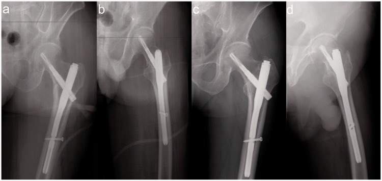
Surgically treatment depends on the individual fracture characteristics, pattern, the size of fractures, the degree of displacement, dislocation, previous fracture at the same place, bone ridge, poor muscle attachment, low blood vessel supply, the location, comminution, the condition of integrity of the soft tissues of the knee joint, the presence of associate bones, multiple fractures, tendon, ligament injuries on the knee joint, comorbidities, and overall functional movement status or your doctor’s decision. [rx]
References
- https://www.ncbi.nlm.nih.gov/pmc/articles/PMC4728702/
- https://www.ncbi.nlm.nih.gov/books/NBK493161/
- https://www.ncbi.nlm.nih.gov/pmc/articles/PMC3834630/
- https://www.ncbi.nlm.nih.gov/pmc/articles/PMC7313392/
- https://www.ncbi.nlm.nih.gov/pmc/articles/PMC6921538/
- https://www.ncbi.nlm.nih.gov/books/NBK559168/
- https://www.ncbi.nlm.nih.gov/books/NBK507803/
- https://www.ncbi.nlm.nih.gov/pmc/articles/PMC7724026/
- https://www.ncbi.nlm.nih.gov/pmc/articles/PMC2903134/
- https://www.ncbi.nlm.nih.gov/pmc/articles/PMC6753548/
- https://www.ncbi.nlm.nih.gov/pmc/articles/PMC6904631/
- https://www.ncbi.nlm.nih.gov/pmc/articles/PMC6205015/
- https://www.ncbi.nlm.nih.gov/pmc/articles/PMC6583643/
- https://www.ncbi.nlm.nih.gov/pmc/articles/PMC3062872/
- https://www.ncbi.nlm.nih.gov/pmc/articles/PMC8013639/
- https://www.ncbi.nlm.nih.gov/pmc/articles/PMC6531999/
- https://www.mdpi.com/2076-3417/11/10/4395
- https://www.cambridge.org/core/journals/journal-of-glaciology/article/novel-method-for-predicting-fracture-in-floating-
- https://www.amazon.com/Biomechanics-Orthopaedic-Trauma-Fracture-Fixation/dp/1853171085
- https://www.amazon.com/Fracture-Management-Primary-Care-Updated/dp/0323546552
- https://online.boneandjoint.org.uk/journal/bjo
- https://www.sciencedirect.com/topics/medicine-and-dentistry/avulsion-fracture
- https://medlineplus.gov/
- https://books.google.com.bd/books?id=XvbsDwAAQBAJ&pg
- https://en.wikipedia.org/wiki/Bone_fracture
- https://en.wikipedia.org/wiki/Fracture
- https://www.ncbi.nlm.nih.gov/pmc/articles/PMC6667981/
- https://www.ncbi.nlm.nih.gov/books/NBK448147/
- https://www.ncbi.nlm.nih.gov/pmc/articles/PMC5903328/
- https://www.ncbi.nlm.nih.gov/pmc/articles/PMC3134017/
- https://www.ncbi.nlm.nih.gov/pmc/articles/PMC5961267/
- https://www.ncbi.nlm.nih.gov/pmc/articles/PMC6379735/
- https://www.ncbi.nlm.nih.gov/books/NBK542250/
- https://www.ncbi.nlm.nih.gov/books/NBK549912/
- https://www.ncbi.nlm.nih.gov/pmc/articles/PMC3445231/
- https://www.ncbi.nlm.nih.gov/books/NBK542221/
- https://www.sciencedirect.com/science/article/pii/S1930043315301862
- https://www.ncbi.nlm.nih.gov/pmc/articles/PMC5903328/
- https://www.ncbi.nlm.nih.gov/pmc/articles/PMC5961267/
- https://www.ncbi.nlm.nih.gov/pmc/articles/PMC5721952/
- https://www.ncbi.nlm.nih.gov/pmc/articles/PMC5961263/
- https://www.ncbi.nlm.nih.gov/pmc/articles/PMC6179081/
- https://www.ncbi.nlm.nih.gov/pmc/articles/PMC5961262/
- https://www.ncbi.nlm.nih.gov/pmc/articles/PMC5481613/
- https://www.ncbi.nlm.nih.gov/pmc/articles/PMC8241249/
- https://www.ncbi.nlm.nih.gov/books/NBK430861/
- https://www.ncbi.nlm.nih.gov/pmc/articles/PMC3360186/
- https://www.ncbi.nlm.nih.gov/pmc/articles/PMC5357522/
- https://www.ncbi.nlm.nih.gov/pmc/articles/PMC6583145/
- https://en.wikipedia.org/wiki/Calcaneal_fracture
- https://www.ncbi.nlm.nih.gov/books/NBK430861/
- https://www.ncbi.nlm.nih.gov/books/NBK430861/figure/article-18765.image.f3/
- https://www.ncbi.nlm.nih.gov/books/NBK77454/
- https://www.ncbi.nlm.nih.gov/pmc/articles/PMC5961258/
- https://www.ncbi.nlm.nih.gov/pmc/articles/PMC2551728/
- https://www.ncbi.nlm.nih.gov/pmc/articles/PMC5017091/
- https://www.ncbi.nlm.nih.gov/pmc/articles/PMC5090255/
- https://www.ncbi.nlm.nih.gov/pmc/articles/PMC6178141/
- https://www.ncbi.nlm.nih.gov/pmc/articles/PMC7330149/
- https://www.ncbi.nlm.nih.gov/pmc/articles/PMC1317258/
- https://www.ncbi.nlm.nih.gov/pmc/articles/PMC7767678/
- https://www.ncbi.nlm.nih.gov/books/NBK482332/
- https://en.wikipedia.org/wiki/Trimalleolar_fracture
- https://www.ncbi.nlm.nih.gov/pmc/articles/PMC7864963/
- https://en.wikipedia.org/wiki/Tillaux_fracture
- https://www.sciencedirect.com/science/article/abs/pii/S1067251696800299
- https://www.ncbi.nlm.nih.gov/books/NBK562254/
- https://www.ncbi.nlm.nih.gov/books/NBK470593/
- https://www.ncbi.nlm.nih.gov/pmc/articles/PMC8152440/
- https://www.ncbi.nlm.nih.gov/books/NBK542324/
- https://www.ncbi.nlm.nih.gov/books/NBK470591/
- https://www.ncbi.nlm.nih.gov/pmc/articles/PMC3427429/
- https://www.ncbi.nlm.nih.gov/pmc/articles/PMC2903122/
- https://www.ncbi.nlm.nih.gov/pmc/articles/PMC6502107/
- https://www.ncbi.nlm.nih.gov/books/NBK535347/
- https://www.ncbi.nlm.nih.gov/books/NBK431095/
- https://www.ncbi.nlm.nih.gov/pmc/articles/PMC5553490/

