
Common Forearm Fractures /The forearm is the part of the arm between the wrist and the elbow. It is made up of two bones: the radius and the ulna. Forearm fractures are common in childhood, accounting for more than 40% of all childhood fractures. About three out of four forearm fractures in children occur at the wrist end of the radius.
Forearm fractures often occur when children are playing on the playground or participating in sports. If a child takes a tumble and falls onto an outstretched arm, there is a chance it may result in a forearm fracture. A child’s bones heal more quickly than an adult’s, so it is important to treat a fracture promptly—before healing begins—to avoid future problems.
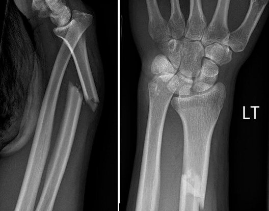
Athlete Forearm Fractures – Types, Classifications
Colles’, Smith’s, Isolated Radial Shaft Fractures, Both Bone Fractures – The Colles’ fracture is the most common fracture of the distal radius in adults. It gets its name from Irish Surgeon, Dr. Abraham Colles, who first described this injury pattern in 1814. The mechanism of injury is classically a FOOSH. It is a metaphyseal fracture that occurs around 1.5 inches proximal to the carpal articulation. Characteristically it presents with dorsal angulation and displacement of the distal fragment of the radius. On X-Ray, the wrist will present with what is known as the “dinner-fork” deformity. Smith’s fracture is essentially the opposite of the Colles’ fracture. It is often referred to as a “reverse Colles’,” and occurs with a fall onto or a direct blow or force to the dorsum of the hand.
Chauffeur’s/Radial Styloid Fracture – The Chauffeur’s fracture is an intra-articular fracture of the radius that includes the radial styloid. The fracture fragment can be variable in size. The injury is often the result of a FOOSH injury with a blow to the back of the wrist causing dorsiflexion and abduction causing the scaphoid to compress against the radial styloid. Patients may have small avulsions of the radial styloid that are not clinically significant, but these injuries are often associated with disruption of the radioscaphocapitate and other collateral ligaments; this can lead to lunate dislocation and scapholunate disruption. These fractures were historically suffered by drivers who would need to start their cars using a hand crank. Occasionally these cranks would backfire and forcefully strike drivers on the back of the wrist.
Die-Punch Fracture – A die punch fracture is an intra-articular fracture involving the lunate facet of the radius. The lunate facet is one of the three articular surfaces of the distal radius. It lies between the ulnar articulation and the scaphoid facet. It connects the distal radius to the lunate bone in the wrist. A die-punch fracture occurs with axial loading of the lunate, which causes an impaction fracture to the lunate facet of the radius. This fracture often occurs in isolation but can have associated injuries.
Galeazzi Fracture-Dislocation – The Galeazzi fracture-dislocation is a fracture of the distal third of the radius with an associated distal radioulnar joint (DRUJ) dislocation. These fractures are typically the result of FOOSH injuries. It is an uncommon injury pattern, and the DRUJ component is easi to miss for clinicians. They are labeled based on the direction of ulnar displacement. For example, if the DRUJ disruption causes volar deviation of the ulna, this is classified as a “Volar Galeazzi.”
Barton’s Fracture – A Barton’s fracture is an intra-articular rim fracture of the distal radius. It can be classified as either dorsal or volar. Dorsal rim fractures are more common and result from forced dorsiflexion and pronation. Volar rim fractures often occur with a fall onto a supinated hand/wrist. These forces disrupt the radiocarpal ligaments and subsequent avulsion fracture of the radial rim. In dorsal fractures, the avulsed fragment migrates dorsally. The opposite is true with volar fractures. These fractures are unstable and often present with a dislocation of the carpal bones. [rx][rx]
Greenstick and Buckle/Torus Fractures – Both Torus and greenstick fractures are incomplete fractures. Pediatric bones are poorly mineralized relative to adults and can bend without frankly breaking. These fractures can occur in any long bone but frequently occur in the metaphysis of the distal radius. Torus fractures occur with axial loading whereas Greenstick fractures result from bending forces. Torus fractures are characterized by buckling of the bony cortex and periosteum without any true fracture lines. There is generally minimal deformity with Torus fractures, and the periosteum and cortex are intact. Greenstick fractures will show bony bending. There will be a fracture of the convex surface with an intact concave surface. These fractures are extremely common in children, and unfortunately frequently missed.
Salter-Harris Type Fractures – A Salter-Harris fracture is a pediatric fracture that involves the epiphyseal plate. These fractures can occur in any bone that has a growth plate but frequently occur in the distal radius. The Salter-Harris scheme was first developed by Doctors William Harris and Robert Salter in 1963 and remains the most common classification system for epiphyseal fractures. Salter-Harris fractures are graded I through IX, with I through V being the most frequently used in clinical practice. Type I is a fracture that runs transversely through the growth plate. Type II runs through the growth plate and the metaphysis. Type III involves the growth plate and epiphysis. Type IV is a fracture of metaphysis, epiphysis, and growth plate. Type V is a complete direct compression fracture of the growth plate. Each of these has a different prognosis and management. [rx][rx][rx]
Torus fracture – This is also called a buckle fracture. The topmost layer of bone on one side of the bone is compressed, causing the other side to bend away from the growth plate. This is a stable fracture, meaning that the broken pieces of bone are still in position and have not separated apart (displaced).
Metaphyseal fracture – The fracture is across the upper or lower portion of the shaft of the bone and does not affect the growth plate.
Growth plate fracture – Also called a physical fracture, this fracture occurs at or across the growth plate. In most cases, this type of fracture occurs in the growth plate of the radius near the wrist. Because the growth plate helps determine the future length and shape of the mature bone, this type of fracture requires prompt attention.
Differential Diagnosis
-
Smith’s fracture
-
Barton fracture
-
Chauffeur’s fracture/radial styloid fracture
-
Isolated distal radial fracture
-
Both bone/radial and ulnar fracture
-
Scaphoid/carpal bone fracture
-
Scaphoid/carpal bone dislocation
-
Distal radioulnar joint dislocation
-
Carpal ligamentous disruption/TFCC injury
-
Die-punch fracture
-
Monteggia fracture
-
Galeazzi fracture
-
Greenstick fracture
-
Torus/Buckle fracture
-
Salter-Harris/growth plate fracture of radius (pediatric)
Do no HARM for 72 hours after injury
- Heat – Heat applied to fracture and injured side by hot baths, electric heat, saunas, heat packs, etc has the opposite effect on the blood flow. Heat may cause more fluid accumulation in the fracture joints by encouraging blood flow. Heat should be avoided when inflammation is developing in the acute stage. However, after about 72 hours, no further inflammation is likely to develop and heat can be soothing.
- Alcohol – stimulates the central nervous system that can increase bleeding and swelling and decrease healing.
- Running and movement – Running and walking may cause further damage, and causes healing delay.
- Massage – A massage also may increase bleeding and swelling. However, after 72 hours of your fracture, you can take a simple message, and applying heat may be soothing the pain.
Medication
The following medications may be considered by your doctor to relieve acute and immediate pain, long term treatment
- Antibiotic – Cefuroxime or Azithromycin, or Flucloxacillin or any other cephalosporin/quinolone, meropenem antibiotic must be used to prevent infection or clotted blood removal to prevent further swelling, inflammation, and edema.
- NSAIDs – Prescription-strength drugs that reduce both pain and inflammation. Pain medicines and anti-inflammatory drugs help to relieve pain and stiffness, allowing for increased mobility and exercise. There are many common over-the-counter medicines called non-steroidal anti-inflammatory drugs (NSAIDs). They include first choice NSAIDs is Ketorolac, then Etoricoxib, then Aceclofenac, naproxen.
- Muscle Relaxants – These medications provide relief from spinal muscle spasms, spasticity. Muscle relaxants, such as baclofen, tolperisone, eperisone, methocarbamol, carisoprodol, and cyclobenzaprine, may be prescribed to control postoperative muscle spasms, spasticity, stiffness, contracture.
- Calcium & vitamin D3 – To improve bone health, blood clotting, helping muscles to contract, regulating heart rhythms, nerve functions, and healing fractures. As a general rule, too absorbed more minerals for men and women age 50 and older should consume 1,200 milligrams of calcium a day, and 600 international units of vitamin D a day to heal back pain, fractures, osteoarthritis.
- Neuropathic Agents – Drugs(pregabalin & gabapentin) that address neuropathic—or nerve-related—pain. This includes burning, numbness, tingling sensation, and paresthesia.
- Glucosamine & Diacerein, Chondroitin sulfate – can be used to tighten the loose tendon, cartilage, ligament, and cartilage, ligament regenerates cartilage or inhabits the further degeneration of cartilage, ligament. The dosage of glucosamine is 15oo mg per day in divided dosage and chondroitin sulfate approximately 500mg per day in different dosages, and diacerein minimum of 50 mg per day may be taken if the patient suffers from osteoarthritis, rheumatoid arthritis, and any degenerative joint disease.[rx]
- Topical Medications and essential oil – These prescription-strength creams, gels, ointments, patches, and sprays help relieve pain and inflammation in acute trauma, pain, swelling, tenderness through the skin. If the fracture is closed and not open fracture then you can use this item.
- Antidepressants – A drug that blocks pain messages from your brain and boosts the effects of endorphins in your body’s natural painkillers. It also helps in neuropathic pain, anxiety, tension, and proper sleep.
- Corticosteroids – Also known as oral steroids, these medications reduce inflammation. To heal the nerve inflammation and clotted blood in the joints.
- Dietary supplement – To eradicate the healing process from fracture your body needs a huge amount of vitamin C, and vitamin E. From your dietary supplement, you can get it, and also need to remove general weaknesses & improved health.
- Cough Syrup – If your doctor finds any chest congestion or fracture-related injury in your chest, dyspnoea, post-surgical breathing problem, then advice you to take bronchodilator cough syrup.
What To Eat and What to avoid
Eat Nutritiously During Your Recovery
All bones and tissues in the body need certain micronutrients in order to heal properly and in a timely manner. Eating a nutritious and balanced diet that includes lots of minerals and vitamins is proven to help heal broken bones and all types of fractures. Therefore, focus on eating lots of fresh food produce (fruits and veggies), whole grains, cereal, beans, lean meats, seafood, and fish to give your body the building blocks needed to properly repair your fracture. In addition, drink plenty of purified mineral water, milk, and other dairy-based beverages to augment what you eat.
- Broken bones or fractures bones need ample minerals (calcium, phosphorus, magnesium, boron, selenium, omega-3) and protein to become strong and healthy again.
- Excellent sources of minerals/protein include dairy products, tofu, beans, broccoli, nuts and seeds, sardines, sea fish, and salmon.
- Important vitamins that are needed for bone healing include vitamin C (needed to make collagen that your body essential element), vitamin D (crucial for mineral absorption, or machine for mineral absorber from your food), and vitamin K (binds calcium to bones and triggers more quickly collagen formation).
- Conversely, don’t consume food or drink that is known to impair bone/tissue healing, such as alcoholic beverages, sodas, fried fast food, most fast food items, and foods made with lots of refined sugars and preservatives.
References
- https://www.ncbi.nlm.nih.gov/books/NBK536916/
- https://www.ncbi.nlm.nih.gov/books/NBK470188/figure/article-22027.image.f1/
- https://en.wikipedia.org/wiki/Forarms_fructure
- https://orthoinfo.aaos.org/en/diseases–conditions/forearm-fractures-in-children
- https://orthoinfo.aaos.org/en/diseases–conditions/adult-forearm-fractures/
- https://www.hopkinsmedicine.org/health/conditions-and-diseases/ulna-and-radius-fractures-forearm-fractures

