
Quadratus Lumborum (QL) – Origin, Nerve Supply, Function

Patient Tools

Read, save, and share this guide

Use these quick tools to make this medical article easier to read, print, save, or share with a family member.

On this page6 sections

Article Summary

Quadratus Lumborum (QL) muscle resides in the deep and posterior, lateral, and inferior areas of the spine, involving the iliac crest, the transverse processes of the lumbar vertebrae, and the 12th rib. The muscular organization is complex, and it is difficult to identify precisely the actions that occur through the contraction of fibers. It is an integral part of the thoracolumbar fascia. There is still...

Key Takeaways

  • This article explains Origin of Quadratus Lumborum (QL) in simple medical language.
  • This article explains Nerves of Quadratus Lumborum (QL) in simple medical language.
  • This article explains Blood Supply of Quadratus Lumborum (QL) in simple medical language.
  • This article explains Function in simple medical language.
Before reading

RX Patient Tools

Use these quick guides before reading the article, or return to them when you need help preparing questions for a doctor.

Start here Choose the right pathway for symptoms, reports, medicines, or urgent warning signs. Disease article roadmap Read this topic step by step: meaning, symptoms, warning signs, diagnosis, treatment, prevention, and follow-up. Treatment planner Prepare questions about treatment choices, benefits, risks, side effects, and follow-up. Family & caregiver guide Organize symptoms, reports, medicines, questions, and follow-up safely. Nutrition & diet guide Prepare food, hydration, supplement, and medicine-timing questions safely. Prevention guide Organize risk factors, protective habits, screening, and warning signs. Recovery guide Prepare a safe plan for activity, rehabilitation, warning signs, and follow-up.
Educational health guideWritten for patient understanding and clinical awareness.
Reviewed content workflowUse writer and reviewer profiles for stronger trust.
Emergency safety firstUrgent warning signs are highlighted below.
Definition

Quadratus Lumborum (QL) muscle resides in the deep and posterior, lateral, and inferior areas of the spine, involving the iliac crest, the transverse processes of the , and the 12th rib. The muscular organization is complex, and it is difficult to identify precisely the actions that occur through the contraction of fibers. It is an integral part of the thoracolumbar . There is still no certainty that an of QL is the primary source of . The QL could potentially act as a crossroad of the forces exerted by the neighboring muscles, influencing the vectors of the different tensions produced. Thanks to its strategic position and the entropic scheme of its fibers, QL is a significant means of access for anesthesia, during surgery on the back, lower limbs or abdominal area. The article reviews the latest anatomical and information on the quadratus lumborum.

The quadratus lumborum muscles originate from the wings of the ; their insertions are on the transverse processes of the upper four lumbar vertebrae plus the lower posterior border of the twelfth rib. Contraction of one of the pair of muscles causes lateral flexion of the , the elevation of the , or both. Contraction of both causes extension of the lumbar spine.
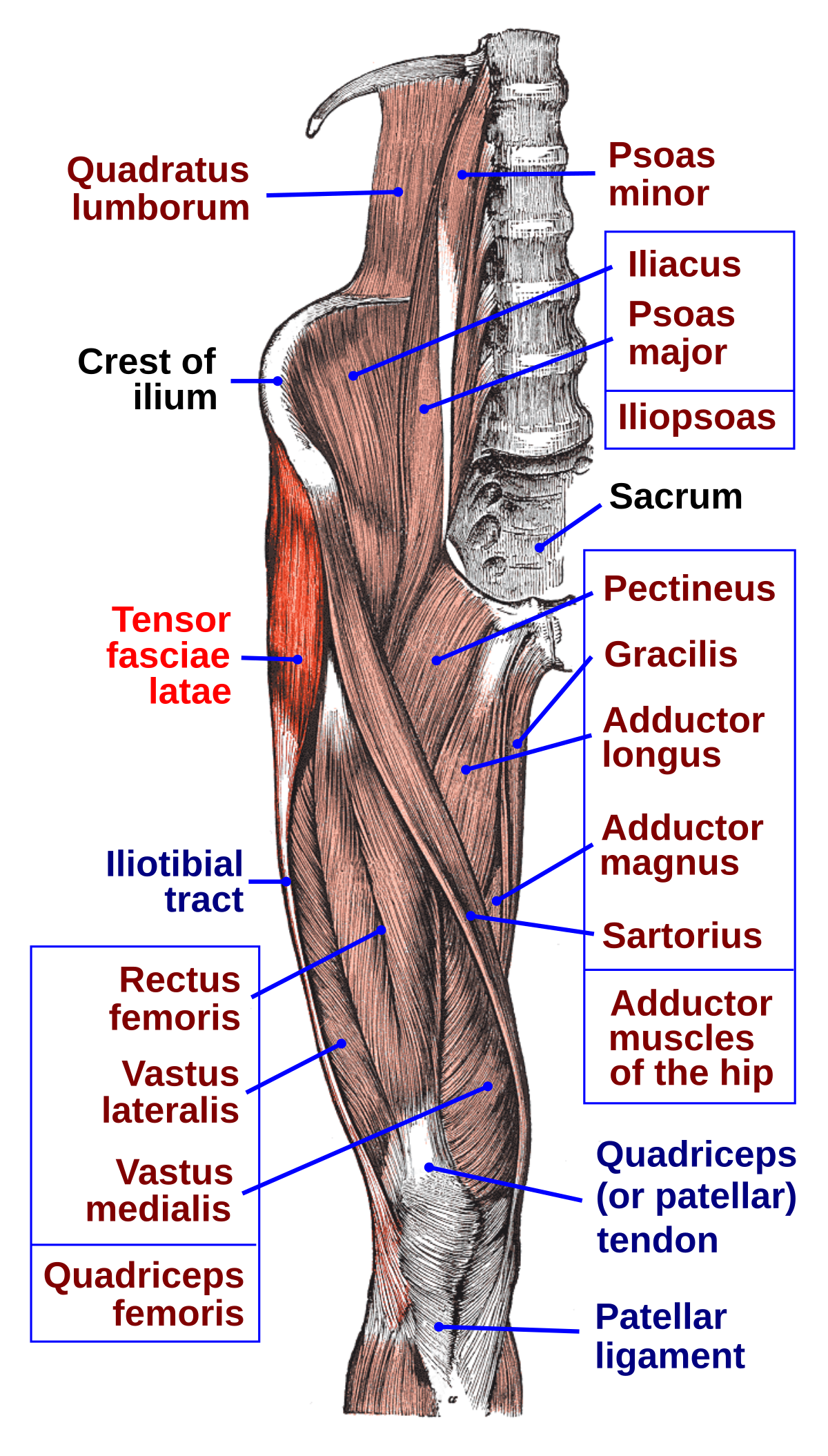
Quadratus Lumborum (QL) - Origin, Nerve Supply, Function

Origin of Quadratus Lumborum (QL)

The quadratus lumborum muscle originates by aponeurotic fibers into the iliolumbar  and the internal lip of the iliac crest for about 5 centimeters (2.0 in). It inserts from the lower border of the last rib for about half its length and by four small from the apices of the transverse processes of the upper four lumbar vertebrae. The QL muscle is flattened and has a quadrangular shape; along with the multifidus and erector spinae muscles, the QL helps to create an antagonist force compared to the muscles of the .

The quadratus lumborum muscle involves the 12th rib (internal surface) and the transverse processes of the lumbar bodies of L1-L4, while it comes from the iliac crest (inner lip) and the iliolumbar ligament.

QL comprises three layers with muscle fibers that have different vectors.

  • The superficial layer is a thin layer, comprising iliocostal muscle fibers (from the iliac crest to the ribs) and iliothoracic (from the iliac crest to the lateral area of the vertebral body of T12), which fibers have a tendinous or even muscular termination.
  • The intermediate layer comprises lumbocostal muscle fibers (from the transverse processes of the lumbar vertebrae to the 12th rib). They are muscle fibers that vary significantly in size, direction, and thickness.
  • The posterior layer consists of lateral iliocostal fibers and medially from iliolumbar fibers (from the iliac crest, connecting to the transverse processes of the lumbar vertebrae). The iliocostal muscle bundles may involve the thoracolumbar fascia, inserting themselves with their endings. This layer is thicker in the caudal area and more tapered in the rostral portion.

Given the arrangement of the highly variable QL muscle fibers, it is not possible to recognize a precise pattern of orientation. Generally, the QL muscle is located medially to the aponeurosis of the transverse abdominal muscle.

Nerves of Quadratus Lumborum (QL)

  • QL receives innervation via the twelfth intercostal nerve, the iliohypogastric nerve, and the ilioinguinal nerve.
  • Anterior branches of the ventral rami of T12 to L4.
  • Subcostal nerve (T12).
  • Iliohypogastric and Ilioinguinal nerve (both from L1).
  • Branches from the ventral rami (L2 and L3).

Blood Supply of Quadratus Lumborum (QL)

QL gets its vascular supply via the lumbar and a lumbar branch of the iliolumbar . The lumbar arteries are four on each side, born from the posterior face of the at the level of the first four lumbar vertebrae. They run posteriorly behind the sympathetic chain and under the arches tendon of the iliopsoas muscle, to finally meet the QL muscle. The first three arteries pass posteriorly to the quadratus lumborum while the fourth passes anteriorly.

  • The iliolumbar artery is the first tributary of the internal iliac artery (or hypogastric artery).
    • Branches of the subcostal artery.
    • Branches of the lumbar arteries.
    • The lumbar branch of the iliolumbar artery.
    • The arteria lumbalis ima from the median sacral artery.
  • The lumbar of each side are joined by longitudinal anastomotic vessels which coalesce to form a small vertical trunk called the ascending lumbar vein. This vessel communicates, at the inferior end, with the iliolumbar and sometimes with the common iliac vein; at the superior aspect, it gives rise to the azygos vein on the right and the left hemizygous vein thus constituting an essential anastomotic pathway between the inferior vena cava system and the superior vena cava system.
  • The lumbar lymph nodes of the , which drain the lymph from the QL, are associated with the inferior vena cava and the abdominal aorta.

Function

The quadratus lumborum can perform four actions

  • Lateral flexion of the vertebral column, with ipsilateral contraction
  • Extension of the lumbar vertebral column, with contraction (based on a line of force passing ~3.5 cm posterior L3 rotation axis[rx])
  • Fixes the 12th rib during forced expiration. The quadratus lumborum assists the in inhalation
  • Elevates the Ilium (bone), with ipsilateral contraction (“hip hiking”)
  • Vertical stabilization of pelvis, lumbar spine, and lumbosacral junction. Prevents collapse of the vertebral column in the frontal plane (i.e. )
  • Reserve mover actions: Anterior pelvic tilt. Contralateral lateral pelvic rotation.
  • The stabilizer of the lumbar area, capable of tilting laterally and capable of acting as an inspiratory accessory muscle.
  • During the extension, the QL exerts a force of 10Nm, compared to 100Nm and 150Nm of the erector spinae muscles and the multifidus. It seems unlikely that it can extend the lumbar area on a sagittal plane with such a small force (10Nm).
  • The QL is positioned laterally, but despite its position during a lateral tilt of the trunk, it exerts only a force of 10Nm. QL participates with less than 10% of the force required for a coronal inclination.
  • The force exerted by the musculature of the erector spinae and multifidus in the lumbar vertebrae is about 1800N and 2800N, respectively. QL exercises the only 200N, and as such it does not appear to play a significant role as a lumbar stabilizer muscle.
  • Regarding its accessory inspiratory muscle function, due to the mobility of the last rib, QL will not be capable of transmitting the contractile force towards the diaphragm.

Actions

  • Quadratus Lumborum fixes the 12th rib to stabilize diaphragm attachments during inspiration.
  • Lateral flexes the vertebral column.
  • Extends lumbar vertebrae.
  • It forms with the contralateral Tensor fascia lata and Gluteus medius a lateral myofascial sling that aims to maintain frontal plane stability of the pelvis.

Functional Contributions

  • The primary antagonist to each Quadratus Lumborum muscle is the Quadratus Lumborum muscle on the other side of the body.[7]
  • The muscle group is considered a postural muscle group.
  • It’s one of the muscles that tends to be tight and overactive.

References

Doctor visit helper

Prepare before seeing a doctor

A simple rural-patient checklist to help you explain symptoms clearly, ask better questions, and avoid unsafe self-treatment.

Safety note: This is not a prescription or diagnosis. For severe symptoms, pregnancy danger signs, children with serious illness, chest pain, breathing difficulty, stroke-like weakness, or major injury, seek urgent care.

Which doctor may help?

Start with a registered doctor or the nearest qualified health center.

What to tell the doctor

  • Write when the problem started and how it changed.
  • Bring old prescriptions, investigation reports, and current medicines.
  • Write allergies, pregnancy status, diabetes, kidney/liver disease, and major past illnesses.
  • Bring one family member if the patient is weak, elderly, confused, or a child.

Questions to ask

  • What is the most likely cause of my symptoms?
  • Which danger signs mean I should go to hospital quickly?
  • Which tests are necessary now, and which can wait?
  • How should I take medicines safely and what side effects should I watch for?
  • When should I come for follow-up?

Tests to discuss

  • Vital signs: temperature, pulse, blood pressure, oxygen saturation
  • Basic physical examination by a clinician
  • CBC, urine test, blood sugar, or imaging only when clinically needed

Avoid these mistakes

  • Do not use antibiotics, steroid tablets/injections, or strong painkillers without proper medical advice.
  • Do not hide pregnancy, kidney disease, ulcer, allergy, or blood thinner use.
  • Do not delay emergency care when danger signs are present.

Medicine safety and first-aid guide

This section is for patient education only. It does not replace a doctor, pharmacist, or emergency care.

Safe first steps

  • Avoid heavy lifting, sudden bending, and prolonged bed rest.
  • Use comfortable posture and gentle movement as tolerated.
  • Discuss physiotherapy, X-ray, or MRI only when clinically needed.

OTC medicine safety

  • For mild back pain, pain-relief medicine may be discussed with a doctor or pharmacist.
  • Avoid repeated painkiller use if you have kidney disease, stomach ulcer, uncontrolled blood pressure, or are taking blood thinners.

Avoid these mistakes

  • Do not start antibiotics without a proper medical decision.
  • Do not use steroid tablets or injections casually for quick relief.
  • Do not delay emergency care because of home remedies.

Get urgent help if

  • Back pain with leg weakness, numbness around private area, loss of urine/stool control, fever, cancer history, or major injury needs urgent care.
Medicine names, dose, and timing must be decided by a qualified clinician or pharmacist after checking age, pregnancy, allergy, other diseases, and current medicines.

For rural patients and family caregivers

Patient health record and symptom diary

Write your symptoms, medicines already taken, test results, and questions before visiting a doctor. This note stays on your device unless you print or copy it.

Doctor to discuss: Orthopedic / spine specialist, physical medicine doctor, or qualified clinician
Tests to discuss with doctor
  • Neurological examination for leg power, sensation, reflexes, and straight leg raise
  • X-ray only if injury, deformity, long-lasting pain, or doctor suspects bone problem
  • MRI discussion if severe nerve symptoms, weakness, bladder/bowel problem, or persistent symptoms
Questions to ask
  • What is the most likely cause of my symptoms?
  • Which warning signs mean I should go to emergency care?
  • Which tests are really needed now?
  • Which medicines are safe for my age, pregnancy status, allergy, kidney/liver/stomach condition, and current medicines?
  • Is physiotherapy, posture correction, or activity modification needed?

Emergency warning signs such as chest pain, severe breathing difficulty, sudden weakness, confusion, severe dehydration, major injury, or loss of bladder/bowel control need urgent medical care. Do not wait for online information.

Safe pathway to proper treatment

Care roadmap for: Quadratus Lumborum (QL) – Origin, Nerve Supply, Function

Use this simple roadmap to understand the next safe steps. It is educational and does not replace examination by a doctor.

Go to emergency care if you notice:
  • Severe or rapidly worsening symptoms
  • Breathing difficulty, chest pain, fainting, confusion, severe weakness, major injury, or severe dehydration
Doctor / service to discuss: Qualified healthcare provider; specialist depends on symptoms and examination.
  1. Step 1

    Check danger signs first

    If danger signs are present, seek emergency care and do not wait for online information.

  2. Step 2

    Record the symptom story

    Write when symptoms started, severity, medicines already taken, allergies, pregnancy status, and test results.

  3. Step 3

    Visit a qualified clinician

    A doctor, nurse, or qualified healthcare provider can examine you and decide which tests or treatment are needed.

  4. Step 4

    Do only useful tests

    Do tests after clinical assessment. Avoid unnecessary tests, random antibiotics, or repeated medicines without diagnosis.

  5. Step 5

    Follow up and return early if worse

    If symptoms worsen, new warning signs appear, or treatment is not helping, return for review quickly.

Rural patient practical tips
  • Take a written symptom diary and all previous prescriptions/test reports.
  • Do not hide medicines already taken, even herbal or over-the-counter medicines.
  • Ask which warning signs mean urgent referral to hospital.

This roadmap is for education. A real diagnosis and treatment plan requires history, examination, and clinical judgment.
