
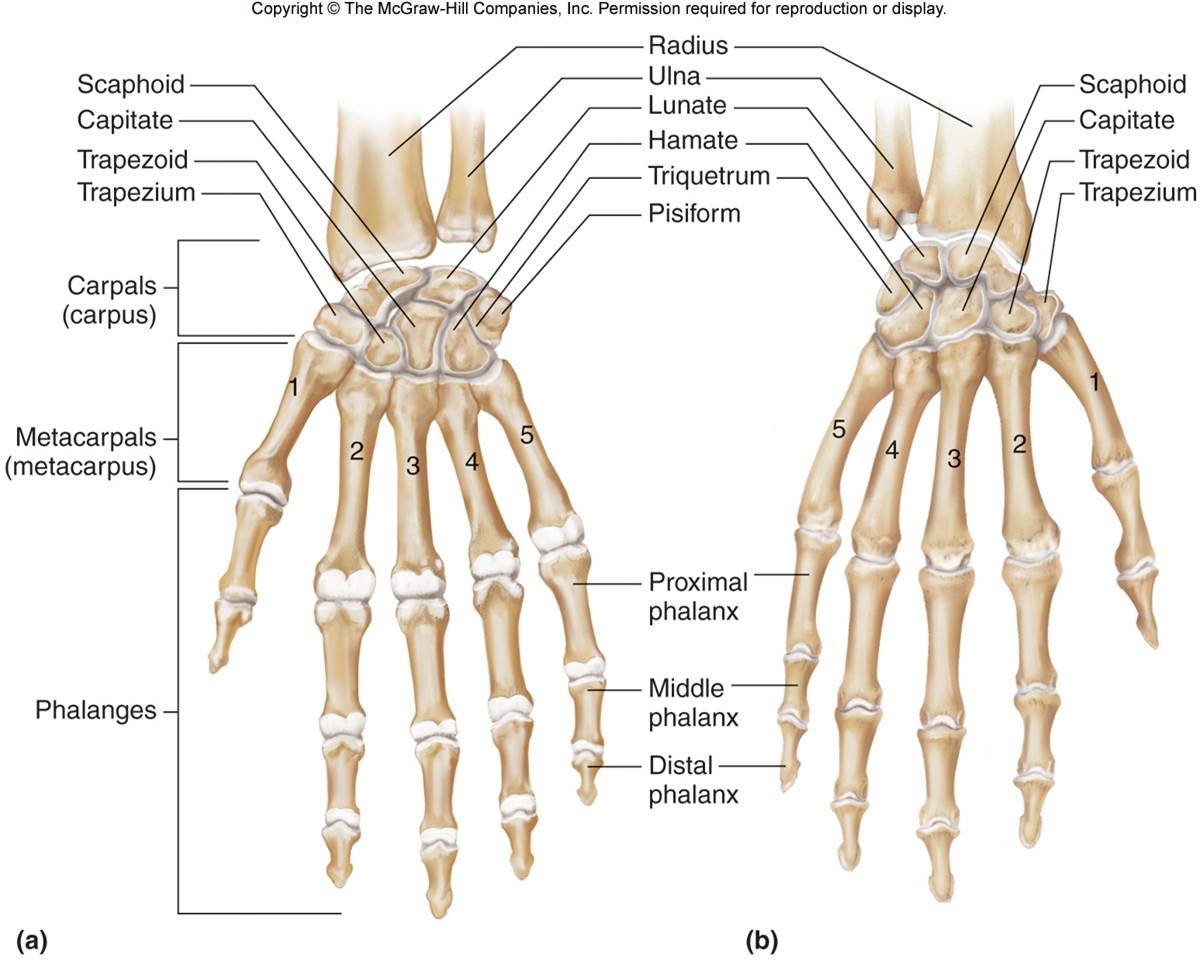
Metacarpophalangeal Joint(MP or MCP), also known as “the knuckle,” is formed by the articulation of the metacarpal head and proximal phalanx. This multiaxial joint allows flexion and extension as well as abduction and adduction motions. The MP joint also dynamically coordinates with the interphalangeal (IP) joint to perform thumb opposition and composite finger flexion. The MP joint has implications in many congenital or acquired deformities, sports-related injuries, and degenerative conditions and sequelae.
Structure and Function of Metacarpophalangeal Joint
The MP joint is responsible for two planes of motion: flexion and extension, as well as abduction and adduction.[rx]
Thumb MP joint
The proximal convex surface of the first metacarpal bone is opposed by the elliptical cavity of the proximal phalanx. Additionally, two sesamoid bones secured by intersesamoid ligaments are present in the palmar aspect of the joint capsule. The first MP joint provides additional flexion to the thumb in opposition, permitting the grasping and contouring objects to facilitate fine motor tasks. Unlike the other digits of the hand, the thumb joint only has one interphalangeal joint which the MP joint coordinates thumb opposition. Compared to the other MP joints of the hand, the MP joint of the thumb has a more restricted range of motion.
Finger MP joints
The finger MP joints have an oval convex metacarpal surface that articulates with a concave, shallow proximal phalangeal surface.[rx] Despite their structural similarities, these joints are much less restricted in motion compared to the first MP joint of the thumb. Extensive musculature stabilizes these joints. On the palmar side, the flexor digitorum superficialis, flexor digitorum profundus, lumbricals, interossei, flexor digiti minimi, flexor pollicis longus, and flexor pollicis brevis muscles support the joint.[rx][rx][x][rx] Dorsally, the extensor digitorum, extensor indicis, extensor digiti minimi, extensor pollicis longus, and extensor pollicis brevis muscles reinforce the joint.[rx]
Various ligamentous structures such as the volar plate, longitudinal fibers of the joint capsule, and transverse metacarpal ligaments (superficial and deep) provide additional stability to the joint.
From deep to superficial, the anatomic structures of the MP joint facilitate the overall dynamic function of the MP joint include[rx]:
-
Volar plate
-
Joint capsule fibers
-
Deep transverse metacarpal ligament
-
Flexor digitorum profundus (FDP) tendon
-
Flexor digitorum superficialis (FDS) tendon
-
Digital tendon sheath
-
A1 annular pulley
The volar plate is a short, transverse thickening of the joint capsule that functions to increase overall joint congruence and prevents MP joint hyperextension.[rx]
The joint capsule is a thin, fibrous, and durable network, reinforced by the flexor pollicis brevis and adductor pollicis brevis muscles, the palmar plate, and various collateral ligament connections. The radial and ulnar collateral ligaments arise proximally from their respective sides on the metacarpal bone, spanning the distal and volar sides of the joint to attach on the proximal phalanx.[rx] Each collateral ligament has its own proper and accessory ligamentous structures.
The volar fan-shaped accessory ligament attaches to the middle of the metacarpal head and extends to the palmar plate and deep transverse metacarpal ligament which tightens during finger extension. The dorsal, cord-shaped, proper ligament attaches to the metacarpal head and extends to the proximal phalanx base, becoming taut at about 30 degrees of finger flexion. The natatory ligament, also known as the superficial transverse metacarpal ligament, originates distal to the MP joint and runs through the webspace to insert on the proximal phalanx. It functions to resist abduction. The deep transverse metacarpal ligament connects the 2nd-5th metacarpal heads together at the volar plate and provides soft tissue support. Dorsal to this ligament, are the ribbon-shaped sagittal bands that centralize the MP joint and stabilize the extensor tendons in motion.[rx]
Blood Supply and Lymphatics of Metacarpophalangeal Joint
The primary blood supply to the hand is through the radial and ulnar arteries. The ulnar artery gives rise to the superficial palmar arch and gives off a deep branch to anastomose with the deep arch. The radial artery gives rise to the deep arch and gives off a superficial branch to anastomose with the superficial arch. This creates an extensive collateral network to prevent ischemia injury. Blood supply to the thumb MP joint may be directly the superficial arch or via a deep branch of the radial artery; the princeps pollicis artery.[rx] The arterial supply to the MP joints of the fingers is by branches from adjacent digital arteries.
The axillary lymph nodes provide lymphatic drainage of the upper limb. The lymphatic system of the upper extremity uses a pathway that originates in the distal fingers tips and palms. The lymphatic system travels on the dorsal aspect of the hand. The lymphatic vessels are typically found just posterior to the cutaneous veins. The lymph drainage continues proximally up the arm, running with the basilic vein until connecting to the lymph nodes in the lateral axillary region. [rx]
Nerves of Metacarpophalangeal Joint
The MP joint receives innervation from the articular branches of the dorsal and palmar digital nerves. The second to fifth MP joints receive innervation from a deep branch of the ulnar nerve that is superficial to the interosseous muscles, but deep and radial to the flexor tendon.[rx]
Muscle Attachment of Metacarpophalangeal Joint
Thenar Muscles
Opponents policies
-
Function: Opposition of the thumb
-
Origin: Flexor retinaculum and tubercle of trapezium
-
Insertion: Lateral aspect of the first metacarpal
-
Innervation: Recurrent branch of the median nerve (C8, T1)
Abductor policies Brevis
-
Function: Abduction of the thumb at the metacarpophalangeal joint
-
Origin: Flexor retinaculum and tubercle of the scaphoid
-
Insertion: Lateral aspect of proximal phalanx of the first finger
-
Innervation: Recurrent branch of the median nerve (C8, T1)
Flexor policies Brevis
-
Function: Flexion of the thumb at the metacarpophalangeal joint
-
Origin: Flexor retinaculum and tubercle of trapezium
-
Insertion: Lateral aspect of proximal phalanx of the first finger
-
Innervation: Recurrent branch of the median nerve (C8, T1)
Adductor Compartment
Adductor policies
-
Function: Adduction of the thumb
-
Origin: Second, third metacarpal, and capitate
-
Insertion: Proximal phalanx and extensor expansion of 1st finger
-
Innervation: Deep branch of the ulnar nerve (C8, T1)
Hypothenar Muscles
Abductor digiti minimi
-
Function: Abduction of the little finger at the metacarpophalangeal joint
-
Origin: Pisiform
-
Insertion: Medial aspect of proximal phalanx of fifth finger
-
Innervation: Deep branch of ulnar nerve (C8, T1)
Flexor digiti minimi brevis
-
Function: Flexion of the little finger at the metacarpophalangeal joint
-
Origin: Flexor retinaculum and hook of hamate
-
Insertion: Medial aspect of proximal phalanx of fifth finger
-
Innervation: Deep branch of ulnar nerve (C8, T1)
Opponens digiti minimi
-
Function: Opposition of the little finger
-
Origin: Flexor retinaculum and hook of hamate
-
Insertion: Medial aspect of fifth metacarpal
-
Innervation: Deep branch of ulnar nerve (C8, T1)
Short Muscles
Lubricants
-
Function: Flexion of the metacarpophalangeal joints with extension of the interphalangeal joints
-
Origin: Arise from tendons of flexor digitorum profundus. First 2 are unipennate, and the third and fourth are bipennate
-
Insertion: Extensor expansions of second, third, fourth, and fifth finger
-
Innervation: Median nerve (C8, T1) for the lateral 2 lumbricals, deep branch of ulnar nerve (C8, T1) for the medial 2 lumbricals
Dorsal interossei
-
Function: Abduction of the second, third, and fourth finger away from the axial line
-
Origin: Adjacent metacarpals
-
Insertion: Extensor expansions and proximal phalanges of the second, third, and fourth fingers
-
Innervation: Deep branch of ulnar nerve (C8, T1)
Palmar interossei
-
Function: Adduction of the second, third, and fourth finger towards the axial line
-
Origin: Palmar surfaces of second, fourth, and fifth metacarpals
-
Insertion: Extensor expansions and proximal phalanges of the second, fourth, and fifth fingers
-
Innervation: Deep branch of ulnar nerve (C8, T1)
References
- https://www.ncbi.nlm.nih.gov/books/NBK551532/
- https://www.ncbi.nlm.nih.gov/books/NBK470428/
- https://www.ncbi.nlm.nih.gov/books/NBK538343/
- https://www.ncbi.nlm.nih.gov/books/NBK535382/
- https://www.ncbi.nlm.nih.gov/books/NBK535382/
- https://www.ncbi.nlm.nih.gov/books/NBK536960/
- https://en.wikipedia.org/wiki/Boxer%27s_fracture

