
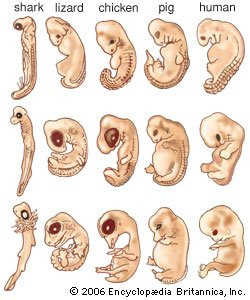
Fetal Development /Fetal Period means the process of prenatal development occurs in three main stages. The first two weeks after conception are known as the germinal stage, the third through the eighth week is known as the embryonic period, and the time from the ninth week until birth is known as the fetal period.
Fetal Period – Anatomy, Mechanism, Stages, Function
Germinal Stage
The germinal stage begins at conception when the sperm and egg cell unite in one of the two fallopian tubes. The fertilized egg is called a zygote. Just a few hours after conception, the single-celled zygote begins making a journey down the fallopian tube to the uterus.
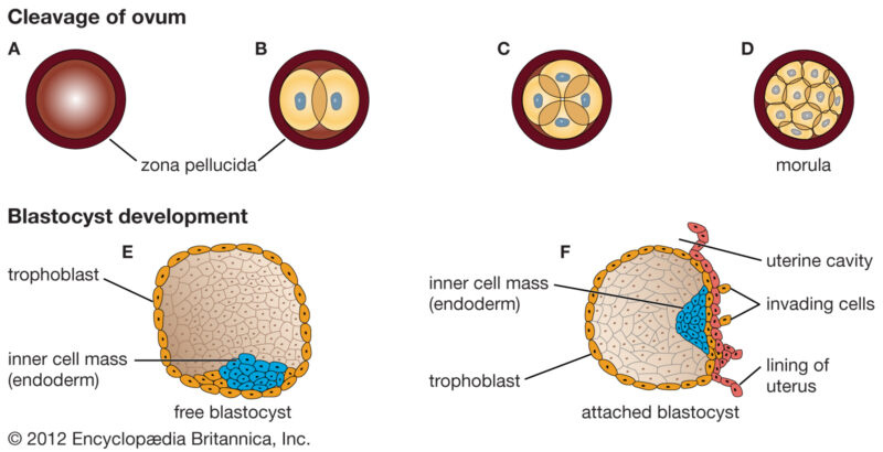
Cell division begins approximately 24 to 36 hours after conception. Through the process of mitosis, the zygote first divides into two cells, then into four, eight, sixteen, and so on. A significant number of zygotes never progress past this early part of cell division, with as many as half of all zygotes surviving less than two weeks.
Once the eight-cell point has been reached, the cells begin to differentiate and take on certain characteristics that will determine the type of cells they will eventually become. As the cells multiply, they will also separate into two distinctive masses: the outer cells will eventually become the placenta, while the inner cells form the embryo.
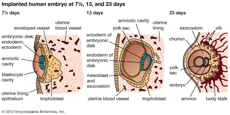
Cell division continues at a rapid rate during the approximately week-long journey from fallopian tube to uterus wall. The cells develop into what is known as a blastocyst. The blastocyst is made up of three layers, each of which develops into different structures in the body.
- Ectoderm: Skin and nervous system
- Endoderm: Digestive and respiratory systems
- Mesoderm: Muscle and skeletal systems
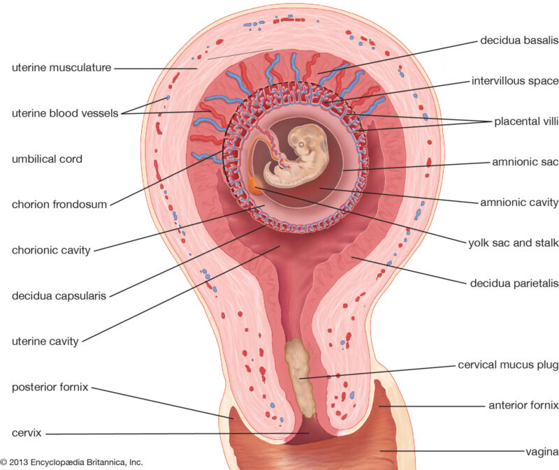
Finally, the blastocyst arrives at the uterus and attaches to the uterine wall, a process known as implantation. Implantation occurs when the cells nestle into the uterine lining and rupture tiny blood vessels. The connective web of blood vessels and membranes that form between them will provide nourishment for the developing being for the next nine months. Implantation is not always an automatic and sure-fire process.
Researchers estimate that approximately 60% of all natural conceptions never become properly implanted in the uterus, which results in the new life ending before the mother is ever aware she is pregnant.
When implantation is successful, hormonal changes halt the normal menstrual cycle and cause a whole host of physical changes. For some people, activities they previously enjoyed such as smoking and drinking alcohol or coffee may become less palatable, possibly part of nature’s way of protecting the growing life inside them.2
Embryonic Stage
At this point, the mass of cells is now known as an embryo. The beginning of the third week after conception marks the start of the embryonic period, a time when the mass of cells becomes distinct as a human. The embryonic stage plays an important role in the development of the brain.
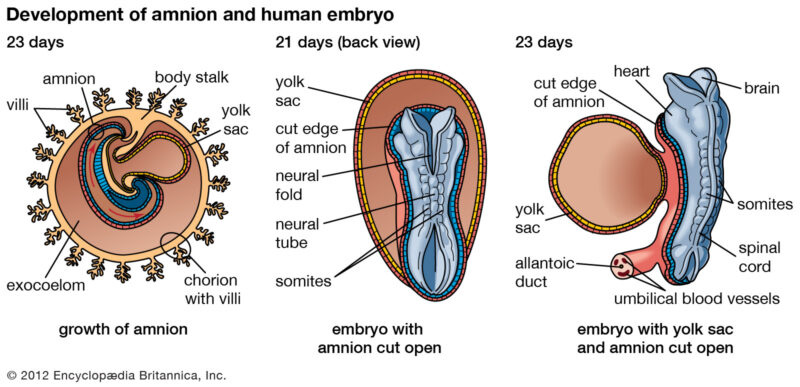
Approximately four weeks after conception, the neural tube forms. This tube will later develop into the central nervous system including the spinal cord and brain. The neural tube begins to form along with an area known as the neural plate. The earliest signs of development of the neural tube are the emergence of two ridges that form along each side of the neural plate.
Over the next few days, more ridges form and fold inward until a hollow tube is formed. Once this tube is fully formed, cells begin to form near the center.3 The tube begins to close and brain vesicles form. These vesicles will eventually develop into parts of the brain, including the structures of the forebrain, midbrain, and hindbrain.
Around the fourth week, the head begins to form, quickly followed by the eyes, nose, ears, and mouth. The blood vessel that will become the heart start to pulse. During the fifth week, buds that will form the arms and legs appear.
By the eighth week of development, the embryo has all of the basic organs and parts except those of the sex organs. At this point, the embryo weighs just one gram and is about one inch in length.
By the end of the embryonic period, the basic structures of the brain and central nervous system have been established. At this point, the basic structure of the peripheral nervous system is also defined.
The production of neurons, or brain cells, begins around day 42 after conception and is mostly complete sometime around the middle of pregnancy.
As neurons form, they migrate to different areas of the brain. Once they have reached the correct location, they begin to form connections with other neural cells, establishing rudimentary neural networks.
Fetal Stage
Once cell differentiation is mostly complete, the embryo enters the next stage and becomes known as a fetus. The fetal period of prenatal develop marks more important changes in the brain. This period of development begins during the ninth week and lasts until birth. This stage is marked by amazing change and growth.
The early body systems and structures established in the embryonic stage continue to develop. The neural tube develops into the brain and spinal cord and neurons continue to form. Once these neurons have formed, they begin to migrate to their correct locations. Synapses, or the connections between neurons, also begin to develop.
Between the ninth and twelfth week of gestation (at the earliest), reflexes begin to emerge. The fetus begins to make reflexive motions with its arms and legs.4
During the third month of gestation, the sex organs begin to differentiate. By the end of the month, all parts of the body will be formed. At this point, the fetus weighs around three ounces. The fetus continues to grow in both weight and length, although the majority of the physical growth occurs in the later stages of pregnancy.
The end of the third month also marks the end of the first trimester of pregnancy. During the second trimester, or months four through six, the heartbeat grows stronger and other body systems become further developed. Fingernails, hair, eyelashes, and toenails form.5 Perhaps most noticeably, the fetus increases about six times in size.
So what’s going on inside the brain during this important period of prenatal development? The brain and central nervous system also become more responsive during the second trimester. Around 28 weeks, the brain starts to mature faster, with an activity that greatly resembles that of a sleeping newborn.
During the period from seven months until birth, the fetus continues to develop, put on weight, and prepare for life outside the womb. The lungs begin to expand and contract, preparing the muscles for breathing.
While development usually follows this normal pattern, there are times when problems with prenatal development occur. Disease, malnutrition, and other prenatal influences can have a powerful impact on how the brain develops during this critical period.
Fetal Development
At the end of the 10th week of gestation, the fetal period begins.
Key Points
Although fetuses can be adversely affect by environmental toxicants, they are less sensitive to them than embryos because the precursors for most organs are already formed.
At the start of the fetal stage, the fetus is typically about 30 millimeters in length from crown to rump, and weighs about eight grams. The head makes up nearly half of the fetus’ size.
The precursors of all the major organs are created by the time the fetal period begins. Therefore, the fetal period is described both by organ and a list of the changes by weeks of gestational age.
Key Terms
- meconium: The earliest stool of a mammalian infant. Unlike later feces, meconium is composed of materials ingested during the time the infant spends in the uterus (intestinal epithelial cells, lanugo, mucus, amniotic fluid, bile, and water).
- gestational age: This relates to the age of an embryo or fetus (or newborn infant). In human obstetrics, this age is often defined as the time elapsed since 14 days prior to fertilization; this is approximately the duration since the woman’s last menstrual period began.
- lanugo: Soft down or fine hair that covers the human fetus.
In humans, the fetal stage of prenatal development starts at the beginning of the 11th week in gestational age, which is the ninth week after fertilization. Since the precursors of all the major organs are created by this time, the fetal period is described both by organ and a list of changes by weeks of gestational age.
All major structures are already formed in the fetus, but they continue to grow and develop. Therefore, the fetus is not as sensitive to damage from environmental exposures as the embryo, though toxic exposures often cause physiological abnormalities, growth retardation, or minor congenital malformations.
At the start of the fetal stage, the fetus is typically about 30 millimeters in length from crown to rump and weighs about eight grams. The head makes up nearly half of the fetus’ size. The four-chamber heart is finishing development, and the embryonic tail goes away. The breathing-like movement of the fetus is necessary for stimulation of lung development, rather than for obtaining oxygen.
- Week 10: Finger nails and hair start to grow. The heart, hands, feet, brain, and other organs are present, but are only at the beginning of development and have minimal operation.
- Week 11: Nearly all structures and organs are formed. Fingers and toes are separated and the genitals begin to take on the proper gender characteristics.
- Week 12: The digestive system and liver function. The pancreas makes insulin.
- Week 13: The fetus begins to get its nourishment from the placenta and the veins and organs are visible through the skin.
- Week 14: The kidneys produce urine, and the liver makes bile. In boys, the prostate gland develops, while in girls the ovaries move from the abdomen to the pelvis.
- Weeks 15 to 16: The heart pumps out 25 quarts of blood a day and the fetal structures are looking more normal.
- Week 17: The fetus start to move its joints and the retina becomes sensitive to light. It weighs about five ounces.
- Week 18: The fetus starts to hear and is startled by noise. Its skin is starting to grow a protective, wax-like layer and tiny air spaces begin to form in the lungs and the vocal cords.
- Week 19: The brain is designating areas for the five senses.
- Week 20: The fetus weighs about 10 ½ oz, swallows more, and produces meconium.
- Week 21: The eyebrows and lids are present, and for the female fetus, the vagina begins to form.
- Week 22: Tiny tooth buds beneath the gums develops. The eyes form, but the irises lack pigment.
- Week 23: The fetus weighs over a pound. It can feel movements and hear sounds. Blood vessels in his/her lungs are developing to prepare for breathing.
- Week 24: The fetus is almost a foot long. The lungs are developing branches of the respiratory tree as well as cells that produce surfactant, a substance that will help the air sacs inflate once born.
- Week 25: The amount of body fat rapidly increases. The lungs are not fully mature. The bones are fully developed, but are still soft and pliable. Thalamic brain connections form that mediate sensory input. Iron, calcium, and phosphorus become more abundant. The fingernails reach the end of the fingertips. The lanugo, or fine hair, begins to disappear, until it is gone (except on the upper arms and shoulders). Small breast buds are present on both sexes. Head hair becomes coarse and thicker.
Birth is imminent and occurs around the 40th week. The fetus is considered full-term between weeks 37 and 40, which means that the fetus is considered sufficiently developed for life outside the uterus.
Teratogens
A teratogen is a compound that permanently deforms the function or structure of a developing embryo or fetus.
Key Points
The effects of a teratogen on the fetus depend on several factors: the potency of the teratogen, the susceptibility of the fetus to the teratogen, the dose and duration of teratogen exposure, the degree of transfer from maternal to fetal circulation, and when during development the exposure occurs.
Approximately 10% of congenital malformations are attributed to environmental factors, and 20% are due to genetic or hereditary factors. The rest have unknown causes or are due to a mix of different factors.
The central nervous and skeletal systems tend to be most affected by teratogens.
Cigarette components, alcohol, cocaine, warfarin, ACE inhibitors, and Accutane are all teratogens that affect fetal development.
Key Terms
- teratogen: Any agent or substance that can cause embryonic malformations or birth defects.
- fetal alcohol syndrome: Any of a spectrum of birth defects that result from excessive alcohol consumption by the mother during pregnancy.
- microcephaly: A neurological disorder in which the person affected has an abnormally small head due to a failure of brain growth.
Characteristics of Teratogens
A teratogen is a compound that permanently deforms the function or structure of a developing embryo or fetus in utero. In general, the degree of teratogenicity depends on:
- The potency of the drug as a mutagen.
- The susceptibility of the fetus to teratogenesis.
- The dose of the teratogen.
- The duration of teratogen exposure.
- The time of exposure.
- The degree of transfer from maternal to fetal circulation.
The global average of all live births complicated by malformation is 6% (Environmental Health Perspectives, (NIH), October 2009). The majority of these complications are due to unknown factors.
The vast majority of recognized etiologies are genetic, with only 10% being attributed to environmental etiologies such as maternal health, infection, and toxicants. In general, the central nervous and skeletal systems are the most affected.
Women may encounter a number of teratogens. Smoking is most likely to cause growth retardation, but has also been implicated in the prelabor rupture of the membranes, preterm labor, abruption of the placenta, spontaneous abortion, perinatal morbidity and mortality, and sudden infant death syndrome. Smoking may exert its effects through competitive binding of carbon monoxide with hemoglobin and/or through the various other components found in cigarettes that cause adverse biological effects.
Example Teratogens
Fetal alcohol syndrome: Alcohol is a teratogen. When consumed in pregnancy, it can result in mothers giving birth to children with fetal alcohol syndrome. The facial characteristics highlighted in the photograph are small eye opening, smooth philtrum, and a thin upper lip.
Alcohol use in pregnancy may result in fetal alcohol ayndrome (FAS). FAS occurs in approximately 1% of all births. Children with FAS present with a flattened and thin upper lip, small palpebral fissures, epicanthal folds, flattened nasal bridge, and a short nose. They may also exhibit microcephaly, mental retardation, and have learning disabilities. It is not clear if there is any safe amount of alcohol consumption in pregnancy.
Cocaine generally produces growth restriction, preterm delivery, microcephaly, spontaneous abortion, placental abruption, limb anomalies, and central nervous system abnormalities. Cocaine appear to exert a number of its effects through peripheral vasoconstriction that leads to fetal hypoxia.
Women with indications for warfarin therapy should either abstain from pregnancy or switch to low molecular weight heparins. Warfarin typically produces mental retardation, growth restriction, nasal hypoplasia, and opthalmic abnormalities.
Angiotensin converting enzyme (ACE) inhibitors will cause fetal renal failure and oligohydramnios that lead to pulmonary hypoplasia and limb contracture. Fetal cranial bone abnormalities are also common.
Isotretinoin (Accutane), used to treat acne, may cause cardiac, oral, otological, thymic, and central nervous system abnormalities. In one quarter of cases, it causes mental retardation.

Thalidomide effects: A photograph of the limbs of baby born to a mother who took thalidomide while pregnant.
Other teratogenic substance classes and conditions include
- Various prescription drugs and nutrient deficiencies (e.g., insufficient
folic acid). - Chemical compounds such as methyl iodide (used in pesticides) and bisphenol A (used in plastics) are suspected teratogens.
- Thalidomide (a sedative previously marketed in Europe to prevent morning sickness) is a classic teratogen that caused limb defects in babies born to women who took this drug in the 1960s.
Prenatal Diagnostic Tests
Prenatal diagnosis is a way to screen a fetus for diseases and/or conditions that may increase its morbidity and/or mortality.
Key Points
Prenatal diagnosis allows: 1) timely medical treatment of a condition before or after birth; 2) parents to make decisions regarding whether to abort a fetus with a diagnosed condition; 3) parents to prepare psychologically, socially, financially, and medically for a baby with a health problem or disability.
The types of conditions commonly screened for include neural tube defects, chromosome abnormalities (e.g., Down syndrome, also called trisomy 21), and genetic abnormalities (e.g., Tay-Sachs disease, sickle cell anemia, and cystic fibrosis ).
Diagnostic screening can be invasive (for example, amniocentesis ) or noninvasive (such as ultrasound). Risks are associated with invasive measures. It is often recommended that women who are at high risk of pregnancy loss due to advanced age or who previously had children with birth defects receive invasive testing.
Early ultrasound scans are done at around six weeks; they are used to confirm the gestational age of the embryo and check for a single or twin pregnancy.
Around weeks 10–11, a nuchal thickness scan (NT) may be offered that can be combined with blood tests that correlate with chromosomal abnormalities. The results of the blood tests are combined with the NT ultrasound measurements, maternal age, and gestational age of the fetus to yield a risk score.
There are ethical issues associated with prenatal tests, including whether parents should elect to abort fetuses with detected abnormalities, the high rate of false positives and false negatives, and whether prenatal screening will lead to the rise of designer babies.
Key Terms
- amniocentesis: A procedure for obtaining amniotic fluid from a pregnant female by inserting a hollow needle through the abdominal wall and into the amniotic sac. It is used to diagnose possible genetic defects and/or obstetric complications.
- chorionic villus sampling: A form of prenatal diagnosis in which part of the placental tissue is tested for genetic abnormalities.
- nuchal thickness scan: A sonographic prenatal screening scan (ultrasound) to help identify any high risks of chromosomal defects, including Down’s Syndrome in a fetus, particularly for older women who have higher risks of such pregnancies.
Prenatal Screening Overview
Prenatal diagnosis and prenatal screening are methods for testing for diseases or conditions in a fetus or embryo before it is born. There are three purposes of prenatal diagnosis:
- To enable timely medical or surgical treatment of a condition before (fetal therapy) or after birth.
- To give the parents the chance to decide to abort a fetus with the diagnosed condition.
- To give parents the chance to prepare psychologically, socially, financially, and medically for a baby with a health problem or disability, or for the likelihood of a stillbirth.
Having this information in advance of the birth means that healthcare staff, as well as parents, can better prepare themselves for the delivery of a child with a health problem.
For example, Down Syndrome is associated with cardiac defects that may need intervention immediately upon birth. The other types of conditions commonly screened for include neural tube defects, chromosome abnormalities, genetic diseases, spina bifida, cleft palate, Tay-Sachs disease, sickle cell anemia, thalassemia, cystic fibrosis, and fragile X syndrome.
Fetal screening is also done to determine characteristics that are not generally considered birth defects, such as determining the sex of the offspring. The primary method for sex determination is prenatal ultrasound. Cell-free fetal DNA testing is also possible, wherein the maternal blood is analyzed to detect the small amount of free circulating fetal DNA.
Invasive and Noninvasive Prenatal Screening
Diagnostic prenatal testing can be done by invasive or noninvasive methods. An invasive method involves probes or needles being inserted into the uterus, e.g., amniocentesis (that can be done from about 14 weeks gestation up to about 20 weeks), and chorionic villus sampling (that can be done earlier: between 9.5 and 12.5 weeks gestation).
Chorionic villus sampling is associated with slightly more risk to the fetus. Since chorionic villus sampling is performed earlier in the pregnancy than amniocentesis, typically during the first trimester, it can reasonably be expected that there will be a higher rate of miscarriage after chorionic villus sampling than after amniocentesis.
Non-invasive techniques include examinations of the woman’s womb through ultrasonography and maternal serum screens. Blood tests for select trisomies based on detecting fetal DNA present in maternal blood are available. Technologies to perform prenatal testing in a noninvasive manner reduce the risk of pregnancy loss that can occur with other methods.
These include the use of maternal blood and DNA sequencing methods such as next-generation sequencing to determine the presence of genetic defects. If an elevated risk of chromosomal or genetic abnormality is indicated by a noninvasive screening test, a more invasive technique may be employed to gather more information. In the case of neural tube defects, a detailed ultrasound can non-invasively provide a definitive diagnosis.

Amniocentesis: An illustration of the amniocentesis procedure.
Because of the risk of miscarriage and fetal damage associated with amniocentesis and chorionic villus sampling procedures, many women prefer to first undergo screening so they can find out if the fetus’ risk of birth defects is high enough to justify the risks of invasive testing. Screening tests yield a risk score that represents the chance that a baby has the birth defect.
The most common threshold for high risk is 1:270. A risk score of 1:300 would be considered low-risk by many physicians. However, the trade-off between the risk of a birth defect and the risk of complications from invasive testing is relative and subjective. Some parents may decide that even a 1:1,000 risk of birth defects warrants an invasive test, while others would not opt for an invasive test even if they had a 1:10 risk score.
The American Congress of Obstetricians and Gynecologists guidelines currently recommend that all pregnant women, regardless of age, be offered invasive testing to obtain a definitive diagnosis of certain birth defects. Therefore, most physicians offer diagnostic testing to all their patients, with or without prior screening, and let the patient decide. Invasive testing is warranted in the following cases:
- Women over the age of 35.
- Women who have previously had premature babies or babies with a birth defect.
- Women who have high blood pressure, lupus, diabetes, asthma, or epilepsy.
- Women who have family histories or ethnic backgrounds prone to genetic disorders, or whose partners have these.
- Women who are pregnant with twins or more.
- Women who have miscarried.
Determination of Gestational Age
At about 6 weeks of pregnancy, an ultrasound dating scan may be offered to help confirm the gestational age of the embryo and check if a single or twin pregnancy exists, but such a scan is unable to detect common abnormalities. Around weeks 10–11, a nuchal thickness scan (NT) may be offered, which can be combined with blood tests that correlate with chromosomal abnormalities.
The results of the blood tests are then combined with the NT ultrasound measurements, maternal age, and gestational age of the fetus to yield a risk score for Down syndrome, trisomy 18, and trisomy 13. Other testing may combine a first trimester blood test with one done in the second trimester to determine if further action needs to be taken.
References
- https://www.ncbi.nlm.nih.gov/books/NBK501922/
- https://www.ncbi.nlm.nih.gov/books/NBK534767/
- https://www.ncbi.nlm.nih.gov/books/NBK235589/
- https://www.ncbi.nlm.nih.gov/books/NBK499981/
- https://www.ncbi.nlm.nih.gov/books/NBK235593/
- https://www.ncbi.nlm.nih.gov/books/NBK538240/
- https://www.ncbi.nlm.nih.gov/books/NBK499981/
- https://www.ncbi.nlm.nih.gov/books/NBK235593/
- https://www.ncbi.nlm.nih.gov/books/NBK235593/
- https://en.wikipedia.org/wiki/Lactation

