
Runner’s Knee may be defined as running injury that is happened during running in competition in football, crickets & athletes that are both great works for the body and hard on the body. It conditions the muscles and joints and it gives you a good aerobic workout, which keeps your heart healthy and weight within a healthy range.
PFP is characterized by pain in the peripatellar/retropatellar area that aggravates with at least one activity that loads the patellofemoral joint during weight bearing on a flexed knee (e.g., squatting, stair climbing, jogging/running and hopping/jumping). Additional nonessential criteria include crepitus or grinding sensation in the patellofemoral joint during knee flexion movements, tenderness on patellar facet palpation, small effusion and pain on sitting, rising on sitting or straightening the knee following sitting.[rx]–[rx]
Runner’s knee may refer to a number of overuse injuries involving pain around the kneecap (patella), such as
- Patellofemoral pain syndrome
- Chondromalacia patellae
- Iliotibial band syndrome
- Plica syndrome
But the relentless impact of your feet against the ground can put you at risk for injuries, particularly in your lower limbs and back. Through a sudden, acute injury—but much more often through repetitive wear and tear—the joints, muscles, and other soft tissues of the back can become damaged or strained. This is especially true for distance runners.
The repetitive impact of running can cause back pain, most commonly low back pain. Whether this pain is caused by strained muscles or by a problem with the spine’s vertebrae or discs may influence treatment and training.
Potential intrinsic risk factors, tests for assessment and their reliability
| Potential risk factor | Tests | Reliability |
|---|---|---|
| Weakness of the quadriceps muscle, especially VMO[rx],[rx]–[rx] | One-legged hop test. The test is performed by jumping and landing on the same foot with the hands behind the back and the hop distance is measured from the toe.[rx] A quotient (%) between the injured and noninjured legs is registered and defined as abnormal if the quotient is <85%[rx] | Good reliability[rx],[rx],[rx] |
| Hip muscle dysfunction (particularly, the abductors and external rotators)[rx],[rx],[rx] | The Trendelenburg test (for hip abductors) to assess the ability to hold the pelvis level, while the subject performs a single-leg stance. Lateral pelvic shift or lowering of one side of the pelvis indicates the weakness of hip abductors[rx],[rx] | Good sensitivity, good to excellent specificity[rx] |
| Poor core muscle endurance[rx],[rx] | Front plank, modified Biering-Sorensen and side bridge for anterior, posterior and lateral core muscle endurance[rx],[rx] | Good reliability[rx] |
| Tightness of hamstring[rx],[rx]–[rx] | Passive knee extension test with goniometric measurement of popliteal angle.[rx],[rx] The test is considered positive when a knee extension angle of >20° is measured[rx],[rx] | Excellent inter-rater and good test-retest reliability[rx] |
| Tightness of iliopsoas and quadriceps[rx],[rx],[rx] | Modified Thomas test.[rx],[rx] Two angles were measured for each limb. Lengths of iliopsoas and quadriceps were determined by measuring the angles of hip and knee flexion, respectively. The inability of the opposite thigh to extend to a neutral position or drop below the horizontal constitutes a positive test for iliopsoas. The angle of knee flexion of <80° determines the quadriceps shortness | Very good to excellent inter-rater and good test-retest reliability[rx] |
| The tightness of iliotibial band[rx]–[rx] | Ober test.[rx]–[rx] A positive test occurs when the leg remains in an abducted position (rests above the horizontal)[rx] | Excellent intra-rater and inter-rater reliability[rx] |
| The tightness of gastrosoleus complex[rx],[rx] | Weight-bearing lunge test.[rx] The test is considered positive when 1) the distance between the wall and the big toe measured is <9 cm or 2) the angle made by the anterior tibia/shin to vertical is <35°[rx] | Excellent intra-rater and inter-rater reliability[rx] |
| Excessive foot pronation[rx],[rx],[rx] | FPI-6.[rx],[rx] Scores of +6 to +9 and +9 to +12 are regarded pronated and highly pronated, respectively[rx] | High intra-rater and inter-rater reliability among PFPS patients[rx],[rx] |
| Limb length discrepancy[rx] | Gauging the distance between the anterior superior iliac spine and the medial malleolus of both legs (average of two measures). Limb length inequalities of >10 mm are considered clinically significant[rx] | High intra-rater and inter-rater reliability[rx],[rx] |
| Patellar malalignment[rx],[rx],[rx,rx] | Patellar tilt and mediolateral glide tests.[rx],[rx],[rx] Tilt occurs when the digit palpating one of the patellar borders is more anterior than the other. The glide is positive when the distance from the mid patella to each femoral epicondyle is not equal[rx] | Fair intra-rater and poor inter-rater reliability[rx],[rx] |
| Patellar hypermobility[rx],[rx] | Patellar mobility test, with the knee, flexed 20°–30° and the quadriceps relaxed.[rx] Patellar mobility of more than three quadrants suggests a hypermobile patella[rx] | Good intra-rater and variable inter-rater reliability[rx] |
| GJL[rx],[rx],[rx] | BHJMI, in which the range of scoring is between 0 and 9, with high scores denoting greater joint laxity | Good to excellent reliability[rx]–[rx] |
| Genu varum[rx] | Goniometric measurement in a standing position and barefoot, with toes placed forward and feet shoulder-width apart[rx] | Correlated well with the angle measured on the full-limb radiograph (gold standard)[rx] |
| Abnormal trochlear morphology[rx] | Measurement of sulcus angle in plain radiography (skyline or tangential patellar views performed in 25° of flexion; normal value 138°, SD 6°)[rx] or evaluation of lateral trochlear inclination, medial trochlear inclination, sulcus angle and trochlear angle on the axial MRI | Excellent intraobserver and interobserver reliability (ICCs of 0.94 and 0.92, respectively),[rx],[rx] but some newer evidence recommended MRI[rx] |
| Abnormal proprioception[rx],[rx] | Measurement of knee joint position sense using five active tests under non-weight-bearing and uni- and bilateral weight-bearing conditions[rx] | Good reliability]rx] |
| Gait abnormalities (heel strike in a less pronated position and there is a rollover more on the lateral side)[rx] | Plantar pressure measurements during walking using a foot scan pressure plate[rx] | Reliable (ICC: 0.75)[rx] |
Abbreviations: BHJMI, Beighton and Horan Joint Mobility Index; FPI-6, Foot Posture Index-Version 6; GJL, generalized joint laxity; ICC, Intraclass Correlation Coefficient; MRI, magnetic resonance imaging; PFPS, patellofemoral pain syndrome; SD, standard deviation; VMO, vastus medialis obliques.
Injury to Back Muscles and Ligaments of Runner’s Knee
Back muscles and ligaments keep the spine upright and help maintain good posture during a run. A runner may experience the following symptoms if these soft tissues become fatigued and strained:
- The back may feel dull and achy
- The affected area may be sore to the touch
- Flexibility may decrease, so that bending over or twisting at the waist is difficult and uncomfortable
Occasionally, pulled back muscles will spasm, causing severe pain that prevents daily activities. In these cases, it is possible for the muscle to squeeze a nerve root and cause radiating pain to the arms or legs, known as radiculopathy or sciatica.
While strained back muscles and ligaments are painful and can be temporarily debilitating, they are relatively benign. When provided adequate rest and treatment, pain should be gone within 2 to 4 weeks.
Causes Injury to the Spine of Runner’s Knee
Injury to the spine is among the top 10 running injuries.5 Both the spine’s vertebrae and intervertebral discs experience extra pressure each time a runner’s foot impacts ground. This pounding can exacerbate an existing or developing back problem.
Examples of these problems include herniated discs, degenerative disc disease, and vertebral stress fractures.
Herniated disc
- The vertebral discs act as shock absorbers between the spine’s vertebrae. When a vertebral disc is squeezed out of its normal space it is called a herniated, bulging or ruptured disc. If a herniated disc pushes against a nearby nerve root or against the spinal cord it can cause significant pain. The most common area for herniated discs is the low back, particularly between the L4-5 vertebrae.
Degenerated disc
- Disc degeneration disease is not actually a disease but the gradual breakdown of one or more intervertebral discs. Over time, a disc’s firm outer layer undergoes wear-and-tear and can weaken. Additionally, a disc’s gelatinous core can lose water content, so the disc is flatter, offers less cushion, and is less flexible. Disc degeneration begins as early as childhood, and by age 60 most people will have some degree of disc degeneration, though not everyone will experience pain.
Vertebral fracture (Compression fracture)
- Typically, healthy vertebrae only break after a serious physical trauma, such as a severe car accident. However, a vertebra that is weakened by osteoporosis, prolonged corticosteroid therapy, infection, ankylosing spondylitis, or certain other diseases can experience a stress fracture. Pain may develop gradually and be more noticeable when standing up. Treatment usually does not require surgery. Among runners, women with a lower than average body-mass index are at the highest risk for spinal injury.
- How much pain a person experiences depends on the nature of the back injury and the individual runner. A person who consistently gets nagging lower back pain after runs or has pain that radiates to the buttocks or legs should seek a medical evaluation.
- As a general rule, running injuries should be treated early on. Runners who try to “run through the pain” may cause their injuries to get worse.
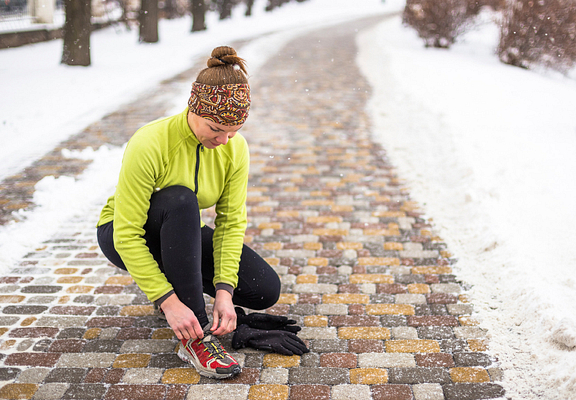
Making sure your shoes are in good condition can be an important part of preventing back pain from running.
The good news is running-related back pain is treatable—many times with a full recovery. Also, you can take steps to prevent back pain from running before it starts.
Suiting up of Runner’s Knee
A running shoe is not just a running shoe. There are several factors that can help determine what the right shoe for you should be, including:
- The height of your arches
- Your pronation style (in other words, how your foot hits the ground and rolls through each stride)
- How much distance you plan to run
A running specialist or doctor can help analyze your gait and make recommendations.
Once you have the right shoes, you still need to take care of them. Don’t put shoes in the dryer, which can deform their internal structure, and replace them every 250 miles or when they show significant wear.
Before you run of Runner’s Knee
Once you have the right shoes, you’re almost ready to hit your stride. But first, take a few minutes before your run to warm up both your core muscles and your leg muscles. You can do this a few different ways:
- Start your run by walking for 1 to 2 minutes, then speed up to a jog before finally hitting your intended speed.
- Do a few minutes of aerobic activities such as jumping jacks, burpees, or push-ups.
- Practice a few yoga poses.
It may also help you foam roll your hip and hamstring muscles before you begin running.
After you run of Runner’s Knee
- The same way you shouldn’t suddenly start running, you also shouldn’t suddenly stop either. A cooldown after your run is an important tactic to prevent back pain.
- The best way to cool down is to reverse the warm-up listed above: slow to a jog, then walk for a few minutes. And don’t forget to stretch—experts agree that post-workout stretching provides the most benefits.
Longer-term considerations of Runner’s Knee
- In addition to maintaining your running shoes in good condition, there are other long-term measures you can take to prevent back pain from running.
In fact, these may be some of the most important tips you can follow, because most back injuries develop over time as a result of repetitive errors in training or form:
- Be careful to avoid overtraining. Run only 3 to 4 days a week, and include a rest day with no workout once a week.
- When you’re ramping up your running program to prepare for a race or event, don’t increase daily running totals by more than 2 miles a week. Also, don’t increase speed and distance simultaneously.
- Have a running coach evaluate your form while running, since factors like how long your stride is or how your heel hits the ground can significantly influence your risk for injury over time.
- Crosstrain by alternating running with strength training and/or other types of exercise. Focus on activities that can strengthen your core, like an exercise ball routine.
By taking a few precautions, you can help prevent back pain as a result of running. But even if back pain develops, there are ways you can keep running despite it. Watch for another blog post coming soon about managing back pain, so you can keep running.
Once the diagnosis and underlying cause of runner’s knee have been established, a physician can prescribe a course of treatment. Most cases are treated without surgery.
Treatment of Runner’s Knee
- R.I.C.E. (rest, icing, compression, and elevation) may be advised to reduce the initial symptoms of runner’s knee. This protocol will be particularly important if the symptoms are manifesting for the first time.
- Over-the-counter or prescribed anti-inflammatory medications.
- Physical therapy to strengthen the muscles surrounding the knee and hip, stretch any tight muscles, and retrain the muscles to contract appropriately during sports and activity.
- Shoe inserts may benefit a subset of individuals with abnormal foot structure or movement patterns while running.
- A patellar brace or taping to maintain proper tracking of the patella during knee movement.
- Correction of training errors, such as the rate of increase in running volume and speed. In general, up to a 10% to 15% increase in either running volume or speed is safe on the patellofemoral joint.
If non-surgical methods fail to resolve the situation, surgery may be recommended.
Surgery for Runner’s Knee
Surgery is rarely indicated for patellofemoral knee pain. If all conservative measures have failed and an individual is unable to participate in desired activities without significant pain, a surgeon may attempt to alter mechanics of the knee through a procedure such as a “lateral release.” In this procedure, the lateral patellofemoral ligament is cut to decrease the pull on the patella to the outside of the knee and attempt to improve tracking along the trochlear groove of the femur. The decision for surgery requires a lengthy discussion with the surgeon, reviewing several factors, including: the medical history of the patient; the likelihood for success of the procedure; and whether or not it will restore the individual’s ability to perform athletically.
Fortunately, with appropriate pelvic and lower limb strengthening and modification of risk factors for patellofemoral pain, the vast majority of cases resolve and individuals can return to their full athletic potential.


