Cervical spondylosis is a generalized disease process affecting all levels of the cervical spine. Cervical spondylosis encompasses a sequence of degenerative changes in the intervertebral discs, osteophytosis of the vertebral bodies, hypertrophy of the facets and laminal arches, and ligamentous and segmental instability. The natural history of cervical spondylosis is associated with the aging process. Senescent and pathologic processes are thus morphologically indistinguishable. Clinical manifestations of cervical spondylosis may arise when morphologic sequelae are superimposed on a developmentally narrow spinal canal. The two clinical syndromes of spondylotic radiculopathy and myelopathy are distinct, yet they may overlap.[rx]
Cervical spondylosis is a term that encompasses a wide range of progressive degenerative changes that affect all the components of the cervical spine (i.e., intervertebral discs, facet joints, joints of Luschka, ligaments Flava, and laminae). It is a natural process of aging and presents in the majority of people after the fifth decade of life.[rx]
Causes of Cervical Spondylosis
A clinical syndrome caused by compression on the spinal cord that is characterized by
- clumsiness in hands
- gait imbalance
- degenerative cervical spondylosis (CSM)
- compression usually caused by anterior degenerative changes (osteophytes, disc osteophyte complex)
- degenerative spondylolisthesis and hypertrophy of ligamentum flavum may contribute
- the most common cause of cervical myelopathy
Congenital stenosis
Symptoms usually begin when congenital narrowing combined with spondylotic degenerative changes in older patients
Neurologic injury
- mechanism of injury can be
- direct cord compression
- ischemic injury secondary to compression of the anterior spinal artery
Associated conditions
- lumbar spinal stenosis
- tandem stenosis occurs in the lumbar and cervical spine in ~20% of patients
- tends to be slowly progressive and rarely improves with nonoperative modalities
- progression characterized by steplike deterioration with periods of stable symptoms
- early recognition and treatment prior to spinal cord damage is critical for good clinical outcomes
Risk Factors for Cervical Spondylosis
The lists below are the factors that you will have a higher risk of getting neck pain and cervical spondylosis:
- Genetics – if your family has a history of neck pain
- Smoking – clearly linked to increased neck pain
- Occupation – jobs with lots of neck motion and overhead work
- Mental health issues – depression/anxiety
- Injuries/trauma – car wreck or on-the-job injury
“Red flag” features and the conditions they may suggest
Malignancy, infection, or inflammation
-
Fever, night sweats
-
Unexpected weight loss
-
History of inflammatory arthritis, malignancy, infection, tuberculosis, HIV infection, drug dependency, or immunosuppression
-
Excruciating pain
-
Intractable night pain
-
Cervical lymphadenopathy
-
Exquisite tenderness over a vertebral body
Myelopathy
-
Gait disturbance or clumsy hands, or both
-
Objective neurological deficit—upper motor neuron signs in the legs and lower motor neuron signs in the arms
-
Sudden onset in a young patient suggests disc prolapse
Other
-
History of severe osteoporosis
-
History of neck surgery
-
Drop attacks, especially when moving the neck, suggest vascular disease
-
Intractable or increasing pain
Symptoms of Cervical spondylosis
The pain can be from minor to major and it becomes worse when looking up or down for a long period of time, such example is reading a book or driving. To improve the pain, most people tend to takes rest or even lay down. The pain usually becomes worse in the morning and at the end of the day.
Symptoms of cervical spondylosis include
- Neck stiffness and pain
- Numbness and weakness in the upper limbs
- Difficulty in walking, losing balance, or weakness in limbs
- Difficulty in turning the head fully or bending the neck, which may hinder drive
- Muscle spasms in neck and shoulders
- Headaches
- Grinding and popping feeling in the neck when rotating the head
- Loss of bladder and bowel control.
- Axial neck pain (oftentimes absent)
- Occipital headache common
- Extremity paresthesias
- Diffuse non-dermatomal numbness and tingling
- Weakness and clumsiness
- Weakness and decreased manual dexterity (dropping object, difficulty manipulating fine objects)
- Gait instability patient feels “unstable” on feet
- Weakness walking up and downstairs
- Gait changes are the most important clinical predictor
- Urinary retention rare and only appear late in disease progression, not very useful in diagnosis due to the high prevalence of urinary conditions in this patient population
- Cervical pain aggravated by movement
- Referred pain (occiput, between the shoulder blades, upper limbs)
- Retro-orbital or temporal pain (from C1 to C2)
- Cervical stiffness—reversible or irreversible
- Vague numbness, tingling, or weakness in upper limbs
- Dizziness or vertigo
- Poor balance
- Rarely, syncope triggers a migraine pseudo-angina
- Poorly localized tenderness
- Limited range of movement (forward flexion, backward extension, lateral flexion, and rotation to both sides)
- Minor neurological changes like inverted supinator jerks (unless complicated by myelopathy or radiculopathy)
Diagnosis of Cervical Spondylosis
Classically, symptomatic cervical spondylosis presents as one or more of the following three primary clinical syndromes
Axial neck pain
-
Commonly complain of stiffness and pain in the cervical spine that is most severe in the upright position and relieved with bed rest when removing the load from the neck
-
Neck motion, especially in hyperextension and side-bending, typically increases the pain
-
In upper and lower cervical spine disease, patients may report radiating pain into the back of the ear or occiput versus radiating pain into the superior trapezius or periscapular musculature, respectively
-
Occasionally, patients can present with atypical symptoms of cervical angina such as jaw pain or chest pain.
Cervical radiculopathy
-
Radicular symptoms usually follow a myotomal distribution depending on the nerve root(s) involved and can present as unilateral or bilateral neck pain, arm pain, scapular pain, paresthesia, and arm or hand weakness
-
Pain is exacerbated by head tilt toward the affected side or by hyperextension and side-bending toward the affected side.
Cervical myelopathy
-
Typically has an insidious onset with or without neck pain (frequently absent)
-
Can initially present with hand weakness and clumsiness, resulting in the inability to complete tasks requiring fine motor coordination (e.g., buttoning a shirt, tying shoelaces, picking up small objects)
-
Frequent reports of gait instability and unexplained falls
-
Urinary symptoms (i.e., incontinence) are rare and typically appear late in disease progression
Physical exam of Cervical spondylosis
-
Other non-specific neck pain lesions—acute neck strain, postural neck ache, or whiplash
-
Fibromyalgia – and psychogenic neck pain
-
Mechanical lesions – disc prolapse or diffuse idiopathic skeletal hyperostosis
-
Inflammatory disease – rheumatoid arthritis, ankylosing spondylitis, or polymyalgia rheumatica
-
Metabolic diseases – Paget’s disease, osteoporosis, gout, or pseudo-gout
-
Infections – osteomyelitis or tuberculosis
-
Malignancy – primary tumors, secondary deposits, or myeloma
Motor signs
-
Weakness in triceps and hand intrinsics
-
Atrophy of intrinsic hand muscles
-
Clumsiness with fine motor skills
-
The proximal weakness of the lower extremities
- weakness usually difficult to detect on physical exam
- lower extremity weakness is more concerning finding
- finger escape sign when the patient holds fingers extended and adducted, the small finger spontaneously abducts due to the weakness of intrinsic muscle grip and release test normally a patient can make a fist and release 20 times in 10 seconds. myelopathic patients may struggle to do this
Upper motor neuron signs
-
Hoffman’s sign (quick flexion of both the thumb and index finger when the middle fingernail is snapped)
-
Inverted radial reflex (flexion of the fingers in response to the brachioradialis reflex)
-
Pathological clonus
-
Babinski sign
Sensory dysfunction
-
Glove-like sensory loss in hands
-
Proprioceptive dysfunction
Proprioception dysfunction
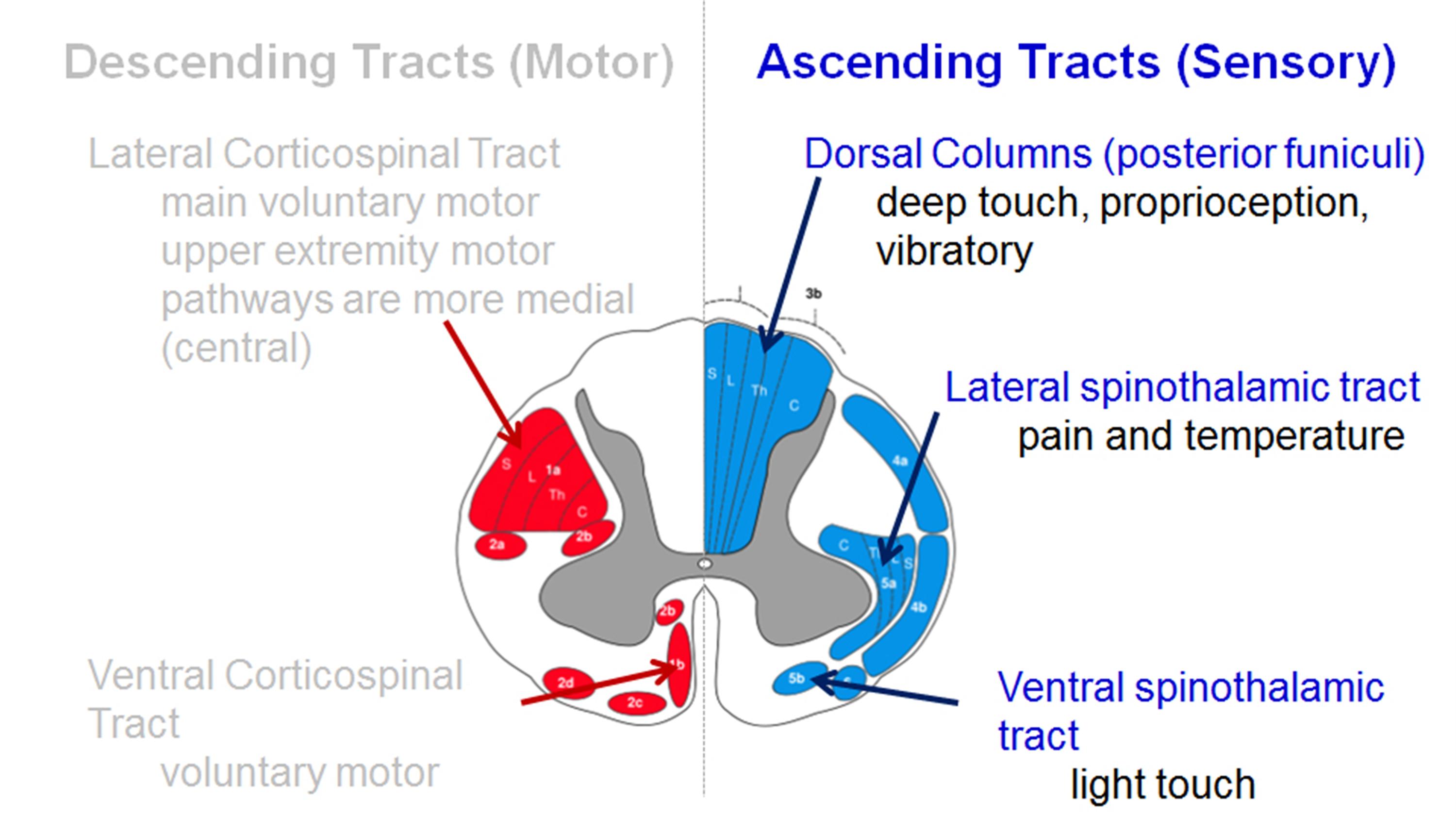
Decreased pain sensation
- pinprick testing should be done to look for a global decrease in sensation or dermatomal changes
- due to the involvement of lateral spinothalamic tract
- vibratory changes are usually only found in the severe case of long-standing myelopathy
Assessment tools
-
Lhermitte sign
-
Romberg test
-
9-Hole peg test
-
Grip and release test (observe a decreasing number of cycles)
-
Timed gait, 30-m walking test
-
Tandem gait
Upper motor neuron signs (spasticity)
- Hyperreflexia – may be absent when there is concomitant peripheral nerve disease (cervical or lumbar nerve root compression, spinal stenosis, diabetes)
- Inverted radial reflex – tapping distal brachioradialis tendon produces ipsilateral finger flexion
- Hoffmann’s sign – snapping patients distal phalanx of the middle finger leads to spontaneous flexion of other fingers
Sustained clonus
- > three beats defined as sustained clonus
- sustained clonus has poor sensitivity (~13%) but high specificity (~100%) for cervical myelopathy
Babinski test
- considered positive with the extension of the great toe
Gait and balance
- toe-to-heel walk patient has difficulty performing
- Romberg test patient stands with arms held forward and eyes closed loss of balance consistent with posterior column dysfunction
- provocative tests: Lhermitte Sign >test is positive when extreme cervical flexion leads to electric shock-like sensations that radiate down the spine and into the extremities
| Motor dysfunction | |
| Upper extremities | |
| 0 | Unable to move hands |
| 1 | Unable to eat with a spoon but able to move hands |
| 2 | Unable to button shirt but able to eat with a spoon |
| 3 | Able to button shirt with great difficulty |
| 4 | Able to button shirt with slight difficulty |
| Lower extremities | |
| 0 | Complete loss of motor & sensory function |
| 1 | Sensory preservation without the ability to move legs |
| 2 | Able to move legs but unable to walk |
| 3 | Able to walk on a flat floor with a walking aid (cane or crutch) |
| 4 | Able to walk up- &/or downstairs w/aid of a handrail |
| 5 | Moderate-to-significant lack of stability but able to walk up &/or downstairs without a handrail |
| 6 | Mild lack of stability but able to walk unaided with smooth reciprocation |
| 7 | No dysfunction |
| Sensory dysfunction | |
| Upper extremities | |
| 0 | Complete loss of hand sensation |
| 1 | Severe sensory loss or pain |
| 2 | Mild sensory loss |
| 3 | No sensory loss |
| Sphincter dysfunction | |
| 0 | Unable to micturate voluntarily |
| 1 | Marked difficulty in micturition |
| 2 | Mild-to-moderate difficulty in micturition |
| 3 | Normal micturition |
[Rx]
Radiographs
- recommended views cervical AP, lateral, oblique, flexion, and extension views
- general findings degenerative changes of uncovertebral and facet joints
- osteophyte formation
- disc space narrowing
- decreased sagittal diameter
- cord compression occurs with canal diameter is < 13mm
- lateral radiograph important to look for the diameter of the spinal canal
- a Pavlov ratio of less than 0.8 suggests a congenitally narrow spinal canal predisposing to stenosis and cord compression
Sagittal alignment
- C2 to C7 alignment determined by tangential lines on the posterior edge of the C2 and C7 body on lateral radiographs in neutral position
- Local kyphosis angle the angle between the lines drawn at the posterior margin of most cranial and caudal vertebral bodies forming the maximum local kyphosis
- Oblique radiograph important to look for foraminal stenosis which often caused by uncovertebral joint arthrosis
- Flexion and extension views important to look for angular or translational instability look for compensatory subluxation above or below the spondylotic/stiff segment
- Sensitivity/specificity changes often do not correlate with symptoms 70% of patients by 70 yrs of age will have degenerative changes seen on plain x -rays
X-ray
- Plain radiographs are an appropriate initial imaging study for neck and upper extremity pain in the absence of “red flag” symptoms. However, degenerative changes seen on imaging often poorly correlate with the presence of neck pain.[rx]
- Common radiographic findings include osteophyte formation, disc space narrowing, endplate sclerosis, degenerative changes of uncovertebral and facet joints, and calcified/ossified soft tissues. AP, lateral, and oblique views of the spine are adequate to evaluate for foraminal stenosis, sagittal alignment, and size of the spinal canal.
- The Torg-Pavlov ratio is obtainable by comparing the sagittal diameter of the spinal canal to the sagittal diameter of the vertebral body. The normal value is 1.0, with a ratio of <0.8 indicating cervical stenosis. Flexion and extension views also merit consideration if there is a concern for ligamentous instability.
MRI
- MRI is a study of choice to evaluate the degree of the spinal cord and nerve root compression effacement of CSF indicates functional stenosis
- Signal changes on T1-weighted images correlate with a poorer prognosis following surgical decompression spinal cord signal changes
- Seen as bright signal on T2 images (myelomalacia) compression ratio of < 0.4 carries poor prognosis CR = smallest AP diameter of cord / largest transverse diameter of the cord
- Sensitivity/specificity has a high rate of false-positive (28% greater than 40 will have findings of HNP or foraminal stenosis)
- CT without contrast can provide complementary information with an MRI and is more useful to evaluate OPLL and osteophytes
- CT myelography more invasive than an MRI but gives excellent information regarding degrees of spinal cord compression
- Useful in patients that cannot have an MRI (pacemaker), or have artifact (local hardware)
- Contrast is given via C1-C2 puncture and allowed to diffuse caudally, or given via a lumbar puncture and allowed to diffuse proximally by putting the patient in Trendelenburg position.
Computed tomography (CT)
- CT provides a good definition of bony structures and is more sensitive than plain radiographs in assessing intervertebral foraminal stenosis in the setting of uncovertebral or facet hypertrophy. However, it is less sensitive than MRI for the evaluation of soft tissues and nerve root compression.
CT myelogram
- CT is most useful when combined with the injection of intrathecal contrast (myelography) to better evaluate the location and amount of neural compression. It is more invasive than an MRI but can be a consideration in patients who have a contraindication to MRI (e.g., pacemaker) or have an artifact from the hardware.
Discogram
- Provocative discography is rarely necessary for cervical spondylosis. It is useful for the evaluation of patients who are experiencing cervical discogenic pain or have multiple herniations in which surgery is a strong possibility. However, the diagnostic procedure remains controversial as it may accelerate the degeneration of normal discs.[rx]
Electromyogram (EMG)
- EMG can be useful in supplementing neuroimaging findings in the diagnosis of cervical radiculopathy. It is especially valuable in differentiating nerve root compression from other possible concomitant neurologic conditions, including peripheral neuropathies, entrapment neuropathies, brachial plexopathies, myopathies, and motor neuron diseases.
Nerve conduction studies high false-negative rate may be useful to distinguish peripheral from the central process (ALS)
- Normal aging mild symptoms of myelopathy often confused with a “normal aging” process
- Stroke
- Movement disorders
- Vitamin B12 deficiency
- Amyotrophic lateral sclerosis (ALS)
- Multiple sclerosis
Treatment of Cervical spondylosis
Nonoperative
Observation, NSAIDs, therapy, and lifestyle modifications
- indications of mild disease with no functional impairment
- the function is a more important determinant for surgery than physical exam finding
- patients who are poor candidates for surgery
- modalities medications (NSAIDS, gabapentin)
- immobilization (hard collar in slight flexion)
- physical therapy for neck strengthening, balance, and gait training
- traction and chiropractic modalities are not likely to benefit and do have some risks
- be sure to watch patients carefully for progression
- outcomes improved nonoperative outcomes associated with patients with larger transverse area of the spinal cord (>70mm2)
- some studies have shown improvement with immobilization in patients with very mild symptoms
Non-Pharmacological Treatments
1. Patient education: Understanding that ordinary wear does not always mean serious damage reduces fear. The purpose is to encourage safe movement and active recovery instead of prolonged bed rest. Education works by improving confidence, self-management and regular participation in rehabilitation.
2. Individual physical therapy: A physiotherapist examines posture, movement, strength and nerve symptoms before creating a programme. Its purpose is to restore comfortable movement and daily function. Exercise gradually improves muscle support around the cervical spine.
3. Deep neck-flexor training: Gentle exercises train the small stabilising muscles at the front of the neck. Better control may reduce excess strain on painful joints and larger muscles. The exercise should be taught correctly because forceful neck bending may worsen symptoms.
4. Neck isometric exercise: During an isometric exercise, the head presses lightly against the hand without visibly moving. It helps develop strength while limiting joint motion. The pressure should remain comfortable and should stop if it produces arm pain, numbness or dizziness.
5. Scapular strengthening: The shoulder-blade muscles help hold the head, shoulders and upper back in a balanced position. Strengthening them may reduce overwork in the upper trapezius and neck muscles while improving posture during sitting and computer work.
6. Range-of-motion exercise: Slow rotation, side bending and flexion may preserve comfortable neck movement. Movements should remain inside a pain-free range rather than forcing a stiff joint. Sudden or repeated end-range movements are avoided when nerve or spinal-cord compression is suspected.
7. Gentle stretching: Stretching tight chest, shoulder and neck muscles may reduce muscle tension and improve upright posture. Each stretch should feel mild rather than sharp. Stretching cannot remove bone spurs, but it may reduce secondary muscular discomfort.
8. Regular walking: Walking maintains general fitness without placing heavy loads on the neck. It supports circulation, sleep, mood and confidence in movement. The distance can be increased gradually according to symptoms and balance.
9. Low-impact aerobic exercise: A stationary bicycle, gentle swimming or another comfortable activity may improve endurance and reduce physical deconditioning. The neck should remain in a neutral position, especially while cycling or swimming.
10. Posture retraining: Posture therapy teaches a relaxed, balanced position rather than holding the neck rigidly. Its purpose is to reduce long periods of forward-head posture and unnecessary muscle loading. Regular position changes are usually more useful than seeking one “perfect” posture.
11. Workplace modification: Raising the screen, supporting the forearms, keeping frequently used objects nearby and taking movement breaks can reduce repeated neck strain. An occupational therapist may also adapt work tasks when pain interferes with employment.
12. Activity pacing: Pacing divides demanding work into shorter periods with planned breaks. It prevents the cycle of doing too much on a good day and becoming inactive after a flare. The aim is steady function, not complete avoidance of movement.
13. Superficial heat: A warm shower or wrapped heat pack may temporarily relax tight muscles and reduce stiffness. Heat is mainly a comfort treatment and does not reverse degeneration. Avoid excessive temperature, sleeping on a heat source or applying it to numb skin.
14. Short cold application: A wrapped cold pack may calm pain after an activity-related flare. It should never be applied directly to the skin. Cold is optional; some people prefer warmth, and neither treatment corrects nerve or spinal-cord compression.
15. Massage: Gentle massage can reduce temporary muscular tension and make movement easier. Its main effect is comfort and relaxation rather than structural correction. Deep pressure should be avoided over painful bones or when severe nerve symptoms, fracture, infection or myelopathy are possible.
16. Manual mobilisation: Carefully selected, low-force joint and soft-tissue techniques may be combined with exercise. Forceful cervical manipulation is unsuitable when there is spinal-cord compression, instability, severe osteoporosis or neurological deterioration because complications can occur.
17. Clinician-supervised traction: Traction gently separates cervical structures and may provide temporary relief for selected nerve-root symptoms. Long-term evidence is limited. It should not be self-prescribed when there is myelopathy, instability, osteoporosis, tumour, infection or worsening neurological function.
18. Short-term soft collar: A soft collar may occasionally reduce movement during a severe, short-lived flare. Prolonged use can weaken muscles and encourage dependence, so it is not routine long-term treatment. It should be used only after clinical assessment.
19. Acupuncture or dry needling: Needling may provide temporary relief for some people with chronic muscular neck pain. Results vary, and it does not remove a compressed nerve or regenerate a disc. Sterile technique and a trained practitioner are essential.
20. Psychological pain management: Cognitive behavioural therapy, relaxation and stress-management techniques can reduce distress, improve sleep and support activity. They do not imply that pain is imaginary; they help control the way persistent pain affects attention, mood and daily life.
Drug Treatments for Cervical Spondylosis
Medicines cannot reverse worn cervical discs, remove bone spurs or release severe spinal-cord compression. They are used to control pain, inflammation, muscle spasm or nerve-related symptoms while rehabilitation continues. Several medicines below are off-label for cervical spondylosis, meaning FDA has approved them for another condition but clinicians may sometimes consider them after assessing the patient. Use the lowest effective treatment for the shortest suitable period, and never combine medicines from the same class without medical supervision.
1. Acetaminophen
Acetaminophen is a non-opioid pain reliever that may help mild or moderate neck pain when inflammation is not the main problem. It acts mainly inside the central nervous system to reduce the perception of pain but has little anti-inflammatory action. It may be considered when an NSAID is unsuitable because of stomach, kidney or cardiovascular risk. Dosage and duration should follow the product label and clinical assessment. Taking several acetaminophen-containing products together can cause severe liver injury, liver failure or death.
2. Ibuprofen
Ibuprofen belongs to the nonsteroidal anti-inflammatory drug, or NSAID, class. It reduces cyclooxygenase activity and lowers prostaglandin production, helping decrease inflammatory pain, tenderness and stiffness during a cervical spondylosis flare. It is normally used for a limited period rather than as an automatic long-term treatment. Possible adverse effects include indigestion, stomach ulcer or bleeding, kidney injury, fluid retention, raised blood pressure, allergic reactions, heart attack and stroke. It should not be combined with another NSAID unless specifically directed by a clinician.
3. Naproxen
Naproxen is an NSAID that may provide longer-lasting relief than some shorter-acting anti-inflammatory medicines. Its purpose is to reduce painful inflammation and improve movement during an acute neck-pain flare. Naproxen blocks cyclooxygenase enzymes and decreases prostaglandin formation. It does not repair discs or nerves. Treatment should use the lowest clinically appropriate exposure, particularly in older adults and people with cardiovascular, kidney or gastrointestinal disease. Side effects include heartburn, nausea, dizziness, stomach bleeding, kidney dysfunction, fluid retention, hypertension and cardiovascular events.
4. Celecoxib
Celecoxib is a selective cyclooxygenase-2, or COX-2, inhibitor. It reduces inflammatory prostaglandins and may relieve pain and stiffness while producing less inhibition of the stomach-protective COX-1 enzyme than traditional NSAIDs. However, it can still cause ulcers, gastrointestinal bleeding, kidney injury, hypertension, swelling, heart attack or stroke. Celecoxib is FDA-approved for several painful arthritic conditions and acute pain, but not specifically for cervical spondylosis. Dose and duration require individual cardiovascular, gastrointestinal, kidney and allergy assessment.
5. Diclofenac
Diclofenac is an NSAID with analgesic and anti-inflammatory effects. Oral diclofenac may occasionally be considered for a painful inflammatory flare when simpler measures are inadequate. It lowers prostaglandin production by inhibiting cyclooxygenase enzymes. Diclofenac does not correct cervical canal narrowing or progressive neurological compression. Its important risks include stomach ulceration or bleeding, kidney injury, liver-enzyme elevation, fluid retention, hypertension and cardiovascular thrombotic events. Oral and topical diclofenac should not be treated as completely separate medicines because both add to total NSAID exposure.
6. Meloxicam
Meloxicam is a prescription NSAID used for inflammatory pain and arthritis symptoms. It decreases prostaglandin synthesis through cyclooxygenase inhibition, which can reduce neck pain, stiffness and movement-related discomfort. It is not a cure for cervical degeneration and should not be combined with ibuprofen, naproxen, diclofenac or another NSAID without clinical direction. Adverse effects include dyspepsia, stomach bleeding, kidney impairment, swelling, increased blood pressure, cardiovascular events and severe allergic or skin reactions. The smallest effective exposure for the shortest suitable duration is preferred.
7. Etodolac
Etodolac is another prescription NSAID that may reduce inflammatory musculoskeletal pain. Its mechanism involves inhibition of cyclooxygenase enzymes and reduced production of prostaglandins that sensitise pain receptors. It may be considered as an alternative rather than an addition to another NSAID. Evidence does not show that etodolac regenerates a cervical disc or reverses spondylosis. Like other NSAIDs, it can cause abdominal pain, ulceration, gastrointestinal bleeding, kidney dysfunction, fluid retention, increased blood pressure, heart attack, stroke and allergic reactions.
8. Nabumetone
Nabumetone is an NSAID prodrug that the body converts into an active anti-inflammatory compound. It is FDA-approved for osteoarthritis and rheumatoid arthritis symptoms rather than specifically for cervical spondylosis. A clinician may occasionally consider it when inflammatory neck pain is prominent and another NSAID is unsuitable. It reduces prostaglandin-mediated pain and inflammation but does not decompress nerves. Possible adverse effects include indigestion, stomach ulcer or bleeding, kidney injury, hypertension, swelling, liver abnormalities and cardiovascular events. It should not be combined with another NSAID.
9. Ketorolac
Ketorolac is a potent NSAID intended only for short-term management of moderately severe acute pain. It is not suitable as routine long-term treatment for chronic cervical spondylosis. Ketorolac inhibits prostaglandin production and can provide strong non-opioid analgesia, but its gastrointestinal and kidney risks rise substantially with inappropriate duration or combination therapy. Important adverse effects include ulceration, serious bleeding, kidney failure, cardiovascular events and allergic reactions. FDA labeling limits total treatment duration, and oral therapy is used only within a tightly controlled clinical plan.
10. Cyclobenzaprine
Cyclobenzaprine is a centrally acting skeletal-muscle relaxant. It may be considered when an acute neck-pain episode includes clear painful muscle spasm. It acts mainly in the brainstem and reduces excessive muscle activity without directly repairing the cervical joints. FDA labeling supports only short-term use as an adjunct to rest and physical therapy because prolonged effectiveness has not been established. Common adverse effects include sleepiness, dizziness, dry mouth, blurred vision and impaired concentration. It may interact dangerously with serotonergic medicines or monoamine oxidase inhibitors.
11. Methocarbamol
Methocarbamol is a centrally acting muscle relaxant used as an adjunct for acute painful musculoskeletal conditions. It may temporarily reduce discomfort associated with protective muscle tightening around the neck. Its exact mechanism is not fully established and may be related mainly to central nervous system depression rather than direct relaxation of skeletal muscle. It should support, not replace, movement and physical therapy. Common adverse effects include drowsiness, dizziness, blurred vision, nausea and reduced coordination. Sedative effects may become stronger when combined with other central nervous system depressants.
12. Tizanidine
Tizanidine is a central alpha-2 adrenergic receptor agonist approved for spasticity. It reduces excitatory signals reaching spinal motor neurons and lowers excessive muscle tone. Its use for ordinary cervical muscle spasm is off-label and requires careful patient selection. Tizanidine may cause pronounced sleepiness, dizziness, weakness, dry mouth, slow heart rate and low blood pressure, especially when standing. Liver monitoring may be necessary, and important interactions occur with certain medicines. It should not be stopped suddenly after sustained treatment because withdrawal effects can occur.
13. Baclofen
Baclofen is a gamma-aminobutyric acid type-B receptor agonist that reduces abnormal motor-neuron activity. It is mainly approved for neurological spasticity, such as spasticity associated with multiple sclerosis or spinal-cord disease. It is not routinely indicated for ordinary mechanical neck spasm from cervical spondylosis. A specialist may consider it when genuine neurological spasticity is present. Adverse effects include sedation, dizziness, weakness, confusion and balance impairment. Abrupt withdrawal can cause serious reactions, so any reduction must be medically supervised.
14. Gabapentin
Gabapentin is an anticonvulsant and neuropathic-pain medicine. FDA approval includes postherpetic neuralgia and certain seizure disorders, not cervical radiculopathy. Its use for burning, shooting or electric arm pain caused by a compressed cervical nerve is therefore off-label. Gabapentin binds to the alpha-2-delta subunit of voltage-gated calcium channels and reduces the release of excitatory neurotransmitters. It may cause dizziness, sleepiness, unsteadiness, swelling and impaired concentration. Kidney function affects drug clearance, and sudden discontinuation after regular use should be avoided.
15. Pregabalin
Pregabalin is a gabapentinoid approved for several neuropathic-pain conditions, including pain related to spinal-cord injury, but not specifically for cervical radiculopathy. It reduces calcium-dependent release of excitatory neurotransmitters by binding to the alpha-2-delta calcium-channel subunit. A clinician may consider it for prominent nerve-related arm pain after confirming the diagnosis. Side effects include dizziness, somnolence, blurred vision, swelling, weight gain and impaired balance. Kidney-based dose adjustment is often necessary, and gradual withdrawal is required after continued treatment.
16. Duloxetine
Duloxetine is a serotonin-norepinephrine reuptake inhibitor, or SNRI. It is FDA-approved for conditions including diabetic peripheral neuropathic pain, fibromyalgia and chronic musculoskeletal pain, but not specifically for cervical spondylosis. It strengthens descending pain-control pathways in the brain and spinal cord and may be considered when chronic pain has a neuropathic or centrally amplified component. Possible adverse effects include nausea, dry mouth, sleep disturbance, dizziness, sweating and increased blood pressure. Liver disease, important drug interactions and serotonin syndrome risk require assessment.
17. Amitriptyline
Amitriptyline is a tricyclic antidepressant sometimes used off-label in carefully selected adults with chronic neuropathic pain or pain-related sleep disturbance. It reduces the reuptake of serotonin and norepinephrine and changes pain processing within the central nervous system. It is not FDA-approved for cervical radiculopathy and may be unsuitable for many older people or patients with cardiac, urinary, eye or cognitive problems. Adverse effects include drowsiness, dry mouth, constipation, blurred vision, urinary retention, low blood pressure, confusion and abnormal heart rhythm.
18. Lidocaine Topical System
A lidocaine patch or topical system is a local anaesthetic treatment approved for postherpetic neuralgia, not cervical spondylosis. Use for a small, superficial area of neuropathic neck or shoulder pain is off-label. Lidocaine temporarily blocks sodium channels in peripheral nerves, reducing the transmission of pain signals from the treated skin. It does not reach a deeply compressed cervical nerve or spinal cord. Possible adverse effects include skin redness, irritation, numbness and, with excessive systemic absorption, neurological or cardiac toxicity.
19. Short-Course Corticosteroid
Prednisone or methylprednisolone is a systemic glucocorticoid with strong anti-inflammatory activity. A specialist may occasionally consider a brief course for severe acute cervical radicular inflammation, although it does not remove the mechanical cause of nerve compression and routine repeated courses are inappropriate. Corticosteroids alter inflammatory gene activity and reduce immune-cell signalling. Adverse effects can include indigestion, mood or sleep changes, raised blood glucose, fluid retention, increased blood pressure and infection risk. Repeated or prolonged exposure can weaken bone and muscle and suppress adrenal function.
20. Tramadol or Another Opioid Analgesic
Opioid analgesics are not routine first-line medicines for cervical spondylosis. They may be considered only for severe pain when safer non-opioid treatments are inadequate, contraindicated or not tolerated, usually within a closely monitored and time-limited clinical plan. Tramadol has opioid activity and also changes serotonin and norepinephrine signalling. Important risks include nausea, constipation, dizziness, sedation, impaired breathing, seizures, serotonin syndrome, tolerance, physical dependence, misuse and overdose. Combining opioids with sedatives can greatly increase respiratory danger.
21. Cartilage-regenerating medicines: No currently approved oral medicine has been shown to rebuild worn cervical facet-joint cartilage or restore a degenerated cervical disc. Pain relievers and anti-inflammatory medicines may improve symptoms, but they cannot replace lost cartilage, dissolve bone spurs or widen a narrowed spinal canal.
22. Glucosamine and chondroitin: These products are dietary supplements rather than FDA-approved cervical-spondylosis drugs. Studies have mainly examined knee osteoarthritis, and their effects on pain and function remain inconsistent. There is very little evidence for joints outside the knee and no reliable evidence that they regenerate cervical cartilage or spinal discs.
23. Collagen, MSM and hyaluronic-acid supplements: These products are sometimes advertised for joint or cartilage repair, but evidence is insufficient to claim that they reverse cervical spondylosis. They should not be described as cartilage-building drugs. A supplement may also interact with medicines or contain variable ingredients, so its use should be reviewed clinically.
Important Consciousness
Cyclobenzaprine for painful muscle spasm: Cyclobenzaprine is a centrally acting skeletal-muscle relaxant that may be used briefly when cervical spondylosis produces significant painful muscle spasm. It reduces muscle overactivity through actions within the central nervous system but does not strengthen muscles or repair cervical structures. Drowsiness, dizziness, dry mouth and reduced alertness can occur.
Methocarbamol for acute muscular discomfort: Methocarbamol is used as an additional treatment alongside rest, physical therapy and other measures for acute painful musculoskeletal conditions. Its action may mainly involve central nervous system sedation rather than direct relaxation of tense muscle fibres. It can cause sleepiness, dizziness, blurred vision and reduced coordination.
Tizanidine for neurological spasticity: Tizanidine is an alpha-2 adrenergic agonist approved for spasticity. It reduces excitatory signals reaching spinal motor neurons. It is not a routine cartilage, tendon or ordinary neck-muscle repair medicine. It may occasionally be considered when true neurological spasticity exists, but it can cause low blood pressure, sleepiness, weakness, dry mouth and liver abnormalities.
Baclofen for genuine spasticity: Baclofen acts on GABA-B receptors and is mainly used for spasticity associated with multiple sclerosis, spinal-cord injury or another neurological disorder. FDA labeling states that it is not indicated for ordinary skeletal-muscle spasm resulting from rheumatic conditions. It should therefore not be routinely described as a muscle medicine for uncomplicated cervical spondylosis.
Medicines should never delay urgent evaluation when cervical spondylosis causes progressive weakness, hand clumsiness, poor balance, difficulty walking, repeated falls or bladder or bowel disturbance. These findings may indicate cervical myelopathy, for which tablets cannot adequately relieve spinal-cord compression.
Dietary Nutrients and Molecular Support Factors
No supplement has been proven to regenerate a cervical disc or cure cervical spondylosis. Nutrients should preferably come from a balanced diet. Supplements are appropriate mainly when dietary intake is inadequate, deficiency is confirmed or a clinician identifies a specific need.
1. Protein: Fish, eggs, dairy foods, beans, lentils, soy and lean meat provide amino acids needed for maintaining muscle. Adequate muscle supports rehabilitation, but unusually high protein intake does not rebuild damaged cervical discs.
2. Calcium: Milk, yoghurt, calcium-set tofu, small fish with edible bones and fortified foods support bone structure. Calcium protects general skeletal health but does not remove existing bone spurs.
3. Vitamin D: Vitamin D supports calcium absorption, bone mineralisation, muscle function and nerve signalling. Testing may be useful when deficiency is suspected. Excess supplement intake can cause high calcium, kidney injury and heart-rhythm problems.
4. Magnesium: Nuts, seeds, beans, whole grains and leafy vegetables provide magnesium for normal muscle, nerve and bone function. Evidence does not show that magnesium supplements cure cervical degeneration.
5. Vitamin B12: Fish, meat, eggs, dairy products and fortified foods provide vitamin B12, which supports healthy blood and nerve cells. A deficiency can produce numbness or weakness that may resemble spinal nerve disease.
6. Omega-3 fats: Oily fish, walnuts, chia and flaxseed provide omega-3 fatty acids. These foods can be part of a heart-healthy dietary pattern, but omega-3 capsules have not been established as a treatment for cervical spondylosis.
7. Vitamin C: Citrus fruit, guava, berries, tomatoes and peppers supply vitamin C, which participates in collagen formation and antioxidant processes. More is not necessarily better, and large supplemental amounts may cause adverse effects.
8. Zinc: Meat, seafood, dairy foods, legumes, nuts and seeds provide zinc for normal immune and cellular functions. Zinc is not an “immune booster” that repairs discs, and excessive supplements can disturb other minerals.
9. Antioxidant-rich plants: Vegetables, fruits, herbs, beans and whole grains provide varied plant compounds. They support general health but should not be described as medicines that dissolve bone spurs or decompress nerves.
10. Water and fibre: Adequate water, vegetables, fruit and whole grains support bowel health, particularly when pain reduces activity. Hydration does not directly rehydrate a degenerated spinal disc, but it supports normal general health.
Immunity, Regenerative and Stem-Cell Treatments
Stem cells, exosomes and regenerative injections: Stem-cell, exosome, amniotic and similar products should not be presented as approved drugs for cervical-disc, tendon or cartilage regeneration. FDA warns that misleading claims are common and that regenerative products marketed for neck pain or orthopaedic conditions may be unapproved.
1. Stem-cell injections: FDA states that no regenerative medicine product is approved for neck pain, back pain or degenerative disc disease. Commercial claims of routine disc regeneration therefore should be treated cautiously.
2. Exosome products: Exosomes are sometimes marketed as cell-repair treatments, but FDA has not approved them for cervical spondylosis. Product contents, effectiveness and long-term risks may be uncertain outside regulated clinical research.
3. Amniotic or umbilical products: Birth-tissue products advertised for neck regeneration are not established treatments for cervical degeneration. They should not be confused with the limited blood-forming stem-cell products approved for particular blood disorders.
4. Platelet-rich plasma: PRP contains concentrated components from the patient’s blood. Evidence remains insufficient to describe it as a proven cervical-disc regenerating treatment, particularly when neurological compression is present.
5. “Immune-boosting” medicines: Cervical spondylosis is primarily degenerative, not an immune-deficiency illness. Stimulating immunity does not remove bone spurs or widen a narrowed spinal canal and could be harmful in some medical conditions.
6. Secret regenerative drugs: No approved medicine currently reverses ordinary cervical spondylosis. Products promising rapid joint or disc repair may contain undeclared drug ingredients; FDA has repeatedly warned about contaminated pain products.
Surgery
1. Anterior cervical discectomy and fusion: The surgeon removes the diseased disc or bone spur from the front of the neck, releases the compressed nerve or spinal cord and joins the vertebrae. It is considered for persistent radiculopathy, weakness, instability or myelopathy.
2. Artificial cervical disc replacement: The painful or compressing disc is removed and replaced with an artificial disc intended to preserve motion. It is suitable only for carefully selected patients because anatomy, instability, arthritis severity and involved levels affect eligibility.
3. Posterior cervical foraminotomy: Through the back of the neck, the surgeon enlarges the opening where a nerve exits. It may relieve one-sided arm pain caused by a compressed nerve while avoiding fusion in selected cases.
4. Cervical laminectomy with or without fusion: Part of the bone covering the spinal canal is removed to create more room for the spinal cord. Fusion may be added when instability, deformity or excessive movement is a concern.
5. Cervical laminoplasty: The lamina is opened like a hinged door rather than completely removed. This expands space for the spinal cord across several levels while preserving some movement. It is mainly used for selected cases of multilevel cervical myelopathy.
Surgical decompression, restoration of lordosis, stabilization
- indications significant functional impairment AND 1-2 level disease lordotic, neutral or kyphotic alignment
- techniques appropriate procedure depends on cervical alignment number of stenotic levels
- medical conditions (e.g., goiter)
- location of compression
- anterior cervical diskectomy/corpectomy and fusion
- posterior laminectomy and fusion
- posterior laminoplasty
- combined anterior and posterior procedure
- cervical disk arthroplasty
- outcomes prospective studies show improvement in overall pain, function, and neurologic symptoms with operative treatment early recognition and treatment prior to spinal cord damage is critical for good clinical outcomes
Goals
optimal surgical treatment depends on the individual. Things to consider include
- number of stenotic levels
- sagittal alignment of the spine
- the degree of existing motion and desire to maintain
- medical comorbidities (eg, dysphasia)
- simplified treatment algorithm
Anterior Decompression and Fusion (ACDF) alone
Indications
- the mainstay of treatment in most patients with single or two level disease
- fixed cervical kyphosis of > 10 degrees anterior procedure can correct kyphosis
- compression arising from 2 or fewer disc segments
- pathology is anterior (OPLL, soft discs, disc osteophyte complexes)
- uses Smith-Robinson anterior approach
Decompression of corpectomy and strut graft may be required for multilevel spondylosis two level corpectomies tend to be biomechanically vulnerable (preferable to combine single-level corpectomy with adjacent level diskectomy)
- 7% to 20% rates of graft dislodgement with cervical corpectomy with associated severe complications, including death, reported.
- fixation anterior plating functions to increase fusion rates and preserve the position of the interbody cage or strut graft
- pros & cons advantages compared to posterior approach lower infection rate blood loss less postoperative pain disadvantages avoid in patients with poor swallowing function
Laminectomy with posterior fusion
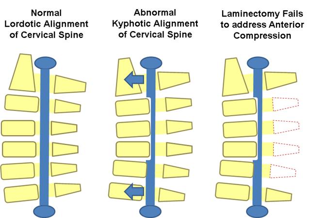
- multilevel compression with kyphosis of < 10 degrees,> 13 degrees of fixed kyphosis is a contraindication for a posterior procedure
- in the flexible kyphotic spine, posterior decompression and fusion may be indicated if kyphotic deformity can be corrected prior to instrumentation
Contraindications
- fixed kyphosis of > 10 degrees is a contraindication to posterior decompression
- will not adequately decompress spinal cord as it is “bowstringing” anterior
Pros & cons
- fusion may improve neck pain associated with degenerative facets
- not effective in patients with > 10 degrees fixed kyphosis
Laminoplasty
- gaining in popularity
- useful when maintaining motion is desired
- avoids complications of fusion so may be indicated in patients at high risk of pseudoarthrosis
- cervical kyphosis > 13 degrees is a contraindication to posterior decompression
will not adequately decompress spinal cord as it is “bowstringing” anterior - severe axial neck pain is a relative contraindication and these patients should be fused
Technique
- the volume of the canal is expanded by hinged-door laminoplasty followed by fusion
usually performed from C3 to C7 - open door technique hinges created unilaterally at the junction of lateral mass and lamina and opening on opposite side opening held open by bone, suture anchors, or special plates
- French door technique hinge created bilaterally and the opening created midline
Pros & cons advantages
- lower complication rate than multilevel anterior decompression especially in patients with OPLL a motion-preserving technique pseudoarthrosis not a concern in patients with poor healing potential (diabetes, chronic steroid users) can be combined with a subsequent anterior procedure
- allows for decompression of multilevel stenotic myelopathy without compromising stability and motion (avoids postlaminectomy kyphosis)
Disadvantages
- higher average blood loss than anterior procedures
- postoperative neck pain
- still associated with loss of motion outcomes equivalent to multilevel anterior decompression and fusion
- Combined anterior and posterior surgery multilevel stenosis in the rigid kyphotic spine
- multi-level anterior cervical corpectomies
- postlaminectomy kyphosis
Laminectomy alone
- indications rarely indicated due to risk of post laminectomy kyphosis
- pros & cons progressive kyphosis 11 to 47% incidence if laminectomy performed alone without fusion
Surgical Infection
- higher rate of surgical infection with posterior approach than anterior approach
- Pseudoarthrosis incidence 12% for single level fusions, 30% for multilevel fusions treatment
- treat with either posterior wiring or plating or repeat anterior decompression and plating if patient has symptoms of radiculopathy
- reported to occur in ~ 4.6% of patients after surgery for cervical compression myelopathy
- no significant differences between patients undergoing anterior decompression and fusion and posterior laminoplasty
- occurs immediately postop to weeks following surgery
Mechanism
- mechanism is controversial
- in laminectomy patients, it is thought to be caused by tethering of nerve root with dorsal migration of spinal cord following removal of posterior elements
Prognosis
- patients with postoperative C5 palsy generally have a good prognosis for functional recovery, but recovery takes time
- Recurrent laryngeal nerve injury approach in the past it has been postulated that the RLN is more vulnerable to injury on the right due to a more aberrant pathway recent studies have shown there is not an increased injury rate with a right sided approach
- treatment if you have a postoperative RLN palsy, watch over time
- if not improved over 6 weeks, then ENT consult to scope patient and inject Teflon
- if you are performing revision anterior cervical surgery, and there is any suspicion of an RLN from the first operation, obtain ENT consult to establish prior injury
- if a patient has prior RLN nerve injury, perform revision surgery on the same as the prior injury/approach to prevent a bilateral RLN injury
- Hardware failure and migration 7-20% with two-level anterior corpectomies two-level corpectomies should be stabilized from behind
- Postlaminectomy kyphosis treat with anterior/posterior procedure
- Postoperative axial neck pain
- Vertebral artery injury
- Esophageal Injury
- Dysphagia & alteration in speech
Keywords: degenerative disc disease, cervical spondylosis, cervical spondylotic myelopathy, cervical spine stenosis, anterior cervical discectomy and fusion, cervical laminoplasty, cervical disk replacement
Complications
In a 2019 cohort study by El-Yahochouchi et al., the overall incidence of immediate and delayed adverse events following an epidural steroid injection was 2.4% and 4.9%, respectively.[rx] Complications include:
-
Neurologic injury
-
Epidural abscess
-
Epidural hematoma
-
Increased pain
-
Vasovagal reactions
-
Central steroid response (e.g., facial flushing, nonpositional headaches)
-
Endocrinologic effects (e.g., hyperglycemia, hypothalamic-pituitary axis suppression, decreased bone density)
Complications from anterior and posterior cervical spine surgery include [18][24]:
-
Injury to spinal cord and nerve roots
-
Infection
-
Dural tear and CSF leak
-
Recurrent laryngeal, superior laryngeal, and hypoglossal nerve injuries
-
Esophageal injury and dysphagia
-
Vertebral and carotid artery injuries
-
Tracheal injury
-
Adjacent segment degeneration
-
Pseudoarthrosis
-
Post-laminectomy kyphosis
Prevention and Flare-Reduction Measures
1. Remain physically active: Regular walking and strengthening help prevent deconditioning and support function.
2. Change position regularly: Avoid keeping the neck bent toward a phone, book or screen for long uninterrupted periods.
3. Arrange the workstation: Keep the screen near eye level and support the forearms to reduce sustained neck loading.
4. Build strength gradually: Sudden heavy exercise can trigger a flare; progressive training is safer than rapid increases.
5. Avoid smoking: Smoking is harmful to overall musculoskeletal, cardiovascular and surgical health and may impair tissue healing.
6. Maintain a healthy weight: Appropriate weight supports mobility and lowers the general physical burden during activity.
7. Use safe lifting technique: Keep loads close, avoid sudden twisting and ask for help with objects that are too heavy.
8. Protect against falls: Good lighting, suitable footwear and treatment of balance problems reduce the risk of neck injury.
9. Sleep comfortably: Use a pillow height that keeps the neck reasonably neutral rather than sharply bent. No special pillow can cure cervical degeneration.
10. Review persistent symptoms early: Timely assessment of progressive numbness, weakness or balance change may prevent delayed recognition of spinal-cord compression.
When to See a Doctor
Arrange medical assessment when pain persists, repeatedly returns, travels into an arm, causes numbness or interferes with sleep and daily activity. Seek urgent care for new or worsening weakness, clumsy hands, poor balance, repeated falls, difficulty walking, loss of bladder or bowel control, fever, unexplained weight loss, cancer history, major trauma or severe night pain.
Foods to Eat and Foods to Limit
1. Eat vegetables and fruit; limit highly processed snacks. Whole foods provide fibre and varied nutrients, while processed snacks often contain excessive salt, sugar and energy.
2. Eat fish, beans, lentils, eggs or lean meat; limit heavily processed meat. These foods provide protein needed for muscle maintenance during rehabilitation.
3. Eat calcium-containing foods; avoid unnecessary high-dose calcium pills. Food sources support bone health, while supplements may be unsuitable for some people.
4. Eat vitamin-D-containing or fortified foods; avoid megadoses. Excess vitamin D can cause dangerous calcium elevation and kidney or heart complications.
5. Eat nuts, seeds and whole grains; limit excessive refined carbohydrates. These foods supply magnesium and fibre, supporting normal muscle, nerve and digestive function.
6. Eat oily fish or plant omega-3 sources; avoid assuming fish-oil capsules cure neck disease. Dietary omega-3 is nutritious but does not decompress nerves.
7. Drink water regularly; limit sugary drinks. Water supports normal body function, while sugary beverages can add substantial energy without treating pain.
8. Choose moderate salt intake; limit very salty packaged foods. This is particularly important for people with high blood pressure, kidney disease or fluid retention.
9. Avoid unlabelled “joint cure” products. Some products promoted for pain have contained undeclared prescription ingredients such as anti-inflammatory drugs or corticosteroids.
10. Avoid crash diets. A balanced eating pattern better supports muscle, bone, energy and participation in rehabilitation than severe food restriction.
Frequently Asked Questions
1. Is cervical spondylosis cancer? No. It is usually age-related degeneration. However, unusual symptoms such as weight loss, fever or severe night pain require assessment for other causes.
2. Can cervical spondylosis be completely cured? Existing wear is not usually reversed, but pain and function often improve greatly with education, exercise, posture management and appropriate medical care.
3. Does an X-ray abnormality always cause pain? No. Degenerative changes can exist without symptoms. Treatment should consider the examination and symptoms, not an imaging report alone.
4. Is exercise safe? Appropriately selected exercise is usually central to treatment. Stop and obtain assessment if exercise causes increasing weakness, loss of balance or severe radiating symptoms.
5. Should I rest in bed? Prolonged rest can reduce strength and confidence. Relative rest during a severe flare may help, but gradual return to comfortable activity is generally preferred.
6. Can massage cure cervical spondylosis? No. Massage may temporarily relax muscles, but it cannot remove bone spurs, restore a worn disc or release serious spinal-cord compression.
7. Is neck cracking safe? Forceful manipulation can be dangerous in myelopathy, instability or certain vascular and bone conditions. Persistent symptoms should be assessed before manipulation.
8. Can cervical spondylosis cause headache? Neck structures can contribute to pain felt around the back or side of the head, but new, severe or unusual headache needs separate medical assessment.
9. Can it cause arm numbness? Yes. A narrowed opening or disc problem may irritate a cervical nerve root, causing arm pain, tingling, numbness or weakness.
10. What is cervical myelopathy? It means pressure or injury affecting the spinal cord in the neck. Warning signs include poor balance, walking difficulty, hand clumsiness and progressive limb weakness.
11. When is MRI needed? MRI may be considered when neurological findings, persistent radicular symptoms, suspected myelopathy, infection, tumour or another serious cause needs investigation.
12. Will I need surgery? Most uncomplicated cases do not require surgery. Surgery is considered when nerve or spinal-cord compression causes persistent pain, progressive weakness, walking difficulty or loss of function.
13. Can food dissolve bone spurs? No food or supplement has been shown to dissolve cervical bone spurs. A balanced diet supports general bone and muscle health but does not replace decompression when severe neurological compression exists.
14. Do stem cells regenerate the cervical spine? This is not an FDA-approved treatment for neck pain or disc disease. Claims of guaranteed regeneration should be approached cautiously.
15. What is the best long-term approach? The safest approach combines an accurate diagnosis, regular appropriate activity, targeted strengthening, ergonomic changes, healthy sleep and diet, symptom monitoring and timely specialist review when neurological warning signs appear.
References
- https://www.ncbi.nlm.nih.gov/pmc/articles/PMC1819511/
- https://www.ncbi.nlm.nih.gov/pmc/articles/PMC5582708/
- https://www.ncbi.nlm.nih.gov/pubmed/17204889
- https://www.ncbi.nlm.nih.gov/pubmed/2536306
- https://www.ncbi.nlm.nih.gov/books/NBK551557/
- https://en.wikipedia.org/wiki/Cervical%20Spondylosis?redirect=no
- https://www.sciencedirect.com/cervical-spondylosis-and-other-disorders-of-the-cervical-spine
- https://www.sciencedirect.com/science/article/pii/B9780750613613500136
- https://www.sciencedirect.com/topics/medicine-and-dentistry/spondylosis
- https://www.sciencedaily.com/releases/2011/04/110411164007.htm
- https://books.google.com/books/about/Manage_and_Cure_Neck_Pain_Cervical_Spond.html?
- https://books.google.com/books/about/Cervical_Spondylosis.html?id=Z6izAAAAIAAJ
- https://books.google.com/books/about/Cervical_Arthrosis.html?id=vhxsAAAAMAAJ
- https://link.springer.com/chapter/10.1007/978-1-59745-106-2_6
- http://link.springer.com/chapter/10.1007/978-3-642-67605-5_21
- https://www.science.gov/topicpages/s/single-level+cervical+spondylosis.html
- https://www.science.gov/topicpages/c/cervical+spondylotic+neuropathy
- https://worldwidescience.org/topicpages/o/onset+cervical+radiculopathy.html