
Cesarean delivery is a surgical procedure that involves delivering a baby through an abdominal incision (laparotomy) and a uterine incision (hysterotomy). The first documented cesarean operation occurred in AD 1020, and the procedure has since evolved significantly.[1] Today, cesarean delivery is the most common surgery performed in the United States, with over 1 million women undergoing cesarean deliveries annually.
The cesarean delivery rate increased from 5% in 1970 to 31.9% in 2016.[2] This sharp increase can be attributed to various factors, including changes in maternal age, medical advancements allowing more complicated pregnancies to proceed, and evolving obstetric practices. In 2022, the United States recorded more than 3.66 million births, most of which resulted from spontaneous or induced labor. Labor dystocia remains the most common indication for primary cesarean delivery. Globally, cesarean delivery rates continue to rise, and reducing unnecessary cesarean procedures remains a priority in the United States, where 32.2% of all births in 2022 were cesarean deliveries.[3]
A C-section delivery is done when it is not possible or safe for the mother to deliver the baby through the vagina.
The procedure is most often done while the woman is awake. The body is numbed from the chest to the feet using epidural or spinal anesthesia.
1. The surgeon makes a cut across the belly just above the pubic area.
2. The womb (uterus) and amniotic sac are opened.
3. The baby is delivered through this opening.
The health care team clears fluids from the baby’s mouth and nose. The umbilical cord is cut. The health care provider will make sure that the infant’s breathing is normal and other vital signs are stable.
The mother is awake during the procedure so she will be able to hear and see her baby. In many cases, the woman is able to have a support person with her during the delivery.
Anatomy and Physiology
Performing a cesarean delivery requires the surgeon to carefully navigate through multiple anatomical layers to access the fetus. The procedure begins with an incision in the skin, followed by the subcutaneous tissue. The fascia covering the rectus abdominis muscles is then exposed. This fascia consists of 2 layers—one is derived from the aponeurosis of the external oblique muscle, and the second is a fused layer formed by the aponeuroses of the transverse abdominis and internal oblique muscles.
After separating the vertically oriented rectus muscles, the surgeon accesses the abdominal cavity by incising the parietal peritoneum. In a gravid patient, the uterus is typically encountered immediately upon entering the abdomen, in contrast to a nongravid patient. If the patient has adhesive disease from previous surgeries, the surgeon may encounter adhesions involving structures such as the omentum, bowel, anterior abdominal wall, bladder, and the anterior aspect of the uterus.[5]
Upon identifying the uterus, the surgeon locates the vesicouterine peritoneum, or vesicouterine serosa, which connects the bladder to the uterus. If a bladder flap has to be created, the vesicouterine peritoneum must be incised. In patients with a history of prior cesarean sections, the bladder may be more challenging to separate from the uterus.
The uterus is composed of 3 layers—the serosal outer layer (perimetrium), the muscular layer (myometrium), and the inner mucosal layer (endometrium). All 3 layers are incised during the uterine incision or hysterotomy. The uterine vessels run bilaterally along the lateral aspects of the uterus, requiring careful attention to avoid damage when making or extending the incision. The uterine arteries branch from the anterior division of the internal iliac artery. During pregnancy, blood flow through the uterine arteries is 8 times faster, with unilateral flow exceeding 300 mL/min at 36 weeks gestation.[6] The uterine arteries cross the ureters anteriorly and enter the uterus at the cardinal ligament. They also anastomose with the ovarian arteries in the broad ligament, which arise from the abdominal aorta.
Ideally, cesarean deliveries are performed during the term period when the lower uterine segment is adequately developed to provide a thinner and less vascular area for the transverse hysterotomy. In preterm deliveries, especially those before 34 weeks gestation, this access point is not available, and a “classical cesarean delivery” may be required. This involves a vertical incision for the hysterotomy, which increases the risk of bleeding and precludes a trial of labor after cesarean delivery in future pregnancies due to the heightened risk of uterine rupture.[7]
Depending on whether the patient’s amniotic membranes are intact or ruptured, the surgeon may encounter the amniotic sac upon incising the uterus. The amniotic sac comprises 2 layers—the chorion and the amnion—which fuse early in pregnancy. If intact, the amniotic sac forms the final barrier between the surgeon and the fetus. Once this layer is breached, the fetus is delivered, achieving the primary goal of the cesarean delivery. The gravid uterus typically obscures much of the reproductive anatomy. However, after fetal delivery, additional structures may become visible, especially if the surgeon exteriorizes the uterus for repair. The surgeon may examine the fallopian tubes and ovaries and perform medically indicated procedures if necessary, such as bilateral salpingectomy in cases of opportunistic salpingectomy or ovarian cystectomy for problematic ovarian cysts.[8][9]
Indications
Several factors can preclude a safe vaginal delivery, necessitating a cesarean delivery.[10][11] For example, cesarean delivery is often recommended for patients with a prior classical cesarean delivery or a history of uterine rupture. Given the potential complications of cesarean delivery, efforts focus on reserving the procedure for medically necessary cases rather than elective or inappropriate indications. However, maternal requests for elective cesarean delivery are not universally denied, as patient autonomy is paramount. Instead, the goal is to ensure that best practices are followed, delivering the most appropriate and beneficial obstetric care through shared decision-making.[12]
Efforts to decrease primary cesarean deliveries are crucial, as many patients who undergo one cesarean delivery will have subsequent deliveries via the same method. Patients may opt for repeat cesareans for various reasons or may not be candidates for vaginal birth after cesarean birth. For example, cervical ripening with medications such as misoprostol is not recommended in patients with an unfavorable cervix at term due to the increased risk of uterine rupture. Common indications for first-time cesarean deliveries include labor dystocia, abnormal fetal heart rate patterns, malpresentation, multiple gestations, and suspected fetal macrosomia.[3][13]
Maternal Indications for Cesarean Section
-
Prior cesarean delivery
-
Maternal request
-
Pelvic deformity or cephalopelvic disproportion
-
Previous perineal trauma
-
Prior pelvic or anal/rectal reconstructive surgery
-
Herpes simplex or HIV infection
-
Cardiac or pulmonary disease
-
Cerebral aneurysm or arteriovenous malformation
-
Pathology requiring concurrent intraabdominal surgery
Uterine/Anatomic Indications for Cesarean Delivery
-
Abnormal placentation (eg, placenta previa and placenta accreta)
-
Placental abruption
-
Prior classical hysterotomy
-
Prior full-thickness myomectomy
-
History of uterine incision dehiscence
-
Invasive cervical cancer
-
Prior trachelectomy
-
Obstructive mass in the genital tract
Fetal Indications for Cesarean Section
-
Nonreassuring fetal status (eg, abnormal umbilical cord on Doppler study or abnormal fetal heart tracing)
-
Umbilical cord prolapse
-
Failed operative vaginal delivery
-
Malpresentation
-
Macrosomia
-
Congenital anomaly
-
Thrombocytopenia
Several conditions make a cesarean delivery more likely. These include:
- Abnormal fetal heart rate. The fetal heart rate during labor is a good sign of how well the fetus is doing. Your healthcare provider will keep track of the fetal heart rate during labor. The normal rate varies between 120 to 160 beats per minute. If the fetal heart rate shows there may be a problem, your provider will take immediate action. This may be giving the mother oxygen, increasing fluids, and changing the mother’s position. If the heart rate doesn’t improve, they may do a cesarean delivery.
- Abnormal position of the fetus during birth. The normal position for the fetus during birth is head-down, facing the mother’s back. Sometimes a fetus is not in the right position. This makes delivery more difficult through the birth canal.
- Problems with labor. Labor that fails to progress or doesn’t progress the way it should.
- Size of the fetus. The baby is too large for your provider to deliver vaginally.
- Placenta problems. This includes placenta previa, in which the placenta blocks the cervix. (Premature detachment from the fetus is known as abruption.)
- Certain conditions in the mother, such as diabetes, high blood pressure, or HIV infection
- Active herpes sores in the mother’s vagina or cervix
- Twins or other multiples
- Previous C-section
Contraindications
Cesarean delivery has no absolute medical contraindications, as it is essential for obstetric emergencies requiring the immediate delivery of the fetus. Although ideal conditions for cesarean delivery include the availability of anesthesia, antibiotics, and appropriate equipment, their absence does not constitute a contraindication when the clinical situation demands it. Obstetricians must be prepared to manage emergencies promptly and skillfully, regardless of resource limitations.[14]
Ethically, a cesarean section is contraindicated if the patient refuses it; maintaining and respecting patient autonomy is always paramount.[12] Adequate education and counseling are essential for informed consent; if the patient does not consent to the procedure, it is their right as an autonomous patient to refuse this mode of delivery. More nuanced situations may require a multidisciplinary discussion, including input from the institution’s risk management team, especially if concerns arise regarding the patient’s decision-making capacity.[15]
Several clinical scenarios may make cesarean delivery less preferable, and these can be considered relative contraindications. For instance, a patient with severe coagulopathy may face significant surgical risks, making vaginal delivery a safer option. Alternatively, a patient with a history of extensive abdominal surgery may not be a suitable candidate for cesarean delivery. In cases of fetal demise, performing a cesarean exposes the patient to surgical risks without any benefit for the fetus. The same considerations apply if the fetus has severe anomalies incompatible with life.[16]
Equipment
The equipment required for a cesarean delivery depends on the clinical scenario.[17] At the most basic level, a cutting instrument is necessary. In an emergency, such as after a motor vehicle accident, a clinician could theoretically perform a perimortem cesarean delivery with a sharp piece of glass. However, such emergency scenarios are exceedingly rare. In most cases, various consumable and reusable items are used to enhance safety for the clinician, the patient, and the fetus. The specific equipment utilized is dictated by the clinical situation.
The surgical suite should be equipped with a surgical bed or table that can be adjusted vertically to meet the clinician’s needs. The surgical table should also have armrests for the patient, a safety strap or belt to prevent the patient from falling off, and a wedge (or rolled blanket) to achieve a left lateral tilt of the gravid patient. Additionally, surgical step stools should be available for the clinician and assistant.
A blanket warmer is typically available in the operating room to provide warmth for both the patient and the neonate. Before the surgery, an indwelling catheter is usually placed in the patient’s bladder. The operating room should also have overhead lighting to adequately illuminate the surgical field. Common consumables used during surgery include sutures, gloves, gowns, wound dressings, and hemostatic agents.
A surgical drape is used to maintain the sterile surgical field once the patient is positioned on the surgical table. The drape may be fenestrated or non-fenestrated around the abdomen and typically includes lateral pouches to collect amniotic or other fluids. The drape is usually secured to 2 poles on either side of the patient’s shoulders, obscuring the view of the surgical field. Clear drapes are also available, allowing the patient to view the surgery and subsequent infant delivery if desired. Many hospitals use a standardized surgical pack for cesarean deliveries, which contains the necessary drapes, surgical towels, bulb suction, umbilical cord clamps, suction tubing, and other consumables specific to the procedure.
The anesthesia equipment, including monitors for vital signs, organizational cabinets, medications for achieving adequate anesthesia, and airway equipment, is positioned at the head of the surgical table. Although cesarean deliveries are typically performed with regional anesthesia, general anesthesia may be required in certain situations. As such, all equipment necessary for securing and maintaining the patient’s airway should be readily available.
Most hospitals have a standardized surgical tray specifically for cesarean sections. This tray includes the surgical instruments traditionally used during the procedure, although its contents may vary by region or hospital. This tray may include various types of scissors (bandage, Metzenbaum, and straight and curved Mayo), clamps (Kelly, Kocher, Allis, and Babcock), ring or sponge forceps, tissue forceps (Adson, Russian, Ferris-Smith, and smooth), retractors (bladder blade, army-navy, and Richardson), knife handles, needle drivers, and suction devices (Yankauer or Poole).
The availability of a standardized surgical pack and cesarean instrument tray can be highly beneficial, particularly in emergency situations, as it reduces the time spent gathering the necessary equipment. In addition to the standard instrument trays for cesarean delivery, a hysterectomy instrument tray should also be readily available. While a peripartum hysterectomy is relatively rare, its incidence is increasing. Having the appropriate instruments on hand can help save valuable time in emergency situations.
Personnel
A cesarean section requires a skilled and coordinated medical team to ensure the safety and well-being of both the patient and the baby. The personnel involved play critical roles throughout the procedure, from preoperative preparation to postoperative care.
The primary personnel for a cesarean section typically include:
-
Surgeon
-
Surgical assistant
-
Anesthesiologist or anesthetist
-
Surgical technician or operating room nurse
-
Circulating or operating room nurse
-
Clinician dedicated to neonatal care
Before any surgery can proceed, the patient must receive appropriate analgesia. Except in rare emergencies, the anesthesia team is responsible for administering this. The team may include an anesthesiologist or a nurse anesthetist. In some institutions, a dedicated obstetric anesthesia team handles these cases, while in others, anesthesia clinicians address patients across all surgical suites, including both the main operating room and the labor floor. In addition to providing analgesia, the anesthesiologist or anesthetist plays a critical role in managing the airway, monitoring vital signs, and overseeing surgical blood loss and urine output. They are also responsible for administering additional medications or blood products, as well as drawing blood for laboratory testing when necessary.
The primary surgeon during a cesarean section may vary depending on the hospital and region. In many hospitals, the primary surgeon is an obstetrician or gynecologist, while in rural settings, a general surgeon may perform the procedure. Family clinicians who practice obstetrics may also be responsible for cesarean sections. The role of the surgical assistant also varies and may be filled by another clinician, such as a practice partner, obstetric hospitalist, trained nurse, certified nurse-midwife, resident physician, or fellow. The surgical technician’s primary responsibility is to provide the surgeon with the necessary instruments, but they may also assist the surgeon if necessary.
The circulating nurse is a non-sterile member of the healthcare team who is responsible for retrieving additional equipment or supplies as needed. They also document and chart relevant information and contribute to ensuring the patient’s safety throughout the procedure. In collaboration with the surgical technician, the circulating nurse plays a key role in verifying that the counts of surgical instruments, needles, and sponges are accurate.
A nurse, advanced clinician, or physician is responsible for caring for the neonate after delivery. They perform the initial resuscitation, including assessment and warming. If the neonate is significantly preterm or requires specialized care (eg, congenital disabilities or drug exposures), additional staff, such as advanced clinicians or neonatal intensive care unit physicians, may be needed. In certain practice settings or situations, the primary surgeon or anesthesiologist may also be called upon to assist in caring for the newborn.[18]
Preparation
According to enhanced recovery protocols, prenatal care should include educating patients and their partners about the potential for a cesarean delivery. Patients should be provided with detailed information regarding what to expect before, during, and after the procedure. If a cesarean delivery is anticipated due to maternal or fetal indications, any maternal comorbidities such as anemia, diabetes, hypertension, or obesity should be optimized preoperatively, if possible.[19]
Aspiration with subsequent pneumonitis is a risk with cesarean delivery. Preoperative antacids (sodium citrate) and a histamine H2 antagonist can be administered to prevent low gastric pH. Traditionally, a patient is asked to be “nil per os,” or NPO after midnight. In a stable patient with an unscheduled cesarean, it is common to ask the patient to fast for 6 hours. Most recently, enhanced recovery protocols have recommended that patients be encouraged to drink clear liquids until 2 hours before the scheduled surgery, and solid food is prohibited for 6 hours prior.
Additionally, carbohydrate fluid supplementation may be offered to non-diabetic patients up to 2 hours before surgery, which may improve outcomes. Oral or mechanical bowel preparation is not recommended. In emergency situations, NPO status may be overridden by urgent maternal or fetal indications.
Preoperative gabapentin has been shown to improve pain control after cesarean delivery. However, preoperative sedation should be avoided due to the risk of impaired psychomotor function post-delivery and potential fetal risks, such as issues with thermogenesis, low Apgar scores, and “floppy baby syndrome.”[19] As with any surgery, a cesarean section carries a risk of infection. The incision is classified as a clean-contaminated surgical wound due to the proximity of the uterus, cervix, and vagina. Cesarean delivery is the most significant risk factor for postpartum infection, with women who undergo a cesarean section being 20 times more likely to develop an infection compared to those who have a vaginal delivery.[20]
Antibiotic prophylaxis can help decrease the risk of infection from cesarean section by 60% to 70%.[20] Prophylactic antibiotics should be administered preoperatively rather than after umbilical cord clamping.[21] The choice of antibiotic depends on the clinical scenario and the patient’s allergy history. Antibiotics should provide coverage against gram-positive and gram-negative bacteria, as well as some anaerobes.[22]
A single intravenous dose of 1 gram of cefazolin is routine for women with a body weight of less than 80 kilograms, while the dose is increased to 2 grams for patients with a body weight of 80 kilograms or more. For women with a body weight of 120 kilograms or more, the cefazolin dose may be increased to 3 grams to ensure adequate tissue concentrations of the antibiotic.[23] For patients with a contraindication to cefazolin, such as a significant allergy, prophylaxis with 900 milligrams of clindamycin and 5 mg/kg of an aminoglycoside is recommended. Allergies should be carefully considered, particularly if symptoms such as urticaria, respiratory distress, angioedema, or anaphylaxis are present. In patients with a history of methicillin-resistant Staphylococcus aureus, adding a single dose of vancomycin is advised.
Due to the nature of the cesarean section, infection risk is increased by both vaginal and skin flora. Women undergoing cesarean delivery after labor or rupture of membranes have heightened exposure to vaginal bacteria. Recent research suggests that, for these women, adding 500 milligrams of intravenous azithromycin to the standard antibiotic prophylaxis can be beneficial in reducing infectious morbidity.[24]
Topical preparations, such as povidone-iodine and chlorhexidine, are commonly used to reduce the risk of infection after cesarean section. Both have been found effective for abdominal skin preparation. While research is mixed and generally of low quality, some evidence suggests that chlorhexidine may be superior to povidone-iodine in reducing infection.[25] However, due to the unclear nature of the data, both options are considered acceptable.
In addition to abdominal skin preparation, vaginal preparation should also be considered. A recent Cochrane review concluded that vaginal preparation likely reduces the risk of endometritis after cesarean section.[26] Both povidone-iodine and chlorhexidine solutions are considered reasonable options for this purpose.
Technique or Treatment
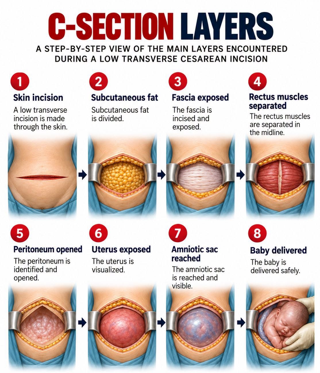
Proper tissue handling, ensuring adequate hemostasis, avoiding tissue ischemia, and preventing infection are essential for wound healing and minimizing adhesion formation. Various methods can be applied at each step or tissue layer during the surgery, with multiple factors influencing a surgeon’s choice of technique. As with all aspects of medical practice, these decisions should be guided by evidence-based recommendations. The 4 primary techniques for cesarean delivery include the Pfannenstiel-Kerr method, the Joel-Cohen method, the Misgav-Ladach method, and the modified Misgav-Ladach method.
The removal of pubic hair before a cesarean is optional. Proponents argue it reduces surgical site contamination and infection, but a Cochrane review found no significant reduction in infection rates with hair removal. Hair removal should be limited to cases where it enhances surgical visualization. This should be performed using clippers, not razors. Patients should also be advised against shaving their pubic area as they near their due date or scheduled cesarean, as shaving can cause microscopic skin breaks, increasing the risk of surgical site infections rather than clipping.
The initial skin incision for a cesarean section can be either suprapubic transverse or midline vertical. A midline vertical incision offers quicker access to the abdominal cavity and disrupts fewer tissue layers and vessels, making it a preferred method in many emergency cesarean deliveries.[27] Additionally, a vertical incision may facilitate visualization away from areas with severe adhesive disease. For a planned cesarean hysterectomy involving a morbidly adherent placenta, a vertical incision may be chosen to provide greater surgical exposure and access to the hypogastric arteries. However, a transverse skin incision is typically preferred in most cases due to its association with better wound healing and greater patient comfort. As most clinicians are more familiar with low-transverse cesarean entries, this technique is commonly used, even in emergency situations. Unplanned cesarean hysterectomies can also be performed through a low-transverse incision. However, patient habitus may influence some surgeons to place the transverse skin incision higher on the abdomen rather than underneath the pannus, although research on this approach remains inconclusive.[28]
The Pfannenstiel skin incision is slightly curved and positioned 2 to 3 cm or 2 fingerbreadths above the symphysis pubis. The midportion of this incision falls within the hair-bearing area of the mons, necessitating hair removal. In contrast, the Joel-Cohen incision is straight rather than curved, and this is located 3 cm below the line connecting the anterior superior iliac spines, placing it more cephalad than the Pfannenstiel incision.[27]
The subcutaneous layer lies beneath the skin and can be dissected either bluntly or sharply. As blood vessels traverse this layer, it is important to minimize blood loss by limiting sharp dissection to the midline until the fascia is exposed, followed by blunt dissection laterally. Alternatively, careful use of cautery can help maintain hemostasis when blood vessels are transected.
The fascia is incised in the midline using a scalpel and extended laterally, either sharply or bluntly. Next, the fascia is dissected away from the underlying rectus muscles. This dissection involves sequentially grasping the superior and inferior edges of the fascia with a clamp, such as a Kocher, and using a combination of blunt dissection and sharp techniques with scissors or cautery. Care must be taken to avoid damaging the rectus muscles. However, in certain clinical scenarios, deliberate incision of the rectus muscles may be necessary to improve surgical access.
A small randomized controlled trial compared dissection and nondissection of the fascia from the rectus muscles. The study found that nondissection was associated with a slower decline in hemoglobin levels postoperatively and less pain, as measured on a visual analog scale. However, surgical time and the difficulty of delivering the fetus were not assessed. As a result, the findings may not provide enough justification to alter current surgical techniques.[27]
After separating the rectus muscles in the midline, the surgeon enters the abdominal cavity by opening the peritoneum, either sharply or bluntly. If using a sharp entry, caution is necessary to avoid injuring underlying structures, such as the bowel. Once the entry is made, the peritoneal incision is typically extended bluntly. Care must be taken to prevent injury to the bladder during the extension of the peritoneal incision.
At this point, a bladder blade is often placed to provide better visualization of the lower uterine segment, or a self-retaining retractor may be used. If necessary, a bladder flap can be created by grasping and incising the peritoneum overlying the bladder and lower uterine segment, and then dissecting the bladder off the lower uterus, either sharply or bluntly. Surgeons may choose to create a bladder flap to reduce surgical injury to the bladder, especially during the repair of the uterine incision.[29] However, several trials have shown that omitting the bladder flap can decrease operative time without increasing complications such as hematuria, pain, or urinary tract infection. Bladder injury remains rare, and existing studies have been underpowered to determine whether omitting the bladder flap changes the incidence of bladder injury.[27] In clinical scenarios where the risk of inferior hysterotomy extension is high, such as in a cesarean for a patient who is fully dilated and actively pushing, creating a bladder flap may be indicated, even if it is not routinely performed.
With adequate visualization, the uterine incision can now be made, regardless of whether a bladder flap has been created. The uterine incision can be either transverse or vertical (classical). For most cesareans, a low transverse incision is preferred. Compared to a classical incision, a low transverse incision results in less bleeding, is easier to repair, and leads to less adhesion formation.[30] However, there may be certain situations where a classical incision is indicated. For example, a fetus in a transverse lie with the fetal back down may require a classical incision. If the lower uterine segment is underdeveloped and does not provide enough room for an adequate transverse incision, a classical hysterotomy may be necessary to facilitate an atraumatic delivery of the fetus.[30] This scenario may arise in early preterm gestations. Additionally, in cases such as severe adhesive disease, where the lower uterine segment may not be accessible, the surgeon must adapt the approach accordingly.
A low vertical hysterotomy may be considered when challenging fetal extraction is anticipated, particularly in cases of breech presentation. A low transverse incision can also be extended vertically, forming a “T,” “U,” or “J” incision to provide additional space. Patients with a history of a transverse or low vertical uterine incision may be candidates for a trial of labor in subsequent pregnancies, whereas a prior classical or “T” incision typically necessitates a repeat cesarean delivery.[2]
Before performing the hysterotomy, the uterus should be palpated to assess for any lateral rotation. Positioning the incision in the midline rather than laterally helps minimize the risk of damaging uterine vessels, particularly when creating a transverse incision. The incision is made cautiously with a scalpel in shallow strokes, often combined with blunt dissection, to avoid injury to the fetus. In patients who have been pushing, positioning the incision higher in the surgical field ensures a low transverse incision while reducing the risk of extension into lateral vessels, the lower uterus, or the cervix.
Upon entering the uterus, the incision can be extended laterally, either bluntly using fingers or sharply with bandage scissors. Blunt extension of the uterine incision is preferred, as sharp extension is associated with increased maternal morbidity and blood loss. Compared to blunt transverse extension, a blunt cephalad-caudad extension of the hysterotomy reduces the risk of unintended extension and significant blood loss.[27] Therefore, a blunt cephalad-caudad extension of the hysterotomy is the preferred approach.
If the uterine myometrium is thick, as seen in earlier gestations or with a classical hysterotomy, bandage scissors may be required. An inadequate hysterotomy increases the risk of difficult fetal extraction, potentially leading to higher neonatal morbidity or mortality. Regardless of the technique used, the primary goal of cesarean delivery is the safe delivery of the fetus.
For a fetus in vertex presentation, delivery involves inserting a hand into the uterine cavity to elevate the fetal head into the hysterotomy. If the elevation of the head is challenging, an assistant may assist by providing additional elevation from below via a hand in the patient’s vagina. Alternatively, a vacuum cup or a single forceps blade may be used to elevate the fetal head. Once the fetal head is elevated into the incision, the bladder blade is removed, and fundal pressure is applied to expel the fetus from the uterus. The surgeon carefully guides the fetal head during delivery, while the surgical assistant often provides most of the fundal pressure. If fundal pressure is insufficient or difficult to achieve—such as in cases of significant maternal obesity—a vacuum cup can be applied to the fetal head to assist with delivery. Forceps may also be used during cesarean delivery while adhering to the same principles when using vacuum or forceps, even in the context of a cesarean section.
For a fetus in breech presentation, the clinician identifies the fetal lie by palpating within the uterine cavity. Several techniques can be employed to deliver a breech fetus, including grasping the feet or hips to guide the fetus into the hysterotomy. Gentle traction is used to deliver the fetus up to the shoulders, often with the assistance of a surgical towel wrapped around the fetus. The bilateral arms are then sequentially swept down and delivered. Fundal pressure is applied to assist in flexing and delivering the fetal head. The Mauriceau-Smellie-Veit maneuver may also be used to flex the fetal head. This technique involves placing the first and third fingers of one hand on the fetal cheekbones while the second finger is positioned in the fetal mouth to gently pull the jaw downward. The application of Piper forceps is rarely required for delivering the fetal head.
After the fetus is delivered, the umbilical cord is doubly clamped and cut. Delayed cord clamping may be considered if the maternal and fetal conditions permit and if the surgeon opts for it. A systematic review of delayed cord clamping in preterm infants showed a reduction in in-hospital mortality and a decrease in low Apgar scores at 1 minute, but no change in scores at 5 minutes. Other outcomes, such as intubation, intraventricular hemorrhage, and necrotizing enterocolitis, remained unchanged. However, there was a potential risk of polycythemia and hyperbilirubinemia.[31] A randomized controlled trial investigating delayed cord clamping in elective cesarean deliveries showed increased neonatal hematocrit levels without an increased need for phototherapy.[32]
After the umbilical cord is cut, cord blood may be collected if needed or desired. The placenta is delivered manually or spontaneously via cord traction and fundal massage. Due to evidence showing reduced operative blood loss and a decrease in infections, spontaneous placental delivery is preferred when the clinical scenario permits it.[33][34] After the placenta is delivered, the uterine cavity is cleaned with moist laparotomy sponges.
The uterus can either be exteriorized or left in situ for the repair of the hysterotomy. Research has shown similar rates of febrile complications and comparable surgical times with both techniques, allowing the decision to be based on clinician preference.[27] For the repair, a delayed absorbable suture is used in a running technique, ensuring that the corners of the incision are incorporated while avoiding the lateral vessels. A running closure is associated with reduced operating time and blood loss compared to an interrupted closure.
The comparison between closing the hysterotomy in 1 versus 2 layers has been studied. Short-term outcomes, such as infectious morbidity, pain, blood transfusion, and hospital readmission, were not significantly different between the 2 techniques.[35] Data on whether a single-layer closure reduces operative time are mixed.[27][29] However, for women desiring a future trial of labor, evidence suggests that a 2-layer closure improves residual myometrial thickness, enhances scar healing, and reduces the risk of uterine rupture in subsequent pregnancies.[27][36][37] An unlocked closure technique may also be preferable to a locked technique.[36][37] A recent study suggested that endometrium-free closure of the uterus was associated with a decrease in placental abnormalities, such as placenta accreta, in subsequent pregnancies.[38] Ongoing research is exploring this specific aspect of uterine closure during cesarean section.
Once the uterus is closed and hemostasis is ensured, the posterior cul-de-sac is cleared of blood and clots using laparotomy sponges or suction. This step may be omitted if the uterus has not been exteriorized. After returning the uterus to the abdomen, the cavity is cleared of blood and clots once again. The use of retractors can help provide exposure to the paracolic gutters. Intrabdominal irrigation before closure has been shown to increase nausea during surgery and does not improve gastrointestinal function or reduce the incidence of infectious morbidity. Once the bladder blade is reinserted, the hysterotomy repair is re-visualized, and hemostasis is confirmed if necessary. The bladder blade is then removed.
The peritoneum can be reapproximated at this time. While closure of the peritoneum may add operative time and potentially increase postoperative fever and length of hospital stay, the decision to close this layer often depends on the surgeon’s interpretation of the literature regarding its impact on adhesion formation. Unfortunately, the data on this topic are mixed, so it remains the surgeon’s prerogative to balance the risks and benefits for the patient.[27]
Before closing the fascia, the rectus muscles and subfascial tissues should be inspected to ensure hemostasis. The rectus muscles can be reapproximated prior to fascial closure. Some clinicians believe suturing the muscles reduces the risk of subsequent diastasis recti and decreases the incidence of intra-abdominal adhesion formation.[39] However, reapproximating the muscles may increase postoperative pain.[39] Over time, the surgeon may involve the patient in shared decision-making regarding this technique.
The fascia is then closed using a delayed-absorbable suture in a running non-locking manner. Historically, some have used an interrupted technique for fascia closure, but this approach is no longer widely practiced. Using a monofilament suture, rather than a braided one, may decrease the risk of infection and should be considered for patients at higher risk of developing this complication.[40] Additionally, monofilament suture may reduce the risk of subsequent hernia formation.[41] Data do not favor either option regarding the closure of the entire incision with a single suture versus 2 sutures meeting at the midline.[29]
The subcutaneous tissues are then irrigated, and hemostasis is ensured. Although wound irrigation has not been shown to reduce infection rates, it may improve visualization of areas requiring cautery. Closure of the subcutaneous space is advised if the thickness is 2 cm or more, as it decreases the risk of hematoma, seroma, wound infection, and wound separation.[27][29] Drain placement in the subcutaneous space is not recommended.[27]
Skin closure can be achieved using various methods, with surgical staples or subcuticular sutures being the most common. Subcuticular absorbable staples and adhesive glues are also options. Research indicates that sutures and staples are similar in terms of cosmesis. Although the data are conflicting, studies suggest that subcuticular suture closure may be superior to staples in preventing wound separation and infection.[27][29] Additionally, a monofilament suture may reduce the risk of infection compared to a braided suture.
Common Operative Methods for Cesarean Delivery
The 4 general operative methods are listed below.
Pfannenstiel-Kerr method
-
Pfannenstiel skin incision
-
Sharp dissection of the subcutaneous layer
-
Sharp extension of the fascial opening
-
Sharp entry into the peritoneum
-
Sharp superficial, then blunt entry into the uterus
-
Manual removal of the placenta
-
Single-layer interrupted closure of the uterus
-
Closure of the peritoneum
-
Interrupted closure of the fascia
-
Continuous suture of the skin
Joel-Cohen method
-
Joel-Cohen skin incision
-
Blunt dissection of the subcutaneous layer
-
Blunt extension of the fascial opening
-
Blunt entry into the peritoneum
-
Sharp superficial, then blunt entry into the uterus
-
Spontaneous removal of the placenta
-
Single-layer interrupted closure of the uterus
-
Nonclosure of the peritoneum
-
Interrupted closure of the fascia
-
Continuous suture of the skin
Misgav-Ladach method
-
Joel-Cohen skin incision
-
Blunt dissection of the subcutaneous layer
-
Blunt extension of the fascial opening
-
Blunt entry into the peritoneum
-
Sharp superficial, then blunt entry into the uterus
-
Manual removal of the placenta
-
Single-layer running closure of the uterus
-
Nonclosure of the peritoneum
-
Continuous closure of the fascia
-
Mattress suture closure of the skin
Modified Misgav-Ladach method
-
Pfannenstiel skin incision
-
Blunt dissection of the subcutaneous layer
-
Blunt extension of the fascial opening
-
Blunt entry into the peritoneum
-
Sharp superficial, then blunt entry into the uterus
-
Spontaneous removal of the placenta
-
Single-layer running closure of the uterus
-
Closure of the peritoneum
-
Continuous closure of the fascia
-
Continuous suture of the skin [27]
Pain Management and Enhaced Recovery after Surgery Protocols
Pain management for cesarean deliveries involves a combination of preoperative anesthesia and an enhanced recovery after surgery (ERAS) protocol, which spans the preoperative, perioperative, and postoperative periods.[42] A regional anesthetic, such as a spinal or epidural block, is the preferred modality of surgical anesthesia and should be administered promptly before surgery. Regional anesthetics help minimize fetal exposure to agents that could suppress respiration or adversely affect the newborn’s transition.
Cesarean deliveries can also be performed under general anesthesia with endotracheal intubation, typically reserved for emergencies where there is insufficient time for regional anesthesia placement. If an anesthesiologist is not readily available in an urgent or emergent case, a cesarean delivery may be performed using a local anesthetic, although this approach is not preferred.
ERAS is a standardized perioperative care program widely implemented across multiple surgical disciplines, including colorectal, urologic, gynecologic, and hepatobiliary surgery. The ERAS protocol refers to a multimodal care pathway aimed at promoting early recovery for patients undergoing major surgery. Cesarean delivery has a unique ERAS protocol tailored to address its dual role as both a perioperative and peripartum procedure.[43][44] Guidelines have been delineated through the ERAS Society for the preoperative, perioperative, and postoperative periods. These evidence-based recommendations help clinical settings develop tailored protocols that best meet the needs of their institutions and patient populations.[19][45][46]
Why the Procedure Is Performed
There are many reasons why a woman may need to have a C-section instead of a vaginal delivery. The decision will depend on your doctor, where you are having the baby, your previous deliveries, and your medical history.
Problems with the baby may include:
- Abnormal heart rate
- Abnormal position in the womb, such as crosswise (transverse) or feet-first (breech)
- Developmental problems, such as hydrocephalus or spina bifida
- Multiple pregnancies (triplets or twins)
Health problems in the mother may include:
- Active genital herpes infection
- Large uterine fibroids near the cervix
- HIV infection in the mother
- Past C-section
- Past surgery on the uterus
- Severe illness, such as heart disease, preeclampsia or eclampsia
Problems at the time of labor or delivery may include:
- Baby’s head is too large to pass through the birth canal
- Labor that takes too long or stops
- Very large baby
- Infection or fever during labor
Problems with the placenta or umbilical cord may include:
- Placenta covers all or part of the opening to the birth canal (placenta previa)
- Placenta separates from the uterine wall ( placenta abruptio )
- Umbilical cord comes through the opening of the birth canal before the baby (umbilical cord prolapse)

Risks
A C-section is a safe procedure. The rate of serious complications is very low. However, certain risks are higher after C-section than after vaginal delivery. These include:
- Infection of the bladder or uterus
- Injury to the urinary tract
- Higher average blood loss. Most of the time, a transfusion is not needed, but risk is higher.
A C-section may also cause problems in future pregnancies. This includes a higher risk for:
- Placenta previa
- Placenta growing into the muscle of the uterus and has trouble separating after the baby is born (placenta accreta)
- Uterine rupture
These conditions can lead to severe bleeding (hemorrhage), which may require blood transfusions or removal of the uterus (hysterectomy).
How do I get ready for a C-section?
- Your healthcare provider will explain the procedure to you and you can ask question.
- You will be asked to sign a consent form that gives your permission to do the procedure. Read the form carefully and ask questions if something is unclear.
- You will be asked when you last had anything to eat or drink. If your C-section is planned and needs general, spinal, or epidural anesthesia, you will be asked to not eat or drink anything for 8 hours before the procedure.
- Tell your healthcare provider if you are sensitive to or are allergic to any medicine, latex, iodine, tape, or anesthesia.
- Tell your healthcare provider of all medicine (prescription and over-the-counter), vitamins, herbs, and supplements that you are taking.
- Tell your healthcare provider if you have a history of bleeding disorders or if you are taking any blood-thinning medicines (anticoagulants), aspirin, or other medicines that affect blood clotting. You may be told to stop these medicines before the procedure.
- You may be given medicine to decrease the acid in your stomach. These also help dry the secretions in your mouth and breathing passages.
- Plan to have someone stay with you after a C-section. You may have pain in the first few days and will need help with the baby.
- Follow any other instructions your provider gives you to get ready.
What happens during a C-section?
A C-section will be done in an operating room or a special delivery room. Procedures may vary depending on your condition and your healthcare provider’s practices.
In most cases, you will be awake for a C-section. Only in rare cases will a birth parent need medicine that puts them into a deep sleep (general anesthesia). Most C-sections are done with a regional anesthesia such as an epidural or spinal. With these, you will have no feeling from your waist down, but you will be awake and able to hear and see your baby as soon as they are born.
Generally, a C-section follows this process:
- You will be asked to undress and put on a hospital gown.
- You will be positioned on an operating or exam table.
- A urinary catheter may be put in if it was not done before coming to the operating room.
- An IV ( intravenous) line will be started in your arm or hand.
- For safety reasons, straps will be placed over your legs to hold you on the table.
- Hair around the surgical site may be shaved. The skin will be cleaned with an antiseptic solution.
- Your belly (abdomen) will be draped with sterile material. A drape will also be placed above your chest to screen the surgical site.
- The anesthesiologist or nurse anesthetist will continuously watch your heart rate, blood pressure, breathing, and blood oxygen level during the procedure.
- Once the anesthesia has taken effect, your provider will make an incision above the pubic bone, either transverse or vertical. You may hear the sounds of an electrocautery machine that seals off bleeding.
- Your provider will make deeper incisions through the tissues and separate the muscles until the uterine wall is reached. They will make a final incision in the uterus. This incision is also either horizontal or vertical.
- Your provider will open the amniotic sac, and deliver the baby through the opening. You may feel some pressure or a pulling sensation.
- They will cut the umbilical cord.
- You will get medicine in your IV to help the uterus contract and expel the placenta.
- Your provider will remove your placenta and examine the uterus for tears or pieces of placenta.
- They will use stitches to close the incision in the uterine muscle and reposition the uterus in the pelvic cavity.
- Your provider will close the muscle and tissue layers with sutures. They will close the skin incision with stitches or surgical staples.
- Finally, your provider will apply a sterile bandage.
What happens after a C-section?
In the hospital
In the recovery room, nurses will watch your blood pressure, breathing, pulse, bleeding, and the firmness of your uterus.
Usually, you can be with your baby while you are in the recovery area. In some cases, babies born by cesarean will first need to be watched in the nursery for a short time. Breastfeeding can start in the recovery area, just as with a vaginal delivery.
After 1 or 2 hours in the recovery area, you will be moved to your room for the rest of your hospital stay.
As the anesthesia wears off, you may get pain medicine as needed. This can be either from the nurse or through a device connected to your IV (intravenous) line called a PCA (patient controlled analgesia) pump. In some cases, pain medicine may be given through the epidural catheter until it is removed.
You may have gas pains as the intestinal tract starts working again after surgery. You will be encouraged to get out of bed. Moving around and walking helps ease gas pains. Your healthcare provider may also give you medicine for this. You may feel some uterine contractions called after-pains for a few days. The uterus continues to contract and get smaller over several weeks.
The urinary catheter is usually removed the day after surgery.
You may be given liquids to drink a few hours after surgery. You can gradually add more solid foods as you can handle them.
You may be given antibiotics in your IV while in the hospital and a prescription to keep taking the antibiotics at home.
At home
You will need to wear a sanitary pad for bleeding. It’s normal to have cramps and vaginal bleeding for several days after birth. You may have discharge that changes from dark red or brown to a lighter color over several weeks.
Don’t douche, use tampons, or have sex until your healthcare provider tells you it’s OK. You may also have other limits on your activity, including no strenuous activity, driving, or heavy lifting.
Take a pain reliever as recommended by your healthcare provider. Aspirin or certain other pain medicines may increase bleeding. So, be sure to take only recommended medicines.
Arrange for a follow-up visit with your healthcare provider. This is usually 2 to 3 weeks after the surgery.
Call your healthcare provider right away if any of these occur:
- Heavy vaginal bleeding
- Foul-smelling drainage from your vagina
- Fever or chills
- Severe belly (abdominal) pain
- Increased pain, redness, swelling, or bleeding or other drainage from the incision
- Leg pain
- Trouble breathing, chest pain, or heart palpitations
Your healthcare provider may give you other instructions, depending on your situation.
Next steps
Before you agree to the test or procedure make sure you know:
- The name of the test or procedure
- The reason you are having the test or procedure
- What results to expect and what they mean
- The risks and benefits of the test or procedure
- What the possible side effects or complications are
- When and where you are to have the test or procedure
- Who will do the test or procedure and what that person’s qualifications are
- What would happen if you did not have the test or procedure
- Any alternative tests or procedures to think about
- When and how you will get the results
- Who to call after the test or procedure if you have questions or problems
- How much you will have to pay for the test or procedure
Outlook (Prognosis)
Most mothers and infants do well after a C-section.
Women who have a C-section may have a vaginal delivery if another pregnancy occurs, depending on:
- The type of C-section done
- Why the C-section was done
Vaginal birth after cesarean (VBAC) delivery is very often successful. However, there is a small risk of uterine rupture, which can harm the mother and the baby. Discuss the benefits and risks of VBAC with your provider.
Berghella V, Mackeen AD, Jauniaux ERM. Cesarean delivery. In: Gabbe SG, Niebyl JR, Simpson JL, et al, eds. Obstetrics: Normal and Problem Pregnancies . 7th ed. Philadelphia, PA: Elsevier; 2017:chap 19.
Hull AD, Resnik R. Placenta previa, placenta accreta, abruptio placentae, and vasa previa. In: Creasy RK, Resnik R, Iams JD, Lockwood CJ, Moore TR, Greene MF, eds. Creasy and Resnik’s Maternal-Fetal Medicine: Principles and Practice . 7th ed. Philadelphia, PA: Elsevier Saunders; 2014:chap 46.

