
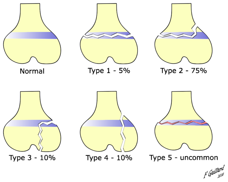
Salter-Harris Type Fractures/ A Salter-Harris fracture is a pediatric fracture that involves the epiphyseal plate. These fractures can occur in any bone that has a growth plate but frequently occur in the distal radius. The Salter-Harris scheme was first developed by Doctors William Harris and Robert Salter in 1963 and remains the most common classification system for epiphyseal fractures. Salter-Harris fractures are graded I through IX, with I through V being the most frequently used in clinical practice. Type I is a fracture that runs transversely through the growth plate. Type II runs through the growth plate and the metaphysis. Type III involves the growth plate and epiphysis. Type IV is a fracture of metaphysis, epiphysis, and growth plate. Type V is a complete direct compression fracture of the growth plate. Each of these has a different prognosis and management. [rx][rx][rx]
A Salter-Harris fracture is a fracture that involves the epiphyseal plate or growth plate of a bone, specifically the zone of provisional calcification.[rx] It is thus a form of child bone fracture. It is a common injury found in children, occurring in 15% of childhood long bone fractures.[rx] This type of fracture and its classification system is named for Robert B. Salter and William H. Harris, who created and published this classification system in the Journal of Bone and Joint Surgery in 1963.[rx]
Types of Salter-Harris Type Fractures
Salter-Harris Fracture Types
There are nine types of Salter-Harris fractures; types I to V as described by Robert B Salter and W Robert Harris in 1963, and the rarer types VI to IX which have been added subsequently:[rx]
- Type I – transverse fracture through the growth plate (also referred to as the “physis”):[rx] 6% incidence
- Type II – A fracture through the growth plate and the metaphysis, sparing the epiphysis:[rx] 75% incidence, takes approximately 12-90 weeks or more in the spine to heal.[rx]
- Type III-A fracture through the growth plate and epiphysis, sparing the metaphysis:[rx] 8% incidence
- Type IV – A fracture through all three elements of the bone, the growth plate, metaphysis, and epiphysis:[rx] 10% incidence
- Type V – A compression fracture of the growth plate (resulting in a decrease in the perceived space between the epiphysis and metaphysis on x-ray):[rx] 1% incidence
- Type VI – Injury to the peripheral portion of the physis and a resultant bony bridge formation which may produce an angular deformity (added in 1969 by Mercer Rang)[rx]
- Type VII – Isolated injury of the epiphyseal plate (VII-IX added in 1982 by JA Ogden)[rx]
- Type VIII – Isolated injury of the metaphysis with possible impairment of endochondral ossification
- Type IX – Injury of the periosteum which may impair intramembranous ossification
SALTER mnemonic for classification
The mnemonic “SALTER” can be used to help remember the first five types.
N.B.: This mnemonic requires the reader to imagine the bones as long bones, with the epiphyses at the base.
- I – S = Slip (separated or straight across). Fracture of the cartilage of the physis (growth plate)
- II – A = Above. The fracture lies above the physis, or Away from the joint.
- III – L = Lower. The fracture is below the physis in the epiphysis.
- IV – TE = Through Everything. The fracture is through the metaphysis, physis, and epiphysis.
- V – R = Rammed (crushed). The physis has been crushed.
Alternatively, SALTER can be used for the first 6 types, as above but adding Type V — ‘E’ for ‘Everything’ or ‘Epiphysis’ and Type VI — ‘R’ for ‘Ring’.
Causes of Salter-Harris Type Fractures
Most of these injuries occur during the time of a child’s growth spurt when physis is the weakest. Active children are the most likely to encounter injuries involving the growth plate as the ligaments and joint capsules surrounding the growth plate tend to be much stronger and more stable. The ligaments and capsules are thereby able to sustain greater external loads to the joint, relative to the growth plate itself.[rx][rx]
Symptoms of Salter-Harris Type Fractures
- Pain and swelling
- Tenderness
- A change in the shape of the injured area that is different than usual
- Not being able to move or put weight on the injured arm or leg
Diagnosis of Salter-Harris Type Fractures
History
Your doctor in the emergency department may ask the following questions
-
How – How was the fracture created, and, if chronic, why is it still open? (underlying etiology)
-
When – How long has this fracture been present? (e.g., chronic less than 1 month or acute, more than 6 months)
- Where – Where on the body parts is it located? Is it in an area that is difficult to offload, complicated, or keep clean? Is it in an area of high skin tension? Is it near any vital organ and structures such as a major artery?
- What is your Past – Has your previous medical history of fracture? Are you suffering from any chronic disease, such as hypertension, blood pressure, diabetes mellitus, previous major surgery? What kind of medicine did you take? What is your food habits, geographic location, Alcohol, tea, coffee consumption habit, anabolic steroid uses for athletes, etc?
Physical
Physical examination is done by your doctor, consisting of palpation of the fracture site, eliciting boney tenderness, edema, swelling. If the fracture is in the dept of a joint, the joint motion, normal movement will aggravate the pain.
- Inspection – Your doctor also check superficial tissue, skin color, involving or not only the epidermal layer or Partial-thickness affects the epidermis and extend into the dermis, but full-thickness also extends through the dermis and into the adipose tissues or full-thickness extends through the dermis, and adipose exposes muscle, bone, evaluate and measure the depth, length, and width of the fracture. Access surrounding skin tissue, fracture margins for tunneling, rolled, undermining fibrotic changes, and if unattached and evaluate for signs and symptoms of infect warm, pain, delayed healing.
- Palpation – Physical examination may reveal tenderness to palpation, swelling, edema, tenderness, worm, temperature, open fracture, closed fracture, microtrauma, and ecchymosis at the site of fracture.
- Motor function – Your doctor may ask the patient to move the injured area to assist in assessing muscle, ligament, and tendon function. The ability to move the joint means only that the muscles and tendons work properly, and does not guarantee bone integrity or stability. The concept that “it can’t be fractured because you can move it” is not correct. The jerk test and manual test are also performed to investigate the motor function.
- Sensory examination – assesses sensations such as light touch, worm, paresthesia, itching, numbness, and pinprick sensations, in its fracture side.
- Range of motion – A range of motion examination of the fracture associate joint and it’s surrounding joint may be helpful in assessing the muscle, tendon, ligament, cartilage stability. Active assisted, actively resisted exercises are performed around the injured area joint.
- Blood pressure and pulse check – Blood pressure is the term used to describe the strength of blood with which your blood pushes on the sides of your arteries as it’s pumped around your body. An examination of the circulatory system, feeling for pulses, blood pressure, and assessing how quickly blood returns to the tip of a toe to heart and it is pressed the toe turns white (capillary refill).
Lab Test
Laboratory tests should be done as an adjunct in overall medical status for surgical treatment.
- CBC, ESR test
- Random blood sugar, glucose, and routine diabetes test if the patient has diabetes mellitus.
- Microscopic urine examination test, and stool test.
- ECG, EKG test for heart abnormality is present
- Ultrasonography test in some cases.
- Normalized hemoglobin, hematocrit test
- Coagulation profile with bleeding time and coagulation time test, prothrombin time (PT) test for surgery if needed,
- Partial thromboplastin time (PTT), and platelet counts will be needed for operative intervention.
- Serum creatinine test,
- Serum lipid profile
- Serum uric acid test
Imaging Findings
- Soft tissue swelling
- Depending on the type of fracture, some displacement of the epiphysis or corner sign (Thurston-Holland fragment)
- Conventional radiography remains study of first choice
- CT with multiplanar reconstruction has been used in problem cases
- Ultrasound can be helpful in infants whose cartilage has not yet ossified
- MRI in problem cases
Plain x-rays – Growth plate fractures are suspected in children who have tenderness and swelling localized over the growth plate or who cannot move or put weight on the affected limb.
Plain x-rays are the diagnostic test of choice – If findings are equivocal, opposite-side comparison x-rays may be helpful. Despite use of comparison views, x-rays may appear normal in Salter types I and V. If x-rays appear normal but a growth plate fracture is suspected, patients are assumed to have a fracture, a splint or cast is applied, and patients are reexamined in several days. Continued pain and tenderness suggest a growth plate fracture.
Treatment of Salter-Harris Type Fractures
Do no HARM for 72 hours after injury
- Heat—hot baths, electric heat, saunas, heat packs, etc has the opposite effect on the blood flow. Heat may cause more fluid accumulation in the fracture joints by encouraging blood flow. Heat should be avoided when inflammation is developing in the acute stage. However, after about 72 hours, no further inflammation is likely to develop and heat can be soothing.
- Alcohol stimulates the central nervous system that can increase bleeding and swelling and decrease healing.
- Running, and walking may cause further damage, and causes healing delay.
- Massage also may increase bleeding and swelling. However, after 72 hours of your fracture, you can take a simple message, and applying heat may be soothing the pain.
Medication
The following medications may be considered by your doctor to relieve acute and immediate pain, long term treatment
- Antibiotic – Cefuroxime or Azithromycin, or Flucloxacillin or any other cephalosporin/quinolone, meropenem antibiotic must be used to prevent infection or clotted blood removal to prevent further swelling, inflammation, and edema.
- NSAIDs – Prescription-strength drugs that reduce both pain and inflammation. Pain medicines and anti-inflammatory drugs help to relieve pain and stiffness, allowing for increased mobility and exercise. There are many common over-the-counter medicines called non-steroidal anti-inflammatory drugs (NSAIDs). They include first choice NSAIDs is Ketorolac, then Etoricoxib, then Aceclofenac, naproxen.
- Muscle Relaxants – These medications provide relief from spinal muscle spasms, spasticity. Muscle relaxants, such as baclofen, tolperisone, eperisone, methocarbamol, carisoprodol, and cyclobenzaprine, may be prescribed to control postoperative muscle spasms, spasticity, stiffness, contracture.
- Calcium & vitamin D3 – To improve bone health, blood clotting, helping muscles to contract, regulating heart rhythms, nerve functions, and healing fractures. As a general rule, too absorbed more minerals for men and women age 50 and older should consume 1,200 milligrams of calcium a day, and 600 international units of vitamin D a day to heal back pain, fractures, osteoarthritis.
- Neuropathic Agents – Drugs(pregabalin & gabapentin) that address neuropathic—or nerve-related—pain. This includes burning, numbness, tingling sensation, and paresthesia.
- Glucosamine & Diacerein, Chondroitin sulfate – can be used to tighten the loose tendon, cartilage, ligament, and cartilage, ligament regenerates cartilage or inhabits the further degeneration of cartilage, ligament. The dosage of glucosamine is 15oo mg per day in divided dosage and chondroitin sulfate approximately 500mg per day in different dosages, and diacerein minimum of 50 mg per day may be taken if the patient suffers from osteoarthritis, rheumatoid arthritis, and any degenerative joint disease.[rx]
- Topical Medications and essential oil – These prescription-strength creams, gels, ointments, patches, and sprays help relieve pain and inflammation in acute trauma, pain, swelling, tenderness through the skin. If the fracture is closed and not open fracture then you can use this item.
- Antidepressants – A drug that blocks pain messages from your brain and boosts the effects of endorphins in your body’s natural painkillers. It also helps in neuropathic pain, anxiety, tension, and proper sleep.
- Corticosteroids – Also known as oral steroids, these medications reduce inflammation. To heal the nerve inflammation and clotted blood in the joints.
- Dietary supplement – To eradicate the healing process from fracture your body needs a huge amount of vitamin C, and vitamin E. From your dietary supplement, you can get it, and also need to remove general weaknesses & improved health.
- Cough Syrup – If your doctor finds any chest congestion or fracture-related injury in your chest, dyspnoea, post-surgical breathing problem, then advice you to take bronchodilator cough syrup.
What To Eat and What to avoid
Eat Nutritiously During Your Recovery
All bones and tissues in the body need certain micronutrients in order to heal properly and in a timely manner. Eating a nutritious and balanced diet that includes lots of minerals and vitamins is proven to help heal broken bones and all types of fractures. Therefore, focus on eating lots of fresh food produce (fruits and veggies), whole grains, cereal, beans, lean meats, seafood, and fish to give your body the building blocks needed to properly repair your fracture. In addition, drink plenty of purified mineral water, milk, and other dairy-based beverages to augment what you eat.
- Broken bones or fractures bones need ample minerals (calcium, phosphorus, magnesium, boron, selenium, omega-3) and protein to become strong and healthy again.
- Excellent sources of minerals/protein include dairy products, tofu, beans, broccoli, nuts and seeds, sardines, sea fish, and salmon.
- Important vitamins that are needed for bone healing include vitamin C (needed to make collagen that your body essential element), vitamin D (crucial for mineral absorption, or machine for mineral absorber from your food), and vitamin K (binds calcium to bones and triggers more quickly collagen formation).
- Conversely, don’t consume food or drink that is known to impair bone/tissue healing, such as alcoholic beverages, sodas, fried fast food, most fast food items, and foods made with lots of refined sugars and preservatives.
Surgery
Salter-Harris I and II fractures can be treated with closed reduction and casting or splinting. The reduction should be performed carefully to avoid damage to or grating of the physis on any metaphyseal bone fragments.[rx][rx][rx]
Salter-Harris III and IV fractures usually require open reduction and internal fixation (avoiding crossing the physis).
Salter V fracture diagnosis may be delayed unless there is a high degree of clinical suspicion and often the diagnosis is not made at the initial presentation. An emergent orthopedic consultation should be obtained if the fracture is recognized. As these fractures involve the germinal matrix, they have a potential for growth arrest.
In all cases, a reexamination in seven to ten days is necessary to monitor proper reduction and healing. This is also important to determine whether any complications, such as growth arrest, have occurred. If clinically indicated, an additional follow-up radiograph at six and 12 months may be obtained to reassess for any growth arrest.
- A cast or splint – may be used to help prevent movement in the injured area until more treatment is done. Some Salter-Harris fractures take up to 14 days before they can be seen on an x-ray. Your child’s injury may need to be put in a cast or splint if a Salter-Harris fracture is known or suspected. This will help prevent more injury to the growth plate and surrounding bone. If the bone is not displaced (moved out of place), your child may get a cast to secure the bone as it heals. Casts are also used after reduction (when the bone is put back into place) or surgery.
- Surgery – may be needed to repair certain types of Salter-Harris fractures. Pins or screws will be placed inside the broken bone. These hold the bone pieces together in the correct places.
Complications of Salter-Harris Type Fractures
- The complications include growth arrest with the potential for deformity and limb length discrepancy. An entrapment of the periosteum within the fracture is a rare complication that requires an MRI scan. Beware that entrapped periosteum can prevent a complete reduction of the fracture.
- In general, the higher the number, the more likely the complication so Salter-Harris types Iv and V have the highest associated complications.
- Greater risk for complication comes with fracture of distal tibia followed by the distal femur
- The primary complication is growth plate disturbance, Early closure, Limb length discrepancy, Closure of only a portion of the plate resulting in angular deformity.
References
- https://www.ncbi.nlm.nih.gov/books/NBK430688/
- https://www.ncbi.nlm.nih.gov/pmc/articles/PMC2908342/
- https://en.wikipedia.org/wiki/Salter%E2%80%93Harris_fracture
- https://www.sciencedirect.com/topics/veterinary-science-and-veterinary-medicine/salter-harris-fracture
- https://www.merckmanuals.com/professional/injuries-poisoning/fractures/pediatric-physeal-growth-plate-fractures
- https://link.springer.com/article/10.1007/s11999-016-4891-3
- https://radiopaedia.org/articles/salter-harris-classification
- https://www.drugs.com/cg/salter-harris-fracture.html
- https://www.rch.org.au/fracture-education/growth_plate_injuries/Physeal_growth_plate_injuries/
- http://learningradiology.com/archives2012/COW%20530-Salter%204%20Fx/salter4correct.html

