Retinal Artery Occlusion – Causes, Symptoms, Diagnosis, Treatment

Patient Tools

Read, save, and share this guide

Use these quick tools to make this medical article easier to read, print, save, or share with a family member.

Patient Mode

Understand this article easily

Switch between simple English and easy Bangla patient notes. This is for education and does not replace a doctor consultation.

Retinal artery occlusion refers to blockage of the retinal artery that carries oxygen to the nerve cells in the retina at the back of the eye. The lack of oxygen and nutrition delivery to the retina may result in severe loss of vision. The artery...

For severe symptoms, danger signs, pregnancy, child illness, or sudden worsening, seek urgent medical care.

বাংলা রোগী নোট এখনো যোগ করা হয়নি। পোস্ট এডিটরে “RX Bangla Patient Mode” বক্স থেকে সহজ বাংলা সারাংশ যোগ করুন।

এই তথ্য শিক্ষা ও সচেতনতার জন্য। এটি ডাক্তারি পরীক্ষা, রোগ নির্ণয় বা প্রেসক্রিপশনের বিকল্প নয়।

Article Summary

Retinal artery occlusion refers to blockage of the retinal artery that carries oxygen to the nerve cells in the retina at the back of the eye. The lack of oxygen and nutrition delivery to the retina may result in severe loss of vision. The artery can re-canalize over time and the edema can clear. However, optic atrophy and spasm lead to permanent loss of vision....

Key Takeaways

  • This article explains Causes in simple medical language.
  • This article explains Symptoms in simple medical language.
  • This article explains Diagnosis in simple medical language.
  • This article explains Treatment and prognosis in simple medical language.
Educational health guideWritten for patient understanding and clinical awareness.
Reviewed content workflowUse writer and reviewer profiles for stronger trust.
Emergency safety firstUrgent warning signs are highlighted below.

Seek urgent medical care if you notice

These warning signs are general safety guidance. Local emergency numbers and clinical judgment should always come first.

  • Sudden vision loss, severe eye pain, new flashes, or many new floaters.
  • Eye symptoms after injury or chemical exposure.
  • Rapidly worsening redness, swelling, or vision changes.
1

Emergency now

Use emergency care for severe, sudden, rapidly worsening, or life-threatening symptoms.

2

See a doctor

Book a professional medical evaluation if symptoms persist, worsen, recur often, affect daily activities, or occur in a high-risk patient.

3

Learn safely

Use this article to understand possible causes, tests, treatment options, prevention, and questions to ask your clinician.

Retinal artery occlusion refers to blockage of the retinal artery that carries oxygen to the nerve cells in the retina at the back of the eye. The lack of oxygen and nutrition delivery to the retina may result in severe loss of vision. The artery can re-canalize over time and the edema can clear. However, optic atrophy and spasm lead to permanent loss of vision. Irreversible damage to neural tissue can occur after approximately 15 minutes of complete blockage to the central retinal artery, but this time may vary from person to person.[rx] Two-thirds of people experience 20/400 vision while only one in six will experience 20/40 vision or better.[rx]

Causes

Retinal artery occlusion occurs due to blockage of the retinal artery, often by an embolus (a small piece of cholesterol that blocks blood flow) or thrombus formation(blood clot). The retinal artery occlusion may be transient and last for only a few seconds or minutes if the blockage breaks up and restores blood flow to the retina, or it may be permanent.

Common risk factors include:

  • Carotid artery disease
  • Atherosclerosis (fatty deposits in the arteries)
  • Faulty heart valves (valvular heart disease)
  • Tumors in the heart (myxoma)
  • Abnormal heart rhythms such as atrial fibrillation
  • insulin is low or not working well. সহজ বাংলা: রক্তে চিনি বেশি থাকার রোগ।" data-rx-term="diabetes" data-rx-definition="Diabetes is a condition where blood sugar stays too high because insulin is low or not working well. সহজ বাংলা: রক্তে চিনি বেশি থাকার রোগ।">Diabetes
  • High blood pressure
  • Intravenous drug abuse
  • Giant-cell arteritis
  • Disorders contributing to blood clot formation, such as sickle cell disease
  • Use of oral contraceptives
  • Homocystinuria (a hereditary disorder that prevents your body from processing the amino acid methionine; this leads to an excess accumulation of homocysteine in the blood and urine)
  • Pregnancy
  • Blood platelet abnormalities

Most retinal artery occlusion patients are in their 60s, and are more commonly men than women. Only 1% to 2% of cases involve both eyes.

Symptoms

Retinal artery occlusion or blockage is usually associated with sudden painless loss of vision in one eye or both eyes. The area of the retina affected by the blocked vessels determines the area and extent of visual loss gradually.

  • The main artery supplying blood to the eye is the ophthalmic artery; when it is blocked, or tear flattened it produces the most damage. A blockage permanently in the main artery in the retina is called central retinal artery occlusion (CRAO), which often results in severe loss of vision. However, about 25% of people who develop CRAO have an extra artery called a cilioretinal artery in their eyes that are supply oxygen. When CRAO occurs, having a cilioretinal artery can greatly lessen the chances of damage to your central vision, as long as the cilioretinal artery is not affected.
  • A blockage in a smaller artery is called branch retinal artery occlusion; this may cause a loss of a section of your visual field, such as your vision to one side. If the affected area is not in the center of the eye or is relatively small, a BRAO may go unnoticed with no symptoms.

Diagnosis

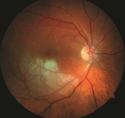
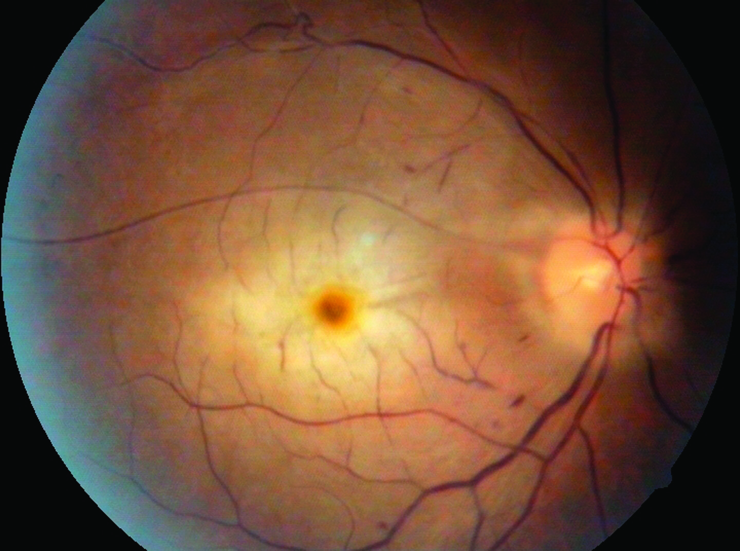
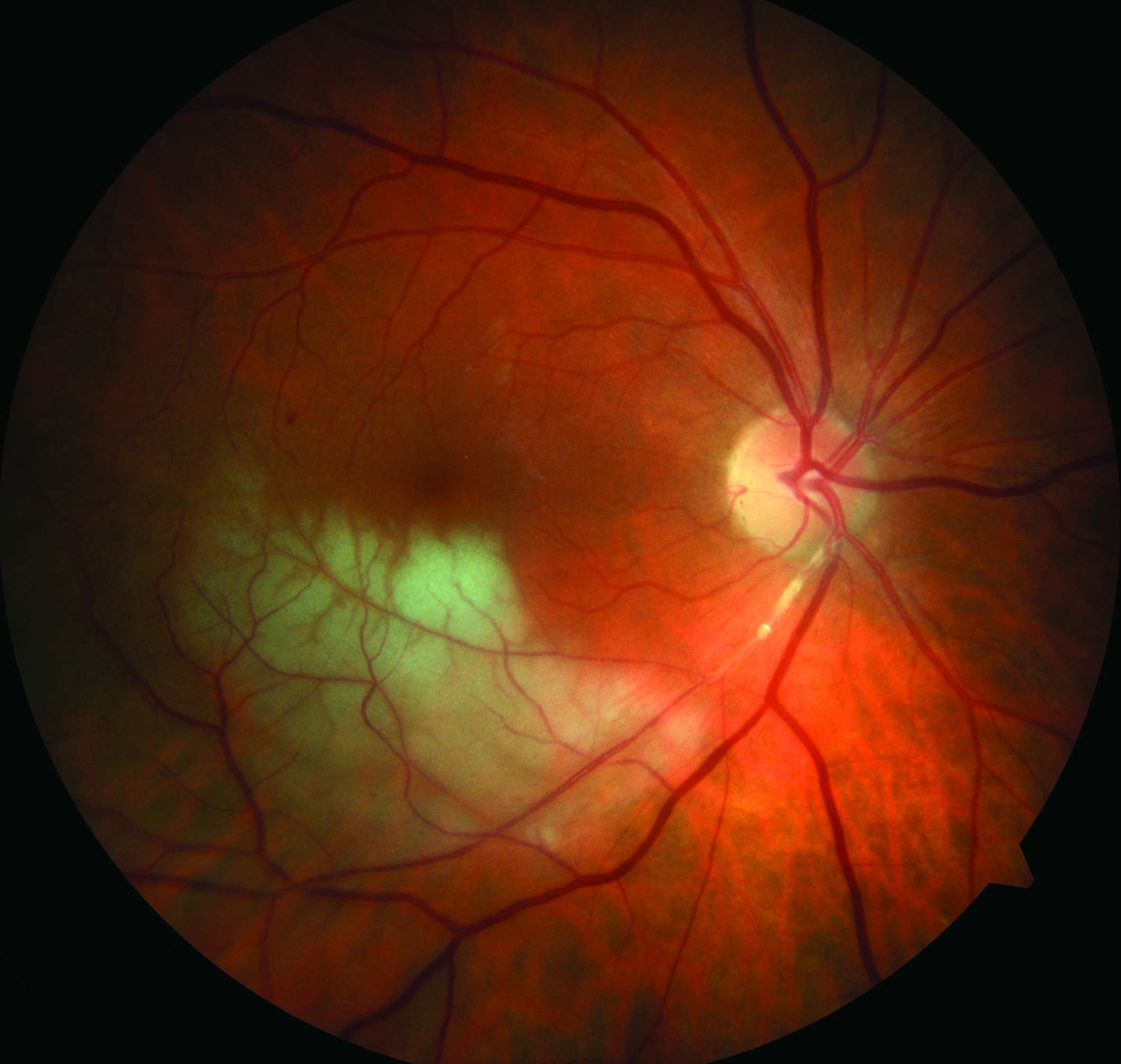
CRAO is usually diagnosed by a dilated eye examination that shows a “cherry red spot,” where the center of the macula appears red, with the surrounding retina pale due to the lack of blood flow. (Figure 1) BRAO appears as an area of superficial retinal whitening along the blocked vessel.(Figure 2)

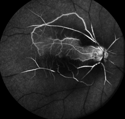
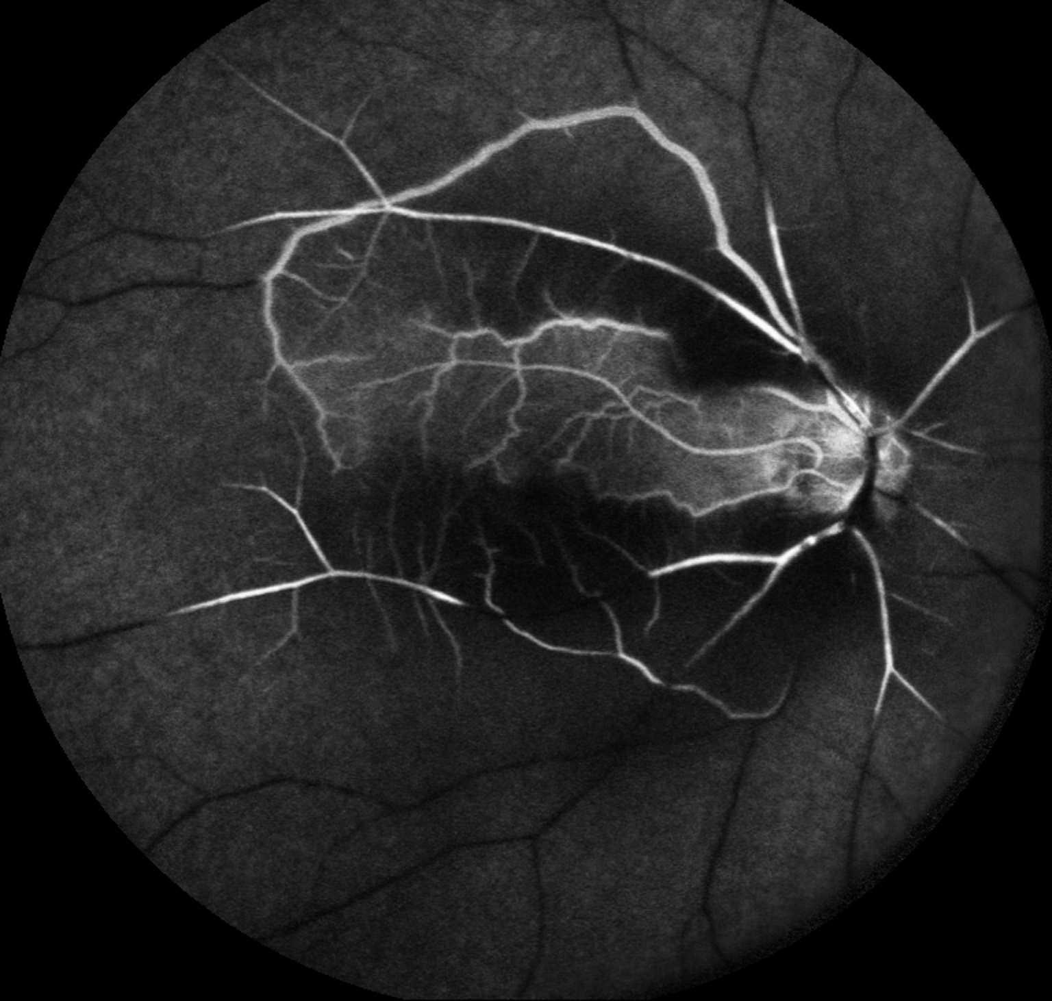
The whitening of the retina generally lasts 4 to 6 weeks before fading. Fluorescein angiography (FA) shows a delay in filling of the retinal arteries. (Figure 3Optical coherence tomography (OCT) provides detailed images of the central retina, and shows swelling in the inner layers of the retina in the affected area, which over time atrophy, becoming much thinner than normal.
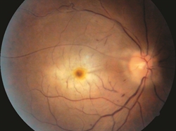
Retinal Artery Occlusion - Causes, Symptoms, Diagnosis, TreatmentFigure 1. Central retinal artery occlusion (CRAO) with cherry red spot. Mehul A. Shah, MD. Retina Image Bank 2014; Image 19815. ©American Society of Retina Specialists.

Retinal Artery Occlusion - Causes, Symptoms, Diagnosis, TreatmentFigure 2. Branch retinal artery occlusion (BRAO) showing retinal whitening in the area of blockage. John S. King, MD. Retina Image Bank 2014; Image 18552. ©American Society of Retina Specialists.

Retinal Artery Occlusion - Causes, Symptoms, Diagnosis, TreatmentFigure 3. Fluorescein angiogram of CRAO showing sparing of the cilioretinal artery. Courtney Crawford, MD. Retina Image Bank 2017; Image 26792. ©American Society of Retina Specialists.

Treatment and prognosis

Unfortunately, there is no clinically proven treatment for CRAO. Several therapies may be used including:

  • While no treatment has been clearly demonstrated to be beneficial for CRAO in large systematic reviews of randomized clinical trials, many of the following are frequently used:[rx]
    • Lowering intraocular pressure;
    • Dilating the CRA;
    • Increasing oxygenation;
    • Isovolemic hemodilution;
    • Anticoagulation;
    • Dislodging or fragmenting thrombus or embolus;
    • Thrombolysis; and
    • Hyperbaric oxygen.

    To achieve the best outcome for a person with CRAO, it is important to identify the condition in a timely manner and to refer to the appropriate specialist.[rx]

  • Hyperventilation—inhaling carbogen, a mixture of 95% oxygen and 5% carbon dioxide, to attempt to dilate the retinal arteries and cause the clot to dislodge
  • Paracentesis (removal of fluid from the front of the eye using a small-gauge needle) to lower
  • the intraocular pressure to try to dislodge the embolus
  • Lowering the intraocular pressure with medication
  • Ocular massage with a thumb to dislodge the clot

However, for any treatment to be potentially effective in CRAO, it must be deployed within a short time window, probably within 4 to 6 hours after symptoms begin. Unfortunately, none of these therapies have been shown to predictably alter the natural history of disease.

Persons with an acute onset CRAO or BRAO should be referred promptly to the emergency room or their primary care doctor to evaluate for stroke risk.

Thrombolytic therapy (“clot-busting” drugs), delivered either intravenously or directly through the ophthalmic artery, has also been tried but clinical trials have not shown this treatment to be effective.

An important aspect of managing retinal artery occlusion is for your doctor to identify and manage risk factors that may lead to other vascular conditions. The risk factors for CRAO are the same atherosclerotic risk factors as for stroke and heart disease; tests are important to try to identify the source of a clot from another part of the body. These tests include:

  • A carotid ultrasound to determine whether there are carotid plaques present
  • Cardiac echocardiography to look for disease in the heart valves

A doctor may also order a test of your blood’s sedimentation rate, and a temporal artery biopsy may be useful if giant cell arteritis is suspected.

Vision loss with CRAO is usually severe. However, CRAOs in patients who  have a cilioretinal artery have better visual prognosis, usually recovering to 20/50 vision or better in over 80% of eyes. Visual field loss in BRAO is usually  permanent, but central visual acuity may recover to 20/40 or better in 80%  of eyes.

Formation of new blood vessels of the retina or iris that are prone to bleed is a rare complication seen after a CRAO or BRAO. Growth of these vessels can further decrease vision by causing vitreous hemorrhage and glaucoma. If this happens, laser photocoagulation therapy is used to create burns in the area of the blocked artery to try

Doctor visit helper

Prepare before seeing a doctor

A simple rural-patient checklist to help you explain symptoms clearly, ask better questions, and avoid unsafe self-treatment.

Safety note: This is not a prescription or diagnosis. For severe symptoms, pregnancy danger signs, children with serious illness, chest pain, breathing difficulty, stroke-like weakness, or major injury, seek urgent care.

Which doctor may help?

Start with a registered doctor or the nearest qualified health center.

What to tell the doctor

  • Write when the problem started and how it changed.
  • Bring old prescriptions, investigation reports, and current medicines.
  • Write allergies, pregnancy status, diabetes, kidney/liver disease, and major past illnesses.
  • Bring one family member if the patient is weak, elderly, confused, or a child.

Questions to ask

  • What is the most likely cause of my symptoms?
  • Which danger signs mean I should go to hospital quickly?
  • Which tests are necessary now, and which can wait?
  • How should I take medicines safely and what side effects should I watch for?
  • When should I come for follow-up?

Tests to discuss

  • Vital signs: temperature, pulse, blood pressure, oxygen saturation
  • Basic physical examination by a clinician
  • CBC, urine test, blood sugar, or imaging only when clinically needed

Avoid these mistakes

  • Do not use antibiotics, steroid tablets/injections, or strong painkillers without proper medical advice.
  • Do not hide pregnancy, kidney disease, ulcer, allergy, or blood thinner use.
  • Do not delay emergency care when danger signs are present.

Medicine safety and first-aid guide

This section is for patient education only. It does not replace a doctor, pharmacist, or emergency care.

Safe first steps

  • Stop activity and seek urgent medical evaluation.
  • Chest pain should not be managed only with home medicine.
  • Discuss ECG and cardiac blood tests with emergency care when appropriate.

OTC medicine safety

  • Do not take random painkillers to hide chest pain before medical evaluation.

Avoid these mistakes

  • Do not start antibiotics without a proper medical decision.
  • Do not use steroid tablets or injections casually for quick relief.
  • Do not delay emergency care because of home remedies.

Get urgent help if

  • Chest pressure, sweating, breathlessness, fainting, pain spreading to arm/jaw/back, or known heart disease needs emergency care.
Medicine names, dose, and timing must be decided by a qualified clinician or pharmacist after checking age, pregnancy, allergy, other diseases, and current medicines.

For rural patients and family caregivers

Patient health record and symptom diary

Write your symptoms, medicines already taken, test results, and questions before visiting a doctor. This note stays on your device unless you print or copy it.

Doctor to discuss: Doctor / qualified healthcare provider
Tests to discuss with doctor
  • Basic vital signs: temperature, pulse, blood pressure, oxygen level if needed
  • Relevant blood, urine, imaging, or specialist tests only after clinical assessment
Questions to ask
  • What is the most likely cause of my symptoms?
  • Which warning signs mean I should go to emergency care?
  • Which tests are really needed now?
  • Which medicines are safe for my age, pregnancy status, allergy, kidney/liver/stomach condition, and current medicines?

Emergency warning signs such as chest pain, severe breathing difficulty, sudden weakness, confusion, severe dehydration, major injury, or loss of bladder/bowel control need urgent medical care. Do not wait for online information.

Safe pathway to proper treatment

Care roadmap for: Retinal Artery Occlusion – Causes, Symptoms, Diagnosis, Treatment

Use this simple roadmap to understand the next safe steps. It is educational and does not replace examination by a doctor.

Go to emergency care if you notice:
  • Severe or rapidly worsening symptoms
  • Breathing difficulty, chest pain, fainting, confusion, severe weakness, major injury, or severe dehydration
Doctor / service to discuss: Qualified healthcare provider; specialist depends on symptoms and examination.
  1. Step 1

    Check danger signs first

    If danger signs are present, seek emergency care and do not wait for online information.

  2. Step 2

    Record the symptom story

    Write when symptoms started, severity, medicines already taken, allergies, pregnancy status, and test results.

  3. Step 3

    Visit a qualified clinician

    A doctor, nurse, or qualified healthcare provider can examine you and decide which tests or treatment are needed.

  4. Step 4

    Do only useful tests

    Do tests after clinical assessment. Avoid unnecessary tests, random antibiotics, or repeated medicines without diagnosis.

  5. Step 5

    Follow up and return early if worse

    If symptoms worsen, new warning signs appear, or treatment is not helping, return for review quickly.

Rural patient practical tips
  • Take a written symptom diary and all previous prescriptions/test reports.
  • Do not hide medicines already taken, even herbal or over-the-counter medicines.
  • Ask which warning signs mean urgent referral to hospital.

This roadmap is for education. A real diagnosis and treatment plan requires history, examination, and clinical judgment.

RX Patient Help

Ask a health question safely

Write your symptom story. A health professional or site editor can review it before any answer is prepared. This box is not for emergency care.

Emergency first: Severe chest pain, breathing trouble, unconsciousness, stroke signs, severe injury, heavy bleeding, or rapidly worsening symptoms need urgent local medical care now.

Frequently Asked Questions

Is this article a replacement for a doctor?

No. It is educational content only. Patients should consult a qualified clinician for diagnosis and treatment.

When should I seek urgent care?

Seek urgent care for severe symptoms, rapidly worsening condition, breathing difficulty, severe pain, neurological changes, or any emergency warning sign.

References

Add references, clinical guidelines, textbooks, journal articles, or trusted medical sources here. You can edit this area from the RX Article Professional Blocks panel.