Muscles in the human body are specialized tissues designed to contract and produce force, which enables movement, maintains posture, and supports various bodily functions. They consist of long, fibrous cells called muscle fibers that work by contracting in response to signals from the nervous system.
The human muscular system is an incredible network that supports movement, posture, and vital functions throughout the body. Muscles are specialized tissues made of fibers capable of contracting, which enables everything from gross movements like walking to the subtle workings of internal organs.
Types of Muscle Tissue
There are three primary types of muscle tissue in the human body:
-
Skeletal Muscle:
These muscles are attached to bones and are under voluntary control. They play a key role in locomotion, posture, and overall movement. Examples include the biceps, quadriceps, and deltoids. -
Cardiac Muscle:
Found only in the heart, cardiac muscle contracts rhythmically and continuously to pump blood throughout the body. It works involuntarily, meaning it functions without conscious control. -
Smooth Muscle:
Present in the walls of internal organs (such as the stomach, intestines, blood vessels, and bladder), smooth muscle contracts slowly and involuntarily, helping to regulate functions like digestion and blood flow.
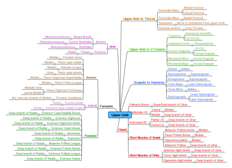
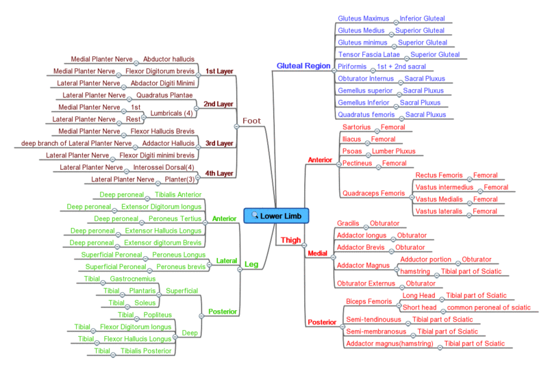
How Muscles Work
Muscles contract through a complex process involving the sliding filament theory, where actin and myosin proteins within muscle fibers slide past each other. This contraction is triggered by electrical signals from the nervous system, resulting in movement or the maintenance of posture. Muscles also play a crucial role in heat production, contributing to body temperature regulation.
Muscles of the head and neck
-
- muscles of the tongue (mnemonic)
- extrinsic muscles of the tongue
- genioglossus muscle
- hyoglossus muscle
- styloglossus muscle
- palatoglossus muscle
- intrinsic muscles of the tongue
- superior longitudinal muscle of the tongue
- inferior longitudinal muscle of the tongue
- transverse muscle of the tongue
- vertical muscle of the tongue
- extrinsic muscles of the tongue
- muscles of mastication
- temporalis muscle
- masseter muscle
- medial pterygoid muscle
- lateral pterygoid muscle
- facial muscles
- epicranius muscle
- occipitofrontalis muscle
- frontalis muscle
- occipitalis muscle
- temporoparietalis muscle
- occipitofrontalis muscle
- circumorbital and palpebral muscles
- orbicularis oculi muscle
- corrugator supercilii muscle
- levator palpebrae superioris muscle
- nasal muscles
- procerus muscle
- nasalis muscle
- compressor naris muscle
- dilator naris muscle
- myrtiformis muscle
- depressor septi nasalis muscle
- levator labii superioris alaeque nasalis muscle
- buccolabial muscles
- elevators, retractors and evertors of the upper lip
- levator labii superioris alaeque nasalis muscle
- levator labii superioris muscle
- zygomaticus major muscle
- zygomaticus minor muscle
- levator anguli oris muscle
- malaris muscle
- risorius muscle
- depressors, retractors and evertors of the lower lip
- depressor labii inferioris muscle
- depressor anguli oris muscle
- mentalis muscle
- compound sphincter
- orbicularis oris muscle
- incisivus labii superioris muscle
- incisivus labii inferioris muscle
- orbicularis oris muscle
- muscle of mastication
- buccinator muscle
- modiolus
- elevators, retractors and evertors of the upper lip
- epicranius muscle
- muscles of the middle ear
- stapedius muscle
- tensor tympani muscle
- orbital muscles
- extraocular muscles
- superior rectus muscle
- inferior rectus muscle
- lateral rectus muscle
- medial rectus muscle
- superior oblique muscle
- inferior oblique muscle
- levator palpebrae superioris muscle
- superior tarsal muscle
- extraocular muscles
- muscles of the soft palate
- tensor veli palatini muscle
- levator veli palatini muscle
- palatopharyngeus muscle
- palatoglossus muscle
- muscle of the uvula
- pharyngeal muscles
- superior pharyngeal constrictor muscle
- Passavant cushion
- middle pharyngeal constrictor muscle
- inferior pharyngeal constrictor muscle
- Killian dehiscence
- stylopharyngeus muscle
- salpingopharyngeus muscle
- superior pharyngeal constrictor muscle
- suprahyoid muscles
- digastric muscle
- geniohyoid muscle
- mylohyoid muscle
- mylohyoid boutonniere
- stylohyoid muscle
- infrahyoid muscles
- sternohyoid muscle
- sternothyroid muscle
- thyrohyoid muscle
- omohyoid muscle
- intrinsic muscles of the larynx
-
Cricothyroid Muscle: Adjusts the tension of the vocal cords for pitch modulation.
-
Thyroarytenoid Muscle: Helps relax or shorten the vocal cords; includes the vocalis muscle which is directly involved in sound production.
-
Posterior Cricoarytenoid Muscle: The only muscle that opens the vocal cords (abducts them) during breathing.
-
Lateral Cricoarytenoid Muscle: Helps close the vocal cords (adducts them) during speech and swallowing.
-
Interarytenoid Muscles: Aid in closing the gap between the arytenoid cartilages, contributing to effective voice production and airway protection.
- Vocalis Muscle
- muscles of the neck
- platysma muscle
- longus colli muscle
- longus capitis muscle
- scalenus anterior muscle
- colliscalene triangle
- scalenus medius muscle
- scalenus posterior muscle
- scalenus pleuralis muscle
- sternocleidomastoid muscle
- suboccipital muscles
- rectus capitis posterior major muscle
- rectus capitis posterior minor muscle
- obliquus capitis superior muscle
- obliquus capitis inferior muscle
- accessory muscles of the neck
- levator glandulae thyroideae muscle
- splenius capitis and
- semispinalis capitis
- muscles of the tongue (mnemonic)
Superficial Neck Muscles
These muscles are generally visible and play major roles in head movement and external appearance.
-
Sternocleidomastoid (SCM):
-
Origin: Medial portion of the manubrium and the medial clavicle.
-
Insertion: Mastoid process of the temporal bone and the lateral portion of the superior nuchal line.
-
Function: Rotates the head to the opposite side and flexes the neck.
-
-
Trapezius:
-
Origin: Occipital bone, ligamentum nuchae, and the spinous processes of the thoracic vertebrae.
-
Insertion: Lateral third of the clavicle, acromion, and spine of the scapula.
-
Function: Elevates, retracts, and rotates the scapula; also helps in neck extension when the scapula is fixed.
-
-
Platysma:
-
Origin: Fascia covering the upper parts of the pectoral and deltoid muscles.
-
Insertion: Lower border of the mandible and skin of the lower face.
-
Function: Tenses the skin of the neck and helps depress the mandible.
-
Deep Neck Flexors
These muscles are found deep in the anterior neck and play critical roles in stabilizing and flexing the cervical spine.
-
Longus Colli:
-
Location: Runs along the anterior vertebral bodies of the cervical and upper thoracic spine.
-
Function: Flexes the neck and contributes to the stabilization of the cervical vertebrae.
-
-
Longus Capitis:
-
Location: Extends from the anterior tubercles of the cervical vertebrae to the base of the skull.
-
Function: Assists in flexion and slight rotation of the head.
-
Lateral Neck Muscles (Scalene Group)
These muscles lie on the side of the neck and are involved in neck movements and assisting respiration by elevating the first and second ribs.
-
Anterior Scalene (Scalenus Anterior):
-
Origin: Transverse processes of cervical vertebrae (C3–C6).
-
Insertion: First rib.
-
Function: Elevates the first rib and aids in lateral flexion of the neck.
-
-
Middle Scalene (Scalenus Medius):
-
Origin: Transverse processes of C2–C7 vertebrae.
-
Insertion: First rib.
-
Function: Works with the anterior scalene to elevate the first rib and laterally flex the neck.
-
-
Posterior Scalene (Scalenus Posterior):
-
Origin: Transverse processes of lower cervical vertebrae.
-
Insertion: Second rib.
-
Function: Elevates the second rib and assists with lateral flexion of the neck.
-
Posterior Neck Muscles
These muscles form the bulk of the neck’s posterior region and are key to extending and rotating the head, as well as maintaining posture.
-
Splenius Capitis:
-
Origin: Lower half of the ligamentum nuchae and spinous processes of C7–T3/T4 vertebrae.
-
Insertion: Mastoid process and lateral third of the superior nuchal line of the occipital bone.
-
Function: Extends and rotates the head.
-
-
Splenius Cervicis:
-
Origin: Spinous processes of T3–T6 vertebrae.
-
Insertion: Transverse processes of the upper cervical vertebrae.
-
Function: Assists in neck extension and lateral flexion.
-
-
Semispinalis Capitis:
-
Location: Lies deep to the splenius muscles.
-
Function: Extends the head and neck, and contributes to rotation.
-
-
Semispinalis Cervicis:
-
Location: Lies adjacent to the vertebral column, extending along the cervical spine.
-
Function: Helps in extending and rotating the cervical spine.
-
-
Longissimus Capitis (part of the Erector Spinae group):
-
Location: Runs along the cervical and upper thoracic spine.
-
Function: Extends and laterally flexes the head.
-
-
Suboccipital Muscles: (A group of four small muscles at the base of the skull, crucial for fine head movements.)
-
Rectus Capitis Posterior Major: Extends and rotates the head.
-
Rectus Capitis Posterior Minor: Assists in head extension.
-
Obliquus Capitis Superior: Helps extend and slightly laterally flex the head.
-
Obliquus Capitis Inferior: Primarily rotates the atlas (and thus the head) on the axis.
-
Muscles Associated with the Hyoid Bone
These muscles are important for actions such as swallowing and speech, as they control the position of the hyoid bone and larynx.
Suprahyoid Muscles:
-
Digastric:
-
Structure: Consists of two bellies (anterior and posterior) connected by an intermediate tendon.
-
Function: Elevates the hyoid bone and assists in depressing the mandible.
-
-
Stylohyoid:
-
Origin: Styloid process of the temporal bone.
-
Insertion: Hyoid bone.
-
Function: Elevates the hyoid during swallowing.
-
-
Mylohyoid:
-
Location: Forms the floor of the oral cavity.
-
Function: Elevates the hyoid bone and the floor of the mouth.
-
-
Geniohyoid:
-
Location: Lies just above the mylohyoid.
-
Function: Pulls the hyoid bone forward (anteriorly) and upward.
-
Infrahyoid Muscles:
-
Sternohyoid:
-
Origin: Manubrium of the sternum and medial end of the clavicle.
-
Insertion: Hyoid bone.
-
Function: Depresses the hyoid bone.
-
-
Omohyoid:
-
Structure: Consists of two bellies (superior and inferior) connected by an intermediate tendon.
-
Function: Depresses and retracts the hyoid bone.
-
-
Sternothyroid:
-
Origin: Manubrium of the sternum.
-
Insertion: Thyroid cartilage.
-
Function: Depresses the thyroid cartilage.
-
-
Thyrohyoid:
-
Origin: Thyroid cartilage.
-
Insertion: Hyoid bone.
-
Function: Elevates the thyroid cartilage and depresses the hyoid bone.
-
Total List Of Muscles in Human body
| Muscle | Location | Origin | Insertion | Artery | Nerve | Action | Antagonist | O | TA |
|---|---|---|---|---|---|---|---|---|---|
| occipitalis | head, occipitofrontalis, back of skull (left/right) | superior nuchal line of occipital bone, mastoid part of temporal bone | epicranial aponeurosis | occipital artery | facial nerve [CNVII], posterior auricular nerve | retracts scalp | ? | 2 | 1 |
| frontalis | head, occipitofrontalis, forehead (left/right) | skin of eyebrow and glabella | epicranial aponeurosis | ophthalmic artery | facial nerve [CNVII], temporal branch | wrinkles eyebrow | procerus, corrugator supercilii, and orbicularis oculi muscles[1] | 2 | 1 |
| orbicularis oculi, orbital part | head, forehead/eyelid (left/right) | frontal bone | lateral palpebral raphe | ophthalmic artery, zygomatico-orbital artery, angular artery | facial nerve [CNVII], zygomatic branch | closes eyelids (voluntary, winking/squeezing) | levator palpebrae superioris | 2 | 3 |
| orbicularis oculi, palpebral part | head, forehead/eyelid (left/right) | medial palpebral ligament | lateral palpebral raphe | ophthalmic artery, zygomatico-orbital artery, angular artery | facial nerve [CNVII], zygomatic branch | closes eyelids (involuntary, blinking/sleeping) | levator palpebrae superioris | 2 | 3 |
| orbicularis oculi, deep palpebral (lacrimal) part | head, forehead/eyelid (left/right) | posterior crest of lacrimal bone | Edges of eyelids | ophthalmic artery, zygomatico-orbital artery, angular artery | facial nerve [CNVII], zygomatic branch | facilitates tear ducts | ? | 2 | 3 |
| corrugator supercilii | head, eye (left/right) | nasal part of frontal bone | intermediate third of skin of eyebrow | ophthalmic artery | facial nerve [CNVII], zygomatic branch | moves skin of forehead medially and inferiorly (towards root of nose) | ? | 2 | 1 |
| depressor supercilii | head, eye (left/right) | nasal part of frontal bone, medial rim of orbit | medial third of skin of eyebrow | ophthalmic artery | facial nerve [CNVII], zygomatic branch | moves skin of eyebrows inferiorly | ? | 2 | 1 |
| levator palpebrae superioris | head, extraocular (left/right) | sphenoid bone | tarsal plate, upper eyelid | ophthalmic artery | oculomotor nerve [CNIII] | retracts and elevates eyelid | orbicularis oculi | 2 | 1 |
| superior tarsal | head, extraocular (left/right) | underside of levator palpebrae superioris | superior tarsal plate of eyelid | ophthalmic artery | sympathetic nervous system | raises upper eyelid | ? | 2 | 1 |
| rectus, superior | head, eye, orbit (left/right) | annulus of Zinn at orbital apex | 7.5 mm superior to corneal limbus | ophthalmic artery | oculomotor nerve [CNIII], superior branch | adducts, elevates, and medially rotates eye | Oblique Superior and inferior | 2 | 1 |
| rectus, inferior | head, eye, orbit (left/right) | annulus of Zinn at orbital apex | 6.5 mm inferior to corneal limbus | ophthalmic artery | oculomotor nerve [CNIII], inferior branch | adducts and depresses eye | Oblique Superior and inferior | 2 | 1 |
| rectus, medial | head, eye, orbit (left/right) | annulus of Zinn at orbital apex | 5.5 mm medial to corneal limbus | ophthalmic artery | oculomotor nerve [CNIII], inferior branch | adducts eye | Oblique Superior and inferior | 2 | 1 |
| rectus, lateral | head, eye, orbit (left/right) | annulus of Zinn at orbital apex | 7 mm temporal to corneal limbus | ophthalmic artery | abducens nerve [CNVI] | abducts eye | Oblique Superior and inferior | 2 | 1 |
| oblique, superior | head, extraocular (left/right) | annulus of Zinn at orbital apex, medial to optic canal | outer posterior quadrant of eyeball | ophthalmic artery, lateral muscular branch | trochlear nerve [CNIV] | abducts, intorts, and depress eye | right medial, superior, and inferior recti (superior and inferior oblique muscles are the synergists) | 2 | 1 |
| oblique, inferior | head, extraocular (left/right) | orbital surface of maxilla, lateral to lacrimal groove | laterally onto eyeball, deep to lateral rectus, by a short flat tendon | ophthalmic artery | oculomotor nerve [CNIII] | abducts, extorts, and elevates eye | right medial, superior, and inferior recti (superior and inferior oblique muscles are the synergists) | 2 | 1 |
| temporoparietalis | head, ear, outer ear (left/right) | auriculares muscles | epicranial aponeurosis | facial nerve [CNVII] | fixes galeal aponeurosis | 2 | 1 | ||
| auricularis anterior | head, ear, auricular, extrinsic (left/right) | temporal fascia | front of helix of ear | posterior auricular artery | facial nerve [CNVII] | pulls auricle forwards | 2 | 1 | |
| auricularis superior | head, ear, auricular, extrinsic (left/right) | epicranial aponeurosis | dorsocranial surface of auricle | posterior auricular artery | facial nerve [CNVII] | pulls auricle upwards | 2 | 1 | |
| auricularis posterior | head, ear, auricular, extrinsic (left/right) | mastoid process of temporal bone, tendon of sternocleidomastoid | dorsal part of auricle | posterior auricular artery | facial nerve [CNVII] | pulls auricle backwards | ? | 2 | 1 |
| helicis major | head, ear, auricular, intrinsic (left/right) | Spina helicis | Anterior border of the helix | Auricular branches of posterior auricular and auricular branch of occipital arteries | Facial nerve | depresses the anterior margin of the ear cartilage | ? | 2 | 1 |
| helicis minor | head, ear, auricular, intrinsic (left/right) | Base of the helical crus | Anterior aspect of the helical crus | Auricular branches of posterior auricular and auricular branch of occipital arteries | Facial nerve, Posterior auricular nerve branch | Adjusts the shape of the anterior margin of the ear cartilage | ? | 2 | 1 |
| tragicus | head, ear, auricular, intrinsic (left/right) | Base of the tragus | Apex of the tragus | Auricular branches of posterior auricular and auricular branch of occipital arteries | Facial nerve | Increase the opening of the external acoustic meatus | ? | 2 | 1 |
| antitragicus | head, ear, auricular, intrinsic (left/right) | Outer part of the antitragus | Cauda helicis and antihelix | Auricular branch of superficial temporal and auricular branches of posterior auricular artery | Facial nerve | Modifies the auricular shape | ? | 2 | 1 |
| auricle, transverse | head, ear, auricular, intrinsic (left/right) | Cranial surface of the eminentia conchae | Cranial surface of the eminentia scaphae | Auricular branches of posterior auricular and auricular branch of occipital arteries | Facial nerve | Flattens the cranial profile outer ear | ? | 2 | 1 |
| auricle, oblique | head, ear, auricular, intrinsic (left/right) | ? | ? | ? | Facial nerve, posterior auricular nerve | ? | ? | 2 | 1 |
| Muscle of terminal notch[2] | head, ear, auricular, intrinsic (left/right) | ? | ? | ? | ? | ? | ? | 0 | 1 |
| stapedius | head, ear, inner ear (left/right) | tip of pyramid of middle ear | neck of stapes | stapedial branch of posterior auricular artery | nerve to the stapedius from facial nerve [CNVII] | reduces movement of stapes, controls amplitude of sound waves to inner ear | 2 | 1 | |
| tensor tympani | head, ear, inner ear (left/right) | Eustachian tube | handle of malleus | superior tympanic artery | medial pterygoid nerve from mandibular nerve [CNV3] | tenses tympanic membrane, controls amplitude of sound waves to inner ear | 2 | 1 | |
| procerus | head, nose (left/right) | fascia over lower part of nasal bone | skin of lower part of forehead between eyebrows | facial artery | facial nerve [CNVII], buccal branch | draws down medial angle of eyebrow (giving expressions of frowning) | 2 | 1 | |
| depressor septi nasi | head, nose (left/right) | incisive fossa of maxilla | nasal septum and back part of alar part of nasalis | superior labial artery | facial nerve [CNVII], buccal branch | depresses nasal septum | 2 | 1 | |
| levator nasolabialis (levator labii superioris alaeque nasi) | head, nose (left/right) | frontal process of maxilla | nostril and upper lip | superior labial artery | facial nerve [CNVII], buccal branch | dilates nostril, elevates upper lip, elevates wing of nose | 2 | 1 | |
| nasalis, transverse part | head, nose, nasalis, transverse part (left/right) | alveolar yoke of canine tooth | lateral nasal cartilage | superior labial artery | facial nerve [CNVII], buccal branch | compresses nostrils | 2 | 2 | |
| nasalis, alar part | head, nose, nasalis, alar part (left/right) | alveolar yoke of lateral incisor tooth, greater and lesser alar cartilages | skin near margin of nostril | superior labial artery | facial nerve [CNVII], buccal branch | dilates nostrils | 2 | 2 | |
| levator anguli oris | head, mouth (left/right) | maxilla | modiolus of mouth | facial artery | facial nerve [CNVII] | elevates angle of mouth (smile) | depressor anguli oris | 2 | 1 |
| depressor anguli oris | head, mouth (left/right) | tubercle of mandible | modiolus of mouth | facial artery | facial nerve [CNVII], mandibular branch | depresses angle of mouth (frown) | levator anguli oris | 2 | 1 |
| transversus menti | head, mouth (left/right) | continuation of depressor anguli oris muscles | other side of depressor anguli oris muscles | facial artery | facial nerve [CNVII], mandibular branch or buccal branch | depresses angle of mouth (frown) | levator anguli oris | 0.6[3] | 1 |
| levator labii superioris | head, mouth (left/right) | medial part of infra-orbital margin of maxilla | skin and muscle of upper lip (labii superioris) | superior labial artery | facial nerve [CNVII], buccal branch | elevates upper lip | 2 | 1 | |
| depressor labii inferioris | head, mouth (left/right) | oblique line of mandible, between symphysis and mental foramen | integument of lower lip, orbicularis oris fibers, its fellow of opposite side | inferior labial artery | facial nerve [CNVII], marginal mandibular branch | depresses lower lip | 2 | 1 | |
| mentalis | head, mouth (left/right) | alveolar yoke of lower, lateral incisor tooth, found on anterior mandible | skin of chin | inferior labial artery | facial nerve [CNVII], marginal mandibular branch | elevates and wrinkles skin of chin, protrudes lower lip | 2 | 1 | |
| bucinator (buccinator) | head, mouth (left/right) | alveolar processes of maxilla and mandible, pterygomandibular raphe | fibres of orbicularis oris | buccal artery | facial nerve [CNVII], buccal branch | compress cheeks against teeth (blowing), mastication | 2 | 1 | |
| orbicularis oris, marginal part | head, mouth (left/right) | maxilla and mandible | skin around lips | superior labial artery, inferior labial artery | facial nerve [CNVII], buccal branch | puckers lips | 2 | 2 | |
| orbicularis oris, labial part | head, mouth (left/right) | maxilla and mandible | skin around lips | superior labial artery, inferior labial artery | facial nerve [CNVII], buccal branch | puckers lips | 2 | 2 | |
| risorius | head, mouth (left/right) | parotid fascia | modiolus of mouth | facial artery | facial nerve [CNVII], buccal branch | draw back angle of mouth | 2 | 1 | |
| zygomaticus major | head, mouth (left/right) | zygomatic bone in region of zygomaticomaxillary suture | modiolus of mouth | facial artery | facial nerve [CNVII], buccal branch | draws angle of mouth upward and laterally | 2 | 1 | |
| zygomaticus minor | head, mouth (left/right) | zygomatic bone in region of zygomaticomaxillary suture | skin of upper lip | facial artery | facial nerve [CNVII], buccal branch | elevates upper lip | 2 | 1 | |
| masseter, superficial part | head, coronal plane (left/right) | anterior two-thirds of inferior margin of zygomatic arch and maxilla | angle of mandible, masseteric tuberosity of mandible | masseteric artery | masseteric nerve from mandibular nerve [CNV3] | elevates and retracts mandible (closes mouth) | platysma | 2 | 2 |
| masseter, deep part | head, coronal plane (left/right) | anterior two-thirds of inferior margin of zygomatic arch and maxilla | angle of mandible, masseteric tuberosity of mandible | masseteric artery | masseteric nerve from mandibular nerve [CNV3] | elevates and retracts mandible (closes mouth) | platysma | 2 | 2 |
| temporalis | head, coronal plane (left/right) | temporal lines on parietal bone of skull | coronoid process of mandible | deep temporal arteries | deep temporal nerves from mandibular nerve [CNV3] | elevates and retracts mandible (closes mouth) | platysma | 2 | 1 |
| pterygoid, lateral | head, coronal plane (left/right) | greater wing of sphenoid and lateral pterygoid process | condyloid process of mandible | maxillary artery, pterygoid branches | external pterygoid nerve from mandibular nerve [CNV3] | depresses mandible (opens mouth) | masseter, temporalis, medial pterygoid | 2 | 1 |
| pterygoideus proprius[4] | head, coronal plane (left/right) | ? | ? | ? | ? | ? | ? | 0 | 1 |
| pterygoid, medial | head, coronal plane (left/right) | medial side of lateral pterygoid plate behind upper teeth (deep head); pyramidal process of palatine bone and maxillary tuberosity (superficial head) | medial angle of mandible | maxillary artery, pterygoid branches | mandibular nerve [CNV3], medial pterygoid nerve | elevates mandible, closes jaw, helps lateral pterygoid in moving jaw from side to side | 2 | 1 | |
| genioglossus, inferior fibers | head, tongue (left/right) | superior part of mental spine of mandible (symphysis menti) | dorsum of tongue, body of hyoid | lingual artery | hypoglossal nerve [CNXII] | protrudes tongue | 2 | 3 | |
| genioglossus, middle fibers | head, tongue (left/right) | superior part of mental spine of mandible (symphysis menti) | dorsum of tongue, body of hyoid | lingual artery | hypoglossal nerve [CNXII] | depresses tongue | 2 | 3 | |
| genioglossus, superior fibers | head, tongue (left/right) | superior part of mental spine of mandible (symphysis menti) | dorsum of tongue, body of hyoid | lingual artery | hypoglossal nerve [CNXII] | draws tip of tongue back and down | 2 | 3 | |
| hyoglossus (ceratoglossus) | head, tongue (left/right) | hyoid | side of tongue | hypoglossal nerve [CNXII] | depresses tongue | 2 | 1 | ||
| chondroglossus | head, tongue (left/right) | lesser cornu and body of hyoid bone | intrinsic muscular fibers of tongue | hypoglossal nerve [CNXII] | depresses tongue (some consider this muscle to be part of hyoglossus) | 2 | 1 | ||
| styloglossus | head, tongue (left/right) | styloid process of temporal bone | tongue | lingual artery, sublingual branch | hypoglossal nerve [CNXII] | elevates and retracts tongue | inferior and middle fibers of genioglossus | 2 | 1 |
| palatoglossus | head, tongue and soft palate (left/right) | palatine aponeurosis | tongue | vagus nerve [CNX], pharyngeal plexus and accessory nerve [CNXI] | raises back part of tongue, aiding in breathing | 2 | 1 | ||
| superior longitudinal lingual | head, tongue (left/right) | close to epiglottis, from median fibrous septum | edges of tongue | lingual artery, tonsilar branch of facial artery, ascending pharyngeal artery | hypoglossal nerve [CNXII] | shortens tongue, turns tip upward, turns lateral margins upward | 2 | 1 | |
| transversus linguae | head, tongue (left/right) | median fibrous septum | sides of tongue | lingual artery, tonsilar branch of facial artery, ascending pharyngeal artery | hypoglossal nerve [CNXII] | narrows tongue with no elongation | 2 | 1 | |
| inferior longitudinal lingual | head, tongue (left/right) | root of tongue | apex of tongue | lingual artery, tonsilar branch of facial artery, ascending pharyngeal artery | hypoglossal nerve [CNXII] | shortens tongue, retracts, pulls tip downward | 2 | 1 | |
| verticalis linguae | head, tongue (left/right) | dorsum of tongue | inferior surface borders of tongue | lingual artery, tonsilar branch of facial artery, ascending pharyngeal artery | hypoglossal nerve [CNXII] | 2 | 1 | ||
| tensor veli palatini | head, soft palate (left/right) | medial pterygoid plate of sphenoid bone | palatine aponeurosis | mandibular nerve [CNV3], medial pterygoid nerve | tenses soft palate, aids in swallowing | ? | 2 | 1 | |
| levator veli palatini | head, soft palate (left/right) | temporal bone, Eustachian tube | palatine aponeurosis | facial artery | vagus nerve [CNX], pharyngeal plexus | elevates soft palate | ? | 2 | 1 |
| palatopharyngeus | head, soft palate (left/right) | palatine aponeurosis and hard palate | upper border of thyroid cartilage (blends with constrictor fibers) | facial artery | vagus nerve [CNX], pharyngeal branch | aids in breathing by pulling pharynx and larynx | 2 | 1 | |
| uvulae | head, soft palate (left/right) | hard palate | soft tissue of uvula | vagus nerve [CNX], pharyngeal branch | moves and changes shape of uvula | 2 | 1 | ||
| stylopharyngeus | head, pharynx (left/right) | styloid process of temporal bone | thyroid cartilage (pharynx) | ascending pharyngeal artery, pharyngeal branches | glossopharyngeal nerve [CNIX] | elevates larynx, elevates pharynx, swallowing | 2 | 1 | |
| salpingopharyngeus | head, pharynx (left/right) | cartilage of Eustachian tube | posterior fasciculus of pharyngopalatinus | vagus nerve [CNX], accessory nerve [CNXI] | raises nasopharynx | 2 | 1 | ||
| pharyngeal constrictor, inferior, thyropharyngeal part | head, pharynx (left/right) | cricoid cartilage, thyroid cartilage | pharyngeal raphe | ascending pharyngeal artery, pharyngeal branches | vagus nerve [CNX], superior laryngeal nerve, external laryngeal branch and recurrent laryngeal nerve | swallowing | 2 | 2 | |
| pharyngeal constrictor, inferior, cricopharyngeal part | head, pharynx (left/right) | cricoid cartilage, thyroid cartilage | pharyngeal raphe | ascending pharyngeal artery, pharyngeal branches | vagus nerve [CNX], superior laryngeal nerve, external laryngeal branch and recurrent laryngeal nerve | swallowing | 2 | 2 | |
| pharyngeal constrictor, middle, chondropharyngeal part | head, pharynx (left/right) | hyoid bone | pharyngeal raphe | ascending pharyngeal artery, pharyngeal branches | vagus nerve [CNX], pharyngeal plexus | swallowing | 2 | 2 | |
| pharyngeal constrictor, middle, ceratopharyngeal part | head, pharynx (left/right) | hyoid bone | pharyngeal raphe | ascending pharyngeal artery, pharyngeal branches | vagus nerve [CNX], pharyngeal plexus | swallowing | 2 | 2 | |
| pharyngeal constrictor, superior, pterygopharyngeal part | head, pharynx (left/right) | medial pterygoid plate, pterygomandibular raphé, alveolar process | pharyngeal raphe, pharyngeal tubercle | ascending pharyngeal artery, tonsilar branch of facial artery | vagus nerve [CNX], pharyngeal plexus | swallowing | 2 | 4 | |
| pharyngeal constrictor, superior, buccopharyngeal part | head, pharynx (left/right) | medial pterygoid plate, pterygomandibular raphé, alveolar process | pharyngeal raphe, pharyngeal tubercle | ascending pharyngeal artery, tonsilar branch of facial artery | vagus nerve [CNX], pharyngeal plexus | swallowing | 2 | 4 | |
| pharyngeal constrictor, superior, mylopharyngeal part | head, pharynx (left/right) | medial pterygoid plate, pterygomandibular raphé, alveolar process | pharyngeal raphe, pharyngeal tubercle | ascending pharyngeal artery, tonsilar branch of facial artery | vagus nerve [CNX], pharyngeal plexus | swallowing | 2 | 4 | |
| pharyngeal constrictor, superior, glossopharyngeal part | head, pharynx (left/right) | medial pterygoid plate, pterygomandibular raphé, alveolar process | pharyngeal raphe, pharyngeal tubercle | ascending pharyngeal artery, tonsilar branch of facial artery | vagus nerve [CNX], pharyngeal plexus | swallowing | 2 | 4 | |
| cricothyroid, straight part | head, larynx (left/right) | anterior and lateral cricoid cartilage | inferior cornu and lamina of thyroid cartilage | cricothyroid branch of superior thyroid artery | vagus nerve [CNX], superior laryngeal nerve, external laryngeal branch | tenses and elongates vocal folds (has minor adductory effect) | 2 | 1 | |
| cricothyroid, oblique part | head, larynx (left/right) | anterior and lateral cricoid cartilage | inferior cornu and lamina of thyroid cartilage | cricothyroid branch of superior thyroid artery | vagus nerve [CNX], superior laryngeal nerve, external laryngeal branch | tenses and elongates vocal folds (has minor adductory effect) | 2 | 1 | |
| arytenoid, transverse | head, larynx (left/right) | arytenoid cartilage on one side | arytenoid cartilage on opposite side | superior laryngeal branch of superior thyroid artery | vagus nerve [CNX], recurrent laryngeal nerve | approximates arytenoid cartilages (closes rima glottidis) | 2 | 1 | |
| arytenoid, oblique | head, larynx (left/right) | arytenoid cartilage on one side | arytenoid cartilage on opposite side | superior laryngeal branch of superior thyroid artery | vagus nerve [CNX], recurrent laryngeal nerve | approximates arytenoid cartilages (closes rima glottidis) | 2 | 1 | |
| arytenoid, oblique, aryepiglottic part | head, larynx (left/right) | apex of arytenoid | lateral border of epiglottis | laryngeal branch of superior thyroid artery | vagus nerve [CNX], inferior laryngeal nerve | closes the laryngeal inlet | ? | 2 | 0 |
| vocalis | head, larynx (left/right) | inner (anterior) surface of thyroid cartilage | anterior surface of arytenoid cartilage | vagus nerve [CNX], recurrent laryngeal nerve | thickens and decreases length of vocal folds, adducts during speech | 2 | 1 | ||
| thyroarytenoid, thyroepiglottic part | head, larynx (left/right) | inner (anterior) surface of thyroid cartilage | anterior surface of arytenoid cartilage | vagus nerve [CNX], recurrent laryngeal nerve | thickens and decreases length of vocal folds, adducts during speech | 2 | 1 | ||
| thyroarytenoid, external part | head, larynx (left/right) | inner (anterior) surface of thyroid cartilage | anterior surface of arytenoid cartilage | vagus nerve [CNX], recurrent laryngeal nerve | thickens and decreases length of vocal folds, adducts during speech | 2 | 1 | ||
| cricoarytenoid, posterior | head, larynx (left/right) | posterior part of cricoid cartilage | muscular process of arytenoid cartilage | vagus nerve [CNX], recurrent laryngeal nerve | abducts and laterally rotates cartilage, pulling vocal ligaments away from midline and forward and so opening rima glottidis | lateral cricoarytenoid | 2 | 1 | |
| cricoarytenoid, lateral | head, larynx (left/right) | lateral part of arch of cricoid cartilage | muscular process of arytenoid cartilage | vagus nerve [CNX], recurrent laryngeal nerve | adducts and medially rotates cartilage, pulling vocal ligaments towards midline and backwards and so closing rima glottidis | posterior cricoarytenoid | 2 | 1 | |
| ceratocricoideus[5][6] | head, larynx (left/right) | ? | ? | ? | ? | ? | ? | 0 | 1 |
| platysma | Neck, Clavicular, Right, left | base of mandible | inferior clavicle and fascia of chest | branches of submental artery, branches of suprascapular artery | cervical branch of facial nerve [CNVII] | tenses skin of neck | masseter, temporalis | 2 | 1 |
| sternocleidomastoid | Neck, Clavicular, Right, left | manubrium sterni (sternal head), medial portion of clavicle (clavicular head) | mastoid process of temporal bone, superior nuchal line | occipital artery, superior thyroid artery | motor: accessory nerve sensory: cervical plexus |
tilts head to its own side, rotates head so face is turned towards opposite side, flexes neck, raises sternum, assists in forced inspiration | ?? | 2 | 1 |
| digastric, anterior belly | Neck, Suprahyoid, Right/left | digastric fossa (mandible) | intermediate tendon (lesser horn of hyoid bone) | submental branch of facial artery | mandibular nerve [CNV3] via mylohyoid nerve | opens jaw when masseter and temporalis are relaxed | ?? | 2 | 2 |
| digastric, posterior belly | Neck, Suprahyoid, Right/left | mastoid process of temporal bone | intermediate tendon (lesser horn of hyoid bone) | occipital artery | facial nerve [CNVII] | opens jaw when masseter and temporalis are relaxed | ?? | 2 | 2 |
| stylohyoid | Neck, Suprahyoid, Right/left | styloid process of temporal bone | greater horn of hyoid bone | occipital artery | facial nerve [CNVII] | elevates hyoid during swallowing | ?? | 2 | 1 |
| mylohyoid | Neck, Suprahyoid, Right/left | mylohyoid line of mandible | pharyngeal raphe | mylohyoid branch of inferior alveolar artery | mylohyoid nerve, from inferior alveolar branch of mandibular nerve [CNV3] | raises oral cavity floor, elevates hyoid, depresses mandible | ?? | 2 | 1 |
| geniohyoid | Neck, Suprahyoid, Right/left | mandibular symphysis | anterior surface of body of hyoid bone | C1 via hypoglossal nerve | elevates hyoid and tongue upward during swallowing | ?? | 2 | 1 | |
| sternohyoid | Neck, Infrahyoid, Right/left | sternum, manubrium | hyoid bone | superior thyroid artery | ansa cervicalis | depresses hyoid | ?? | 2 | 1 |
| sternothyroid | Neck, Infrahyoid, Right/left | sternum, manubrium | thyroid cartilage | superior thyroid artery | ansa cervicalis | depresses larynx, may slightly depress hyoid | ?? | 2 | 1 |
| thyrohyoid | Neck, Infrahyoid, Right/left | thyroid cartilage | hyoid bone | superior thyroid artery | C1 | depress hyoid | ?? | 2 | 1 |
| omohyoid, superior belly | Neck, Infrahyoid, Right/left | upper border of scapula | hyoid bone | inferior thyroid artery | ansa cervicalis | depresses larynx, depresses and moves to side hyoid | ?? | 2 | 2 |
| omohyoid, inferior belly | Neck, Infrahyoid, Right/left | upper border of scapula | hyoid bone | inferior thyroid artery | ansa cervicalis | depresses larynx, depresses and moves to side hyoid | ?? | 2 | 2 |
| levator glandulae thyroideae | Neck, Infrahyoid, Right/left | thyrohyoid muscle | thyroid isthmus | ? | ansa cervicalis | ? | ? | 0 | 1 |
| longus colli | Neck, Anterior | transverse processes of vertebrae C3, C4, C5, and C6 | anterior arch of atlas | ascending pharyngeal artery, vertebral artery | C2, C3, C4, C5, C6 | flexes neck and head | ?? | 2 | 1 |
| longus capitis | Neck, Anterior | anterior tubercles of transverse processes of vertebrae C3, C4, C5, and C6 | basilar part of occipital bone | ?? | C1, C2, C3/C4 | flexes neck at atlanto-occipital joint | ?? | 2 | 1 |
| rectus capitis, anterior | Neck, Anterior | atlas | occipital bone | ?? | C1 | flexes neck at atlanto-occipital joint | ?? | 2 | 1 |
| rectus capitis, lateralis | Neck, Anterior/Lateral, Right/left | upper surface of transverse process of atlas (C1) | under surface of jugular process of occipital bone | ?? | C1 | laterally flexes neck, stabilises sidebens at atlanto-occipital joint | ?? | 2 | 1 |
| scalenus anterior | Neck, Lateral, Right/left | cervical vertebrae C3-C6 | first rib | inferior thyroid artery, ascending cervical artery | ventral ramus of C5, C6 | when neck is fixed, elevates first rib to aid in breathing; when rib is fixed, bends neck forward and sideways and rotates it to opposite side | ?? | 2 | 1 |
| scalenus medius | Neck, Lateral, Right/left | cervical vertebrae C2-C6 | first rib | inferior thyroid artery, ascending cervical artery | ventral rami of third to eighth cervical spinal nerves | elevates first rib, rotates neck to opposite side | ?? | 2 | 1 |
| scalenus posterior | Neck, Lateral, Right/left | cervical vertebrae C4 – C6, transverse processes | second rib | ascending cervical artery and superficial cervical artery | C6, C7, C8 | elevates second rib, tilts neck to same side | ?? | 2 | 1 |
| scalenus minimus | Neck, Lateral, Right/left | cervical vertebrae C7 | first rib | superficial cervical artery | C7, C8 | elevates first rib | ?? | 0 | 1 |
| levator scapulae | Neck/Upper Limb, Verterbral Column, Lateral, Right/left | posterior tubercles of transverse processes of C1 – C4 vertebrae | superior part of medial border of scapula | dorsal scapular artery | cervical nerve (C3, C4) and dorsal scapular nerve (C5) | elevates scapula, tilts glenoid cavity inferiorly by rotating scapula | serratus anterior | 2 | 1 |
| obliquus capitis, superior | Neck, Posterior/Lateral, Right/left | lateral mass of atlas | lateral half of inferior nuchal line | ?? | suboccipital nerve | flexes head to same side | ?? | 2 | 1 |
| obliquus capitis, inferior | Neck, Posterior/Lateral, Right/left | spinous process of axis | lateral mass of atlas | ?? | suboccipital nerve | rotates neck | ?? | 2 | 1 |
| rectus capitis, posterior minor | Neck, Posterior | tubercle on posterior arch of atlas (C1) | medial part of inferior nuchal line of occipital bone and surface between it and foramen magnum | ?? | a branch of dorsal primary division of suboccipital nerve | sensory organ for neck position, also extends head at neck | ?? | 2 | 1 |
| rectus capitis, posterior major | Neck, Posterior | spinous process of axis (C2) | inferior nucheal line of occipital bone | occipital artery | dorsal ramus of C1 (suboccipital nerve) | rotates and extends head to same side | ?? | 2 | 1 |
| rectus abdominis | Torso, Abdomen, right/left | pubic symphysis, pubic crest | xiphoid process, costal cartilages of ribs 5-7 | inferior epigastric artery, superior epigastric artery; contributions from posterior intercostal arteries, subcostal arteries, and deep circumflex arteries | intercostal nerves, subcostal nerve | flexes trunk, compresses abdominal organs, aids in expiration | erector spinae | 2 | 1 |
| external abdominal oblique | Torso, Abdomen, right/left | external surfaces of ribs 5-12 | linea alba, pubic tubercle, anterior half of iliac crest | lower posterior intercostal arteries, subcostal artery, deep circumflex iliac artery | motor: intercostal nerves (T7-T11), subcostal nerve sensory: iliohypogastric nerve | flexes trunk to same side, rotates trunk to opposite side, expiration, compresses abdominal organs | 2 | 1 | |
| internal abdominal oblique | Torso, Abdomen, right/left | anterior two-thirds of iliac crest, iliopectineal arch, thoracolumbar fascia | inferior borders of ribs 10–12, pubic crest | lower posterior intercostal arteries, subcostal artery, superior epigastric artery, inferior epigastric artery, superficial circumflex iliac artery, deep circumflex iliac artery, posterior lumbar arteries | intercostal nerves (T7-T11), subcostal nerve, iliohypogastric nerve, ilioinguinal nerve | flexes trunk to same side, rotates trunk to opposite side, expiration, compresses abdominal organs | 2 | 1 | |
| cremaster | Torso, Abdomen, inguinal canal and scrotum | Internal oblique | Tunica vaginalis | Cremasteric artery | Genital branch of genitofemoral nerve | raise and lower the scrotum (involuntary) | ? | 0-1(M) | 1 |
| transversus abdominis | Torso, Abdomen, right/left | internal surface of costal cartilage of ribs 7–12, thoracolumbar fascia, anterior two-thirds of iliac crest, iliopectineal arch | linea alba, aponeurosis of internal oblique muscle, pubic crest | lower posterior intercostal arteries, subcostal artery, superior epigastric artery, inferior epigastric artery, superficial circumflex iliac artery, deep circumflex iliac artery, posterior lumbar arteries | intercostal nerves T7-T11, subcostal nerve, iliohypogastric nerve, ilioinguinal nerve | rotates trunk to same side, compresses abdominal organs, expiration | 2 | 1 | |
| pyramidalis | Torso, Abdomen, right/left | pubic symphysis, pubic crest | linea alba | inferior epigastric artery | subcostal nerve | tenses linea alba | 2 | 1 | |
| quadratus lumborum | Torso, Abdomen, right/left | iliac crest, iliolumbar ligament | inferior border of rib 12, transverse processes of L1-L4 | lumbar, median sacral, iliolumbar, subcostal arteries | subcostal nerve, anterior rami of spinal nerves L1-L4 | flexes trunk to same side, fixes rib 12 during inspiration, extends trunk | 2 | 1 | |
| iliocostalis colli | Torso, Back, right/left | sacrum, iliac crest, thoracolumbar fascia, spinous processes | ribs | lateral sacral artery | posterior branch of spinal nerve | extends/flexes vertebral column | rectus abdominis | 2 | 1 |
| iliocostalis lumborum | Torso, Back, right/left | sacrum, iliac crest, thoracolumbar fascia, spinous processes | The lower six or seven ribs | lateral sacral artery | posterior branch of spinal nerve | extends/flexes vertebral column | rectus abdominis | 2 | 1 |
| iliocostalis thoracis | Torso, Back, right/left | sacrum, iliac crest, thoracolumbar fascia, spinous processes(the lower six ribs) | upper six ribs and the transverse process of CVII | lateral sacral artery | posterior branch of spinal nerve | extends/flexes vertebral column | rectus abdominis | 2 | 1 |
| longissimus capitis | Torso/Neck, Back, right/left | articular processes of C4-C7; transverse processes of T1-T5 | posterior margin of mastoid process | lateral sacral artery | posterior branch of spinal nerve | extends/flexes vertebral column, flexes head and neck to same side | rectus abdominis | 2 | 1 |
| longissimus colli | Torso, Back, right/left | transverse process | transverse process | lateral sacral artery | posterior branch of spinal nerve | extends/flexes vertebral column | rectus abdominis | 2 | 1 |
| longissimus thoracis | Torso, Back, right/left | transverse process | transverse process | lateral sacral artery | posterior branch of spinal nerve | extends/flexes vertebral column | rectus abdominis | 2 | 1 |
| longissimus thoracis, lumbar part | Torso, Back, right/left | transverse process | transverse process | lateral sacral artery | posterior branch of spinal nerve | extends/flexes vertebral column | rectus abdominis | 2 | 0 |
| spinalis capitis | Torso, Back, right/left | spinous process | spinous process | lateral sacral artery | posterior branch of spinal nerve | extends/flexes vertebral column | rectus abdominis | 2 | 1 |
| spinalis colli | Torso, Back, right/left | spinous process | spinous process | lateral sacral artery | posterior branch of spinal nerve | extends/flexes vertebral column | rectus abdominis | 2 | 1 |
| spinalis thoracis | Torso, Back, right/left | spinous process | spinous process | lateral sacral artery | posterior branch of spinal nerve | extends/flexes vertebral column | rectus abdominis | 2 | 1 |
| latissimus dorsi | Torso/Upper Limb, Back, Vertebral column, right/left | spinous processes of thoracic T6-T12, thoracolumbar fascia, iliac crest and inferior 3 or 4 ribs | floor of intertubercular groove of humerus | subscapular artery, dorsal scapular artery | thoracodorsal nerve(C6 to C8) | pulls forelimb dorsally and caudally(Extends, medially rotates and adducts humerus) | deltoid, trapezius | 2 | 1 |
| semispinalis thoracis | Torso, Back, right/left | transverse processes of sixth to tenth thoracic vertebrae | spinous processes of upper four thoracic vertebrae and lower two cervical vertebrae | posterior branch of spinal nerve | extends/rotates vertebral column | 2 | 1 | ||
| semispinalis colli (cervicis) | Torso, Back, right/left | transverse processes of upper five or six thoracic vertebrae | spinous processes, cervical, from axis to fifth | posterior branch of spinal nerve | extends/rotates vertebral column | 2 | 1 | ||
| semispinalis capitis | Torso/Neck, Back, right/left | articular processes of C4-C6; transverse processes of C7 and T1-T7 | spinous process, occipital bone, area between superior nuchal line and inferior nuchal line | greater occipital nerve | extends head | 2 | 1 | ||
| multifidus lumborum | Torso, Back, right/left | sacrum, erector spinae aponeurosis, PSIS, and iliac crest | spinous process | posterior branch of spinal nerve | stabilizes vertebrae in local movements of vertebral column | 2 | 1 | ||
| multifidus thoracis | Torso, Back, right/left | sacrum, erector spinae aponeurosis, PSIS, and iliac crest | spinous process | posterior branch of spinal nerve | stabilizes vertebrae in local movements of vertebral column | 2 | 1 | ||
| multifidus colli | Torso, Back, right/left | sacrum, erector spinae aponeurosis, PSIS, and iliac crest | spinous process | posterior branch of spinal nerve | stabilizes vertebrae in local movements of vertebral column | 2 | 1 | ||
| rotatores, long 1-11 | Torso, Back, thoracis, right/left | transverse process | spinous process | spinous process | posterior branch | 22 | 1 | ||
| rotatores, short 1-11 | Torso, Back, thoracis, right/left | transverse process | spinous process | spinous process | posterior branch | 22 | 1 | ||
| rotatores lumborum | Torso, Back, right/left | transverse process | spinous process | spinous process | posterior branch | 0 | 1 | ||
| rotatores colli | Torso, Back, right/left | transverse process | spinous process | spinous process | posterior branch | 0 | 1 | ||
| interspinales lumborum 1-4 | Torso, Back, right/left | spinous process | spinous process | posterior rami of spinal nerves | extends, flexes, and rotates vertebral column | 8 | 1 | ||
| interspinales thoracis 1-3 | Torso, Back, right/left | spinous process | spinous process | posterior rami of spinal nerves | extends, flexes, and rotates vertebral column | 6 | 1 | ||
| interspinales colli 1-6 | Torso, Back, right/left | spinous process | spinous process | posterior rami of spinal nerves | extends, flexes, and rotates vertebral column | 12 | 1 | ||
| intertransversarii, anterior colli 1-7 | Torso, Back, right/left | transverse process | transverse process above | posterior rami of spinal nerves | laterally flexes trunk | 14 | 1 | ||
| intertransversarii, lateral posterior colli 1-7 | Torso, Back, right/left | transverse process | transverse process above | posterior rami of spinal nerves | laterally flexes trunk | 14 | 1 | ||
| intertransversarii, medial posterior colli 1-7 | Torso, Back, right/left | transverse process | transverse process above | posterior rami of spinal nerves | laterally flexes trunk | 14 | 1 | ||
| intertransversarii, lateral lumborum, dorsal parts 1-5 | Torso, Back, right/left | transverse process | transverse process above | posterior rami of spinal nerves | laterally flexes trunk | 10 | 2 | ||
| intertransversarii, lateral lumborum, ventral parts 1-5 | Torso, Back, right/left | transverse process | transverse process above | posterior rami of spinal nerves | laterally flexes trunk | 10 | 2 | ||
| intertransversarii, medial lumborum 1-5 | Torso, Back, right/left | transverse process | transverse process above | posterior rami of spinal nerves | laterally flexes trunk | 10 | 1 | ||
| intertransversarii, thoracis 1-9 | Torso, Back, right/left | transverse process | transverse process above | posterior rami of spinal nerves | laterally flexes trunk | 18 | 1 | ||
| splenius capitis | Torso/Neck, Back, right/left | nuchal ligament, spinous process of C7-T6 | mastoid process of temporal bone, occipital bone | ?? | C3, C4 | extends, rotates, and laterally flexes head | 2 | 1 | |
| splenius colli | Torso, Back, right/left | spinous processes of T3-T6 | transverse processes of C1, C2, C3 | C5, C6 | 2 | 1 | |||
| external intercostal 1-11 | Torso, Chest (left/right) | inferior border of rib (above) | superior border of rib (below) | intercostal arteries | intercostal nerves | inhalation | internal | 22 | 1 |
| internal intercostal 1-11 | Torso, Chest (left/right) | inferior border of rib (above) | superior border of rib (below) | intercostal arteries | intercostal nerves | holds ribs steady | external | 22 | 1 |
| innermost intercostal 1-11 | Torso, Chest | inferior border of rib (above) | superior border of rib (below) | intercostal arteries | intercostal nerves | elevates ribs, expiration | 22 | 1 | |
| subcostales 1-n | Torso, Chest | inner surface of one rib | inner surface of second or third rib above, near its angle | intercostal arteries | intercostal nerves | 11[7] | 1 | ||
| transversus thoracis | Torso, Chest | costal cartilages of last 3–4 ribs, body of sternum, xiphoid process | ribs/costal cartilages 2–6 | intercostal arteries | intercostal nerves | depresses ribs | 2 | 1 | |
| levatores costarum, longi 1-4 | Torso, Chest | transverse processes of C7 to T12 vertebrae | superior surfaces of ribs immediately inferior to preceding vertebrae | dorsal rami – C8, T1, T2, T3, T4, T5, T6, T7, T8, T9, T10, T11 | assists in elevation of thoracic rib cage | 8 | 1 | ||
| levatores costarum, breves 1-12 | Torso, Chest | transverse processes of C7 to T12 vertebrae | superior surfaces of ribs immediately inferior to preceding vertebrae | dorsal rami – C8, T1, T2, T3, T4, T5, T6, T7, T8, T9, T10, T11 | assists in elevation of thoracic rib cage | 24 | 1 | ||
| serratus posterior inferior | Torso, Chest | vertebrae T11 – L3 | inferior borders of 9th through 12th ribs | intercostal arteries | intercostal nerves | depresses lower ribs(IX to XII), aiding in expiration | 2 | 1 | |
| serratus posterior superior | Torso, Chest | nuchal ligament (or ligamentum nuchae) and spinous processes of vertebrae C7 through T3 | upper borders of 2nd through 5th ribs | intercostal arteries | 2nd through 5th intercostal nerves | elevates ribs(II to V), aiding in inspiration | 2 | 1 | |
| hemidiaphragm, lumbar/costal/sternal part | Torso, Chest, Left/right | septum transversum, pleuroperitoneal folds, inner abdominal wall | pericardiacophrenic artery, musculophrenic artery, inferior phrenic arteries | phrenic and lower intercostal nerves | breathing | 2 | 1 | ||
| coccygeus | Torso, Pelvis | sacrospinous ligament | coccyx | sacral nerves: S4, S5 or S3-S4 | closes back part of pelvic outlet | 2 | 1 | ||
| iliococcygeus | Torso, Pelvis, Levator ani | ischial spine, posterior part of tendinous arch of pelvic fascia | coccyx and anococcygeal raphe | inferior gluteal artery | levator ani nerve (S4)
|
supports organs in pelvic cavity | 2 | 1 | |
| pubococcygeus | Torso, Pelvis, Levator ani | back surface of pubis, anterior part of obturator fascia | coccyx and sacrum | inferior gluteal artery | levator ani nerve (S4)
|
controls urine flow, contracts during orgasm | 2 | 1 | |
| puboanalis (puborectalis) | Torso, Pelvis, Levator ani | lower part of pubic symphysis | – | inferior gluteal artery | S3, S4. levator ani nerve | inhibits defecation | 2 | 1 | |
| puboperinealis | Torso, Pelvis, Levator ani | ? | ? | ? | ? | ? | ? | 2 | 1 |
| puboprostaticus (males) / pubovaginalis (females) | Torso, Pelvis, Levator ani | ? | ? | ? | ? | ? | ? | 2(F)(M) | 1 |
| external anal sphincter, subcutaneous, superficial, and deep parts | Torso, Perineum | – | – | inferior rectal artery | S4 and twigs from inferior anal nerves of pudendal nerve | keeps anal canal and anus closed, aids in expulsion of feces | – | 1 | 1 |
| superficial transverse perineal | Torso, Perineum, Superficial perineal pouch | anterior surface of ischial tuberosity | central point of perineum | perineal artery | pudendal nerve | constricts urethra (maintains urinary continence), constricts vagina (women) | 2 | 1 | |
| bulbospongiosus | Torso, Perineum, Superficial perineal pouch | perineal raphe | – | perineal artery | pudendal nerve | empties urethra (men), clenches vagina (women) | 2 | 1 | |
| ischiocavernosus | Torso, Perineum, Superficial perineal pouch | ischial tuberosity | crus of penis (men), crus of clitoris (women) | perineal artery | pudendal nerve | assists bulbospongiosus | 2 | 1 | |
| Deep transverse perineal | Torso, Perineum, Deep perineal pouch | inferior ramus of ischium | its fellow of opposite side | pudendal nerve | constricts urethra (maintains urinary continence), constricts vagina (women) | 0-2(M) | 1 | ||
| compressor urethrae | Torso, Perineum, Deep perineal pouch | ? | ? | ? | ? | ? | ? | 0-1(F) | 1 |
| sphincter urethrovaginalis | Torso, Perineum, Deep perineal pouch | ? | ? | ? | ? | ? | ? | 0-1(F) | 1 |
| external urethral sphincter | Torso, Perineum, Deep perineal pouch | junction of inferior rami of pubis and ischium about 1.25 – 2 cm, and from neighboring fascia | its fellow of opposite side | perineal branch of pudendal nerve (S2, S3, S4) | constricts urethra, maintains urinary continence | 2 | 1 | ||
| trapezius, descending part | Upper limb, Vertebral column, Right/Left | down midline, external occipital protuberance, nuchal ligament, medial part of superior nuchal line, spinous processes of vertebrae C7-T12 | at shoulders, lateral third of clavicle, acromion of scapula, spine of scapula | transverse cervical artery | motor: accessory nerve [CNXI]sensory: cervical nerves C3 and C4 | retracts and assists in rotating scapula(lower fibers depress scapula, upper fibers elevate it and middle fibers adduct ) | serratus anterior | 2 | 3 |
| trapezius, transverse part | Upper limb, Vertebral column, Right/Left | down midline, external occipital protuberance, nuchal ligament, medial part of superior nuchal line, spinous processes of vertebrae C7-T12 | at shoulders, lateral third of clavicle, acromion of scapula, spine of scapula | transverse cervical artery | motor: accessory nerve [CNXI]sensory: cervical nerves C3 and C4 | retracts and elevates scapula | serratus anterior | 2 | 3 |
| trapezius, ascending part | Upper limb, Vertebral column, Right/Left | down midline, external occipital protuberance, nuchal ligament, medial part of superior nuchal line, spinous processes of vertebrae C7-T12 | at shoulders, lateral third of clavicle, acromion of scapula, spine of scapula | transverse cervical artery | motor: accessory nerve [CNXI]sensory: cervical nerves C3 and C4 | retracts and elevates scapula | serratus anterior | 2 | 3 |
| transversus nuchae | Upper limb, Vertebral column, Right/Left | ? | ? | ? | ? | ? | ? | 0 | 1 |
| rhomboid major | Upper limb, Vertebral column, Right?/Left? | spinous processes of T2 to T5 vertebrae | medial border of scapula, inferior to insertion of rhomboid minor | dorsal scapular artery | dorsal scapular nerve (C4 and C5) | retracts scapula and rotates it to depress glenoid cavity, fixes scapula to thoracic wall | serratus anterior | 2 | 1 |
| rhomboid minor | Upper limb, Vertebral column, Right?/Left? | nuchal ligaments and spinous processes of C7- to T1 vertebrae | medial border of scapula, superior to insertion of rhomboid major | dorsal scapular artery | dorsal scapular nerve (C4 and C5) | retracts (adducts) scapula and rotates it to depress glenoid cavity, fixes scapula to thoracic wall | serratus anterior | 2 | 1 |
| sternalis | Upper limb,Thoracic walls, Right?/Left? | manubrium of sternum or clavicle | xiphoid process, pectoral fascia, lower ribs, costal cartilages or rectus sheath | ? | various | ? | ? | 0 | 1 |
| pectoralis major | Upper limb,Thoracic walls, Right?/Left? | clavicular head: anterior surface of medial half of clavicle sternocostal head: anterior surface of sternum, superior six costal cartilages | intertubercular groove of humerus | pectoral branch of thoracoacromial artery | lateral pectoral nerve, medial pectoral nerve clavicular head: C5 and C6 sternocostal head: C7, C8 and T1 | adducts and medially rotates humerus, draws scapula anteriorly and inferiorly clavicular head: flexes humerus sternocostal head: extends humerus | 2 | 1 | |
| pectoralis major, abdominal part | Upper limb,Thoracic walls, Right?/Left? | clavicular head: anterior surface of medial half of clavicle sternocostal head: anterior surface of sternum, superior six costal cartilages | intertubercular groove of humerus | pectoral branch of thoracoacromial artery | lateral pectoral nerve, medial pectoral nerve clavicular head: C5 and C6 sternocostal head: C7, C8 and T1 | adducts and medially rotates humerus, draws scapula anteriorly and inferiorly clavicular head: flexes humerus sternocostal head: extends humerus | 0 | 0 | |
| pectoralis minor | Upper limb,Thoracic walls, Right?/Left? | 3rd to 5th ribs, near their costal cartilages | medial border and superior surface of coracoid process of scapula | pectoral branch of thoracoacromial artery | medial pectoral nerve (C8, T1) | stabilizes scapula by drawing it inferiorly and anteriorly against thoracic wall | 2 | 1 | |
| subclavius | Upper limb,Thoracic walls, Right?/Left? | first rib | subclavian groove of clavicle | thoracoacromial artery, clavicular branch | subclavian nerve | depresses clavicle | 2 | 1 | |
| serratus anterior | Upper limb,Thoracic walls, Right?/Left? | fleshy slips from outer surface of upper 8 or 9 ribs | costal surface of medial margin of scapula | lateral thoracic artery (upper part), thoracodorsal artery (lower part) | long thoracic nerve (from roots of brachial plexus C5, C6, C7) | protracts and stabilises scapula, assists in upward rotation | rhomboid major, rhomboid minor, trapezius | 2 | 1 |
| deltoid, clavicular/acromial/scapular spinal parts | Upper limb, Shoulder, Right/left | clavicle, acromion, spine of scapula | deltoid tuberosity of humerus | primarily posterior circumflex humeral artery | axillary nerve | abducts, flexes, and extends shoulder | latissimus dorsi | 2 | 1 |
| teres major | Upper limb, Shoulder, Right/left | posterior surface of inferior angle of scapula | medial lip of intertubercular sulcus of humerus | subscapular artery, circumflex scapular artery | lower subscapular nerve (segmental levels C5 and C6) | internally rotates humerus | 2 | 1 | |
| supraspinatus | Upper limb, Shoulder, rotator cuff, Right/left | supraspinous fossa of scapula | superior facet of greater tubercle of humerus | suprascapular artery | suprascapular nerve | abducts and stabilises humerus | infraspinatus, teres minor, pectoralis major, latissimus dorsi | 2 | 1 |
| infraspinatus | Upper limb, Shoulder, rotator cuff, Right/left | infraspinous fossa of scapula | middle facet of greater tubercle of humerus | suprascapular artery, circumflex scapular artery | suprascapular nerve | laterally rotates, adducts, and stabilises humerus | subscapularis, pectoralis major, latissimus dorsi | 2 | 1 |
| teres minor | Upper limb, Shoulder, rotator cuff, Right/left | lateral border of scapula | inferior facet of greater tubercle of humerus | posterior circumflex humeral artery, circumflex scapular artery | axillary nerve | laterally rotates and adducts humerus | subscapularis, pectoralis major, latissimus dorsi | 2 | 1 |
| subscapularis | Upper limb, Shoulder, rotator cuff, Right/left | subscapular fossa of scapula | lesser tubercle of humerus | subscapular artery | upper subscapular nerve, lower subscapular nerve (C5, C6) | medially rotates humerus, stabilizes shoulder | infraspinatus, teres minor | 2 | 1 |
| coracobrachialis | Upper Limb, Arm, Anterior compartment, right/left | coracoid process of scapula | medial surface of humerus | brachial artery | musculocutaneous nerve | flexes and adducts shoulder | 2 | 1 | |
| biceps brachii | Upper Limb, Arm, Anterior compartment, right/left | short head: coracoid process of scapula long head: supraglenoid tubercle |
radial tuberosity, bicipital aponeurosis | brachial artery | musculocutaneous nerve (lateral cord: C5, C6, C7) | flexes elbow, supinates forearm | triceps brachii | 2 | 1 |
| brachialis | Upper Limb, Arm, Anterior compartment, right/left | anterior surface of humerus (mainly distal half) | coronoid process of ulna, tuberosity of ulna | radial recurrent artery | musculocutaneous nerve | flexes elbow | triceps brachii | 2 | 1 |
| triceps brachii | Upper Limb, Arm, Posterior compartment, right/left | long head: infraglenoid tubercle of scapula lateral head: posterior humerus (above radial sulcus) medial head: posterior humerus – (below radial sulcus) |
olecranon of ulna | deep artery of arm | radial nerve | extends forearm, adducts shoulder (long head).medial head does not function at shoulder. | biceps brachii, brachialis | 2 | 1 |
| articularis cubiti | Upper Limb, Arm, Posterior compartment, right/left | deep distal surface of medial head of triceps | posterior capsule of elbow joint | deep artery of arm | radial nerve | lifts capsule away from joint | ? | 2 | 1 |
| anconeus | Upper Limb, Arm, Posterior compartment, right/left | lateral epicondyle of humerus | lateral surface of olecranon, superior part of posterior ulna | deep artery of arm, interosseous recurrent artery | radial nerve (C7, C8, and T1) | partly blended with triceps, extends forearm, stabilises elbow, abducts ulna during pronation | 2 | 1 | |
| pronator teres | Upper limb, Forearm, Anterior compartment, Superficial, Right/left | humeral head: medial epicondyle of humerus (common flexor tendon) ulnar head: coronoid process of ulna |
pronator tuberosity of radius | ulnar artery, radial artery | median nerve | pronates forearm, flexes elbow | supinator | 2 | 1 |
| flexor carpi radialis | Upper limb, Forearm, Anterior compartment, Superficial, Right/left | medial epicondyle of humerus (common flexor tendon) | bases of second metacarpal, base of third metacarpal | radial artery | median nerve | flexes and abducts wrist | extensor carpi radialis brevis, extensor carpi radialis longus | 2 | 1 |
| palmaris longus | Upper limb, Forearm, Anterior compartment, Superficial, Right/left | medial epicondyle of humerus (common flexor tendon) | palmar aponeurosis | ulnar artery | median nerve | flexes wrist | extensor carpi radialis brevis, extensor carpi radialis longus, extensor carpi ulnaris | 2 | 1 |
| flexor carpi ulnaris | Upper limb, Forearm, Anterior compartment, Superficial, Right/left | medial epicondyle of humerus (common flexor tendon) | pisiform, hook of hamate, base of fifth metacarpal | ulnar artery | muscular branches of ulnar nerve | flexes and adducts wrist | extensor carpi ulnaris | 2 | 1 |
| flexor digitorum superficialis | Upper limb, Forearm, Anterior compartment, Superficial, Right/left | medial epicondyle of humerus (common flexor tendon), parts of radius and ulna | bases of middle phalanges 2, 3, 4, and 5 | ulnar artery | median nerve | flexes fingers (primarily at proximal interphalangeal joints) | extensor digitorum | 2 | 1 |
| pronator quadratus | Upper limb, Forearm, Anterior compartment, Deep, Right/left | medial anterior surface of ulna | lateral anterior surface of radius | anterior interosseous artery | anterior interosseous nerve (median nerve) | weakly pronates forearm | supinator | 2 | 1 |
| flexor digitorum profundus | Upper limb, Forearm, Anterior compartment, Deep, Right/left | ulna | distal phalanges | anterior interosseous artery | lateral belly: anterior interosseous nerve (median nerve)medial belly: muscular branches of ulnar nerve | flexes wrist, flexes interphalangeal joints | extensor digitorum | 2 | 1 |
| flexor pollicis longus | Upper limb, Forearm, Anterior compartment, Deep, Right/left | middle half of volar surface of radius, interosseus membrane | base of distal phalanx of thumb | anterior interosseous artery | anterior interosseous nerve (median nerve) (C8, T1) | flexes thumb | extensor pollicis longus, extensor pollicis brevis | 2 | 1 |
| extensor digitorum | Upper limb, Forearm, Posterior compartment, Superficial, Right/left | lateral epicondyle of humerus (common extensor tendon) | 2nd–5th phalanges | posterior interosseous artery | posterior interosseous nerve (C7, C8) | extends hand, extends fingers | flexor digitorum superficialis, flexor digitorum profundus | 2 | 1 |
| extensor digiti minimi | Upper limb, Forearm, Posterior compartment, Superficial, Right/left | anterior surface of lateral epicondyle of humerus (common extensor tendon) | extensor expansion, located at base of proximal phalanx on dorsal side | posterior interosseous artery | posterior interosseous nerve (C7, C8) | extends little finger at all joints | flexor digiti minimi brevis (hand) | 2 | 1 |
| extensor carpi ulnaris | Upper limb, Forearm, Posterior compartment, Superficial, Right/left | lateral epicondyle of humerus (common extensor tendon), ulna | 5th metacarpal | ulnar artery | posterior interosseous nerve (C7, C8) | extends and adducts wrist | flexor carpi ulnaris | 2 | 1 |
| brachioradialis | Upper limb, Forearm, mobile wad, Right/left | lateral supracondylar ridge of humerus | radial styloid process (distal radius) | radial recurrent artery | radial nerve | flexes forearm, pronates forearm when supine, supinates forearm when prone | 2 | 1 | |
| extensor carpi radialis longus | Upper limb, Forearm, mobile wad, Right/left | lateral supracondylar ridge of humerus | 2nd metacarpal | radial artery | radial nerve | extends wrist joint, abducts hand at wrist | flexor carpi radialis | 2 | 1 |
| extensor carpi radialis brevis | Upper limb, Forearm, mobile wad, Right/left | anterior surface of lateral epicondyle of humerus (common extensor tendon) | base of 3rd metacarpal | radial artery | posterior interosseus nerve | extends wrist joint, abducts hand at wrist | flexor carpi radialis | 2 | 1 |
| supinator | Upper limb, Forearm, Posterior compartment, Deep, Right/left | lateral epicondyle of humerus, supinator crest of ulna, radial collateral ligament, annular ligament | lateral proximal shaft of radius | radial recurrent artery | posterior interosseus nerve (C7, C8) | supinates forearm | pronator teres, pronator quadratus | 2 | 1 |
| extensor indicis | Upper limb, Forearm, Posterior compartment, Deep, Right/left | ulna | index finger (extensor hood) | posterior interosseous nerve (C7, C8) | extends index finger, wrist | 2 | 1 | ||
| abductor pollicis longus | Upper limb, Forearm, anatomical snuffbox, Right/left | ulna | first metacarpal | posterior interosseous artery | posterior interosseous nerve (C7, C8) | abducts and extends thumb | adductor pollicis | 2 | 1 |
| extensor pollicis brevis | Upper limb, Forearm, anatomical snuffbox, Right/left | radius, interosseous membrane of forearm | proximal phalanx of thumb | posterior interosseous artery | posterior interosseous nerve (C7, C8) | extends thumb at metacarpophalangeal joint | flexor pollicis longus, flexor pollicis brevis | 2 | 1 |
| extensor pollicis longus | Upper limb, Forearm, anatomical snuffbox, Right/left | ulna, interosseous membrane of forearm | distal phalanx of thumb | posterior interosseous artery | posterior interosseous nerve (C7, C8) | extends thumb (metacarpophalangeal and interphalangeal) | flexor pollicis longus, flexor pollicis brevis | 2 | 1 |
| opponens pollicis | Upper limb, Hand, Lateral volar, Thenar | trapezium, transverse carpal ligament | metacarpal bone of thumb on its radial side | superficial palmar arch | median nerve | opposes thumb | 2 | 1 | |
| flexor pollicis brevis | Upper limb, Hand, Lateral volar, Thenar | trapezoid, flexor retinaculum | thumb, proximal phalanx | superficial palmar arch | median nerve, deep branch of ulnar nerve (medial head) | flexes thumb | extensor pollicis longus, extensor pollicis brevis | 2 | 1 |
| abductor pollicis brevis | Upper limb, Hand, Lateral volar, Thenar | flexor retinaculum of hand, scaphoid and trapezium | radial base of proximal phalanx of thumb and thumb extensors | superficial palmar arch | median nerve | abducts thumb | adductor pollicis | 2 | 1 |
| adductor pollicis | Upper limb, Hand, Lateral volar, Thenar | transverse head: anterior body of third metacarpal oblique head: bases of second and third metacarpals and adjacent trapezoid and capitate bones |
medial side of base of proximal phalanx of thumb and ulnar sesamoid | deep palmar arch | deep branch of ulnar nerve (T1) | adducts thumb at carpometacarpal joint | abductor pollicis longus, abductor pollicis brevis | 2 | 1 |
| palmaris brevis | Upper limb, Hand, Medial volar | flexor retinaculum (medial), palmar aponeurosis | palm | palmar metacarpal artery | superficial branch of ulnar nerve | wrinkle skin of palm | 2 | 1 | |
| abductor digiti minimi (hand) | Upper limb, Hand, Medial volar, hypothenar | pisiform | base of proximal phalanx of 5th digit on ulnar or medial side | ulnar artery | deep branch of ulnar nerve | abducts little finger | 2 | 1 | |
| flexor digiti minimi (hand) | Upper limb, Hand, Medial volar, hypothenar | hamate bone | little finger | ulnar artery | deep branch of ulnar nerve | flexes little finger | extensor digiti minimi | 2 | 1 |
| opponens digiti minimi (hand) | Upper limb, Hand, Medial volar, hypothenar | hook of hamate, flexor retinaculum | medial border of 5th metacarpal | ulnar artery | deep branch of ulnar nerve (C8 and T1) | draws 5th metacarpal anteriorly and rotates it, bringing little finger (5th digit) into opposition with thumb | 2 | 1 | |
| lumbricals of the hand 1-4 | Upper limb, Hand, Intermediate | flexor digitorum profundus | extensor expansion | superficial palmar arch, common palmar digital arteries, deep palmar arch, dorsal digital artery | deep branch of ulnar nerve, median nerve | flex metacarpophalangeal joints, extend interphalangeal joints | 8 | 1 | |
| dorsal interossei (hand) 1-4 | Upper limb, Hand, Intermediate | metacarpals | proximal phalanges | dorsal metacarpal artery, palmar metacarpal artery | deep branch of ulnar nerve | abduct fingers | palmar interossei | 8 | 1 |
| palmar interossei 1-3 | Upper limb, Hand, Intermediate | metacarpals | proximal phalanges | palmar metacarpal artery | deep branch of ulnar nerve | adduct fingers | dorsal interossei | 6 | 1 |
| pollical palmar interosseus | Upper limb, Hand, Intermediate | metacarpals | proximal phalanges | palmar metacarpal artery | deep branch of ulnar nerve | adduct fingers | dorsal interossei | 1.6 | 1 |
| psoas major | Lower Limb, Iliac region, iliopsoas | transverse processes, bodies and intervertebral discs of T12-L5 vertebrae | lesser trochanter of femur | iliolumbar artery | lumbar plexus via anterior branches of L1, L2, L3[8] | flexes and rotates laterally thigh | gluteus maximus | 2 | 1 |
| psoas minor | Lower Limb, Iliac region, iliopsoas | side of T11+L1 and IV intervertebral disc | Pectineal line and iliopectineal eminence | iliolumbar artery, lumbar arteries | L1 | weakly flexes trunk flexor | gluteus maximus | 0.8 | 1 |
| iliacus | Lower Limb, Iliac region, iliopsoas | iliac fossa | lesser trochanter of femur | medial femoral circumflex artery, Iliolumbar artery | femoral nerve (L2, L3[8]) | flexes hip[9] | gluteus maximus | 2 | 1 |
| tensor fasciae latae | Lower limb, Gluteal, Left/right | iliac crest | iliotibial tract | primarily lateral circumflex femoral artery, superior gluteal artery | superior gluteal nerve (L4, L5) | flexes thigh, medially rotates thigh, stabilises torso | 2 | 1 | |
| gluteus maximus | Lower limb, gluteal, Left/right | gluteal surface of ilium, lumbar fascia, sacrum, sacrotuberous ligament | gluteal tuberosity of femur, iliotibial tract | superior gluteal artery, inferior gluteal artery | inferior gluteal nerve (L5, S1, S2 nerve roots) | externally rotates and extends hip joint, supports extended knee through iliotibial tract, chief antigravity muscle in sitting | Iliacus, psoas major, psoas minor | 2 | 1 |
| gluteus medius | Lower limb, gluteal, Left/right | gluteal surface of ilium, under gluteus maximus | greater trochanter of femur | superior gluteal artery | superior gluteal nerve (L4, L5, S1 nerve roots) | abduction of hip; preventing adduction of hip Medial rotation of thigh | lateral rotator group | 2 | 1 |
| gluteus minimus | Lower limb, gluteal, Left/right | gluteal surface of ilium, under gluteus medius | greater trochanter of femur | superior gluteal artery | superior gluteal nerve (L4, L5, S1 nerve roots) | abduction of hip; preventing adduction of hip Medial rotation of thigh | lateral rotator group | 2 | 1 |
| piriformis | Lower limb, gluteal, lateral rotator group, Left/right | sacrum | greater trochanter | inferior gluteal artery, lateral sacral artery, superior gluteal artery | piriformis nerve (S1 and S2 nerve roots)[10] | laterally rotates (outward) thigh | 2 | 1 | |
| obturator externus | Lower limb, gluteal, lateral rotator group, Left/right | obturator foramen and obturatory membrane | medial surface of greater trochanter of femur | obturator artery | posterior branch of obturator nerve (L3, L4) | adduct thigh, rotate laterally thigh | 2 | 1 | |
| superior gemellus | Lower limb, gluteal, lateral rotator group, Left/right | ischial spine | nerve to obturator internus (L5, S1, S2) | 2 | 1 | ||||
| obturator internus | Lower limb, gluteal, lateral rotator group, Left/right | ischiopubic ramus, obturator membrane | medial surface of greater trochanter of femur | nerve to obturator internus (L5, S1, S2) | abducts & rotates laterally thigh, stabilises hip during walking | 2 | 1 | ||
| inferior gemellus | Lower limb, gluteal, lateral rotator group, Left/right | ischial tuberosity | obturator internus tendon | nerve to quadratus femoris (L4, L5, S1) | laterally rotates thigh | 2 | 1 | ||
| quadratus femoris | Lower limb, gluteal, lateral rotator group, Left/right | ischial tuberosity | intertrochanteric crest | inferior gluteal artery | nerve to quadratus femoris (L4, L5, S1) | 2 | 1 | ||
| articularis genus | Lower limb, Thigh, Anterior compartment | femur | suprapatellar bursa | femoral artery | femoral nerve | pulls suprapatellar bursa during extension of knee | 2 | 1 | |
| sartorius | Lower limb, Thigh, Anterior compartment | superior to anterior superior iliac spine | medial side of upper tibia in pes anserinus | femoral artery | femoral nerve | flexes, laterally rotates, and abducts thigh, flexes and medially rotates leg | 2 | 1 | |
| rectus femoris | Lower limb, Thigh, Anterior compartment | anterior inferior iliac spine and exterior surface of bony ridge which forms iliac portion of acetabulum | patella and tibial tuberosity via patellar tendon | femoral artery | femoral nerve | knee extension; hip flexion | hamstring | 2 | 1 |
| vastus lateralis | Lower limb, Thigh, Anterior compartment | greater trochanter, intertrochanteric line, and linea aspera of femur | patella and tibial tuberosity via patellar tendon | femoral artery | femoral nerve | extends knee | hamstring | 2 | 1 |
| vastus intermedius | Lower limb, Thigh, Anterior compartment | anterior surface of femur | patella and tibial tuberosity via patellar tendon | femoral artery | femoral nerve | extends knee | hamstring | 2 | 1 |
| vastus medialis | Lower limb, Thigh, Anterior compartment | anteromedial surface of femur | patella and tibial tuberosity via patellar tendon | femoral artery | femoral nerve | extends knee | hamstring | 2 | 1 |
| biceps femoris | Lower limb, Thigh, Posterior compartment/hamstring | ischial tuberosity (long head), linea aspera of femur (short head)[11] | head of fibula[11] articulating with back of lateral tibial condyle | inferior gluteal artery, perforating arteries, popliteal artery | medial (tibial) part of sciatic nerve (long head), lateral (common fibular) part of sciatic nerve (short head)[11] | flexes knee joint, laterally rotates leg at knee (when knee is flexed), extends hip joint (long head only)[11] | quadriceps femoris | 2 | 1 |
| semitendinosus | Lower limb, Thigh, Posterior compartment/hamstring | ischial tuberosity[11] | pes anserinus | inferior gluteal artery, perforating arteries | sciatic nerve[11] (tibial, L5, S1, S2) | flexes knee, extends hip, medially rotates leg at knee[11] | quadriceps femoris | 2 | 1 |
| semimembranosus | Lower limb, Thigh, Posterior compartment/hamstring | ischial tuberosity[11] | medial surface of tibia[11] | profunda femoris, gluteal artery | sciatic nerve[11] | flexes knee, extends hip, medially rotates leg at knee[11] | quadriceps femoris | 2 | 1 |
| gracilis | Lower limb, Thigh/Hip, Medial compartment (adductor muscles) | inferior pubic ramus[12] | tibia (pes anserinus) | obturator artery | anterior branch of obturator nerve[12] | adducts hip, flexes hip, medially rotates knee[12] | gluteus medius, gluteus minimus | 2 | 1 |
| pectineus | Lower limb, Thigh/Hip, Medial compartment (adductor muscles) | superior pubic ramus[12] | lesser trochanter, linea aspera | obturator artery | femoral nerve and obturator nerve (medial compartment)[12] | adducts and flexes hip[12] | gluteus medius, gluteus minimus | 2 | 1 |
| adductor brevis | Lower limb, Thigh/Hip, Medial compartment (adductor muscles) | anterior surface of inferior pubic ramus[12] | lesser trochanter and linea aspera of femur | obturator artery | anterior branch of obturator nerve[12] | adducts hip[12] | gluteus medius, gluteus minimus | 2 | 1 |
| adductor longus | Lower limb, Thigh/Hip, Medial compartment (adductor muscles) | pubic body just below pubic crest | middle third of linea aspera | obturator artery | anterior branch of obturator nerve[12] | adducts and medially rotates hip[12] | gluteus medius, gluteus minimus | 2 | 1 |
| adductor magnus, adductor part | Lower limb, Thigh/Hip, Medial compartment (adductor muscles) | pubis[12] | femur, adductor tubercle of femur | obturator artery | posterior branch of obturator nerve (adductor) and tibial part of sciatic nerve (vertical head)[12] | adducts hip[12] | gluteus medius, gluteus minimus | 2 | 3 |
| adductor minimus | Lower limb, Thigh/Hip, Medial compartment (adductor muscles), part of adductor magnus | Inferior ramus | Linea aspera of the femur | Obturator artery and the medial circumflex femoral artery of the femoral artery. | Obturator and tibial nerves | Adduction and lateral rotation of thigh | gluteus medius, gluteus minimus | 2 | 3 |
| adductor magnus, ischiocondylar part | Lower limb, Thigh/Hip, Medial compartment (adductor muscles) | pubis[12] | femur, adductor tubercle of femur | obturator artery | posterior branch of obturator nerve (adductor) and tibial part of sciatic nerve (vertical head)[12] | adducts hip[12] | gluteus medius, gluteus minimus | 2 | 3 |
| tibialis anterior | Lower limb, Leg, Anterior Compartment | body of tibia | medial cuneiform and first metatarsal bones of foot | anterior tibial artery | deep fibular nerve | dorsiflexes and inverts foot | fibularis longus, gastrocnemius, soleus, plantaris, tibialis posterior | 2 | 1 |
| extensor hallucis longus | Lower limb, Leg, Anterior Compartment | middle portion of anterior surface of fibula, anterior surface of interosseous membrane | dorsal side of base of distal phalanx of hallux | anterior tibial artery | deep fibular nerve | extends big toe, assists in dorsiflexion of foot at ankle, weakly inverts foot | flexor hallucis longus, flexor hallucis brevis | 2 | 1 |
| extensor digitorum longus | Lower limb, Leg, Anterior Compartment | lateral condyle of tibia, superior ¾ of interosseous membrane | middle and distal phalanges of lateral four digits | anterior tibial artery | deep fibular nerve | extension of toes and ankle | flexor digitorum longus, flexor digitorum brevis | 2 | 1 |
| fibularis tertius | Lower limb, Leg, Anterior Compartment | distal anterior surface of fibula | dorsal surface of fifth metatarsal | anterior tibial artery | deep fibular nerve | dorsi flexes and everts foot | 2 | 1 | |
| gastrocnemius | Lower Limb, Leg, Calf | medial condyle and lateral condyle of femur | calcaneus | sural arteries | tibial nerve from sciatic nerve, specifically, nerve roots S1, S2 | plantarflexes ankle, flexes knee (minor) | tibialis anterior | 2 | 1 |
| soleus | Lower Limb, Leg, Calf | fibula, medial border of tibia (soleal line) | tendo calcaneus | sural arteries | tibial nerve, specifically, nerve roots L5–S2 | plantarflexes ankle | tibialis anterior | 2 | 1 |
| plantaris | Lower Limb, Leg, Posterior compartment, Superficial | lateral supracondylar ridge of femur above lateral head of gastrocnemius | calcaneal tendon (medial side, deep to gastrocnemius tendon) | sural arteries | tibial nerve | plantarflexes ankle, flexes knee | tibialis anterior | 2 | 1 |
| popliteus | Lower Limb, Leg, Posterior compartment, Deep | middle facet of lateral surface of lateral femoral condyle | posterior tibia under tibial condyles | popliteal artery | tibial nerve | medially rotates and flexes knee | 2 | 1 | |
| flexor hallucis longus | Lower Limb, Leg, Posterior compartment, Deep, tarsal tunnel | posterior surface of upper 1/3 of fibula | base of distal phalanx of hallux | fibular artery (peroneal branch of posterior tibial artery | tibial nerve, S1, S2 nerve roots | flexes all joints of big toe, plantarflexes ankle | extensor hallucis longus | 2 | 1 |
| flexor digitorum longus | Lower Limb, Leg, Posterior compartment, Deep, tarsal tunnel | medial tibia | distal phalanges of lateral four digits | posterior tibial artery | tibial nerve | flexes toes | extensor digitorum longus, extensor digitorum brevis | 2 | 1 |
| tibialis posterior | Lower Limb, Leg, Posterior compartment, Deep, tarsal tunnel | tibia, fibula | navicular, medial cuneiform | posterior tibial artery | tibial nerve | inverts foot, plantarflexes foot at ankle | tibialis anterior | 2 | 1 |
| fibularis longus | Lower Limb, Leg, Lateral compartment | fibula | first metatarsal, medial cuneiform | fibular artery | superficial fibular nerve | plantarflexes and everts ankle | tibialis anterior | 2 | 1 |
| fibularis brevis | Lower Limb, Leg, Lateral compartment | fibula | fifth metatarsal | fibular artery | superficial fibular nerve | plantarflexes and everts ankle | tibialis posterior | 2 | 1 |
| extensor digitorum brevis | Lower limb, Foot, Dorsal, left/right | calcaneus | toes | deep fibular nerve | extends digits 2, 3, and 4 | flexor digitorum longus, flexor digitorum brevis | 2 | 1 | |
| extensor hallucis brevis | Lower limb, Foot, Dorsal, left/right | calcaneus | base of proximal phalanx of big toe | deep fibular nerve | extends big toe | flexor hallucis brevis | 2 | 1 | |
| dorsal interossei (foot) 1-4 | Lower limb, Foot, Dorsal / Fourth layer, left/right | metatarsals | proximal phalanges | lateral plantar nerve(fourth interosseous space: superficial branch others: deep branch), first and second interossei: lateral branch of deep fibular nerve | abducts toes | plantar interossei | 8 | 1 | |
| abductor hallucis | Lower limb, plantar, first layer, left/right | medial process of calcaneus, flexor retinaculum, plantar aponeurosis | medial side of base of proximal phalanx of first digit | medial plantar nerve | abducts hallux | adductor hallucis | 2 | 1 | |
| flexor digitorum brevis | Lower limb, plantar, first layer, left/right | medial process of calcaneus, plantar aponeurosis, intermuscular septa | middle phalanges of digits 2–5 | medial plantar nerve | flexes lateral four toes | extensor digitorum longus, extensor digitorum brevis | 2 | 1 | |
| abductor digiti minimi (foot) | Lower limb, plantar, first layer, left/right | plantar aponeurosis | phalanges of fifth toe | lateral plantar artery | lateral plantar nerve (S1, S2) | flexes and abducts fifth toe | flexor digiti minimi brevis (foot) | 2 | 1 |
| Abductor of fifth metatarsal abductor ossis metatarsi quinti (foot) | Lower limb, plantar, first layer, left/right | calcaneus | fifth toe | lateral plantar artery | lateral plantar nerve (S1, S2) | abducts fifth toe | flexor digiti minimi brevis (foot) | 0 | 1 |
| quadratus plantae | Lower limb, plantar, second layer, left/right | calcaneus | tendons of flexor digitorum longus | lateral plantar nerve (S1, S2) | flexes distal interphalangeal joints (assists flexor digitorum longus) | 2 | 1 | ||
| lumbricals of the foot 1-4 | Lower limb, plantar, second layer, left/right | tendons of flexor digitorum longus | medial surface of extensor expansion of proximal phalanges of lateral four toes | lateral plantar artery, plantar arch, four plantar metatarsal arteries | lateral plantar nerve (lateral three lumbricals) and medial plantar nerve (first lumbrical) | maintain extension of digits at interphalangeal joints | 8 | 1 | |
| flexor hallucis brevis | Lower limb, plantar, Third layer, left/right | plantar surface of cuneiforms, plantar calcaneocuboid ligament, long plantar ligament | medial head: medial sesamoid bone of metatarsophalangeal joint, proximal phalanx of great toelateral head: lateral sesamoid bone of metatarsophalangeal joint, proximal phalanx of great toe | medial plantar nerve | flexes big toe | extensor hallucis longus | 2 | 1 | |
| adductor hallucis | Lower limb, plantar, Third layer, left/right | oblique head: proximal ends of middle 3 metatarsalstransverse head: metatarsophalangeal joints, ligaments of lateral 3 toes | lateral side of base of proximal phalanx of big toe, sesamoid | lateral plantar nerve | adducts big toe | abductor hallucis | 2 | 1 | |
| flexor digiti minimi brevis (foot) | Lower limb, plantar, Third layer, left/right | fifth metatarsal bone | phalanx of fifth toe | lateral plantar nerve (superficial branch) | extends and adducts fifth toe | abductor digiti minimi | 2 | 1 | |
| opponens digiti minimi muscle of foot (opponens digiti quinti) | Lower limb, plantar, first layer, left/right | ? | middle phalanges of digit 5 | ? | ? | ? | 0 | 1 | |
| plantar interossei 1-3 | Lower limb, plantar, Fourth layer, left/right | tendons of plantar Interossei | proximal phalanges III-V – muscles cross the metatarsophalangeal joint of toes III-V so the insertions correspond with the origin and there is no crossing between toes | plantar arch, dorsal metatarsal artery | lateral plantar nerve | adducts toes 3 – 5, strengthens transverse arch | dorsal interossei | 6 | 1 |