Iliotibial Band – Origin, Nerve Supply, Function

Patient Tools

Read, save, and share this guide

Use these quick tools to make this medical article easier to read, print, save, or share with a family member.

On this page6 sections

Article Summary

Iliotibial Band/Iliotibial band Muscle or IT band (ITB) is a longitudinal fibrous sheath that runs along the lateral thigh and serves as an important structure involved in lower extremity motion. The ITB is also sometimes known as Maissiat's band.  The ITB spans the lower extremity on its lateral aspect before inserting on Gerdy's tubercle on the proximal/lateral tibia. The iliotibial tract or iliotibial band (also known as Maissiat's band or IT band)...

Key Takeaways

  • This article explains Anatomy of Iliotibial Band Muscle in simple medical language.
  • This article explains Structure and Function of Iliotibial Band Muscle in simple medical language.
  • This article explains Blood Supply of Iliotibial Band Muscle in simple medical language.
  • This article explains Nerves in simple medical language.
Before reading

RX Patient Tools

Use these quick guides before reading the article, or return to them when you need help preparing questions for a doctor.

Start here Choose the right pathway for symptoms, reports, medicines, or urgent warning signs. Disease article roadmap Read this topic step by step: meaning, symptoms, warning signs, diagnosis, treatment, prevention, and follow-up. Treatment planner Prepare questions about treatment choices, benefits, risks, side effects, and follow-up. Family & caregiver guide Organize symptoms, reports, medicines, questions, and follow-up safely. Nutrition & diet guide Prepare food, hydration, supplement, and medicine-timing questions safely. Prevention guide Organize risk factors, protective habits, screening, and warning signs. Recovery guide Prepare a safe plan for activity, rehabilitation, warning signs, and follow-up.
Educational health guideWritten for patient understanding and clinical awareness.
Reviewed content workflowUse writer and reviewer profiles for stronger trust.
Emergency safety firstUrgent warning signs are highlighted below.
Definition

Iliotibial Band/Iliotibial band Muscle or IT band (ITB) is a longitudinal fibrous sheath that runs along the lateral thigh and serves as an important structure involved in lower extremity motion. The ITB is also sometimes known as Maissiat’s band.  The ITB spans the lower extremity on its lateral aspect before inserting on Gerdy’s tubercle on the proximal/lateral .

The iliotibial tract or iliotibial band (also known as Maissiat’s band or IT band) is a longitudinal fibrous reinforcement of the  lata. The action of the ITB and its associated muscles is to extend, abduct, and laterally rotate the hip. In addition, the ITB contributes to lateral knee stabilization. During knee extension, the ITB moves anterior to the lateral condyle of the , while ~30 degrees knee flexion, the ITB moves posterior to the lateral condyle.

of Iliotibial Band Muscle

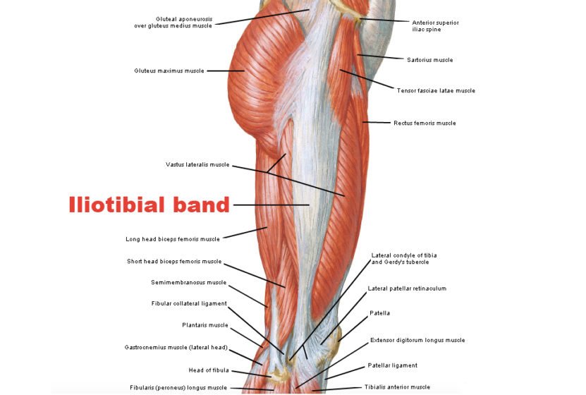
Proximally in the thigh, the ITB receives fascial contributions from the deep fascia of the thigh, gluteus maximus, and tensor fascia lata (TFL).  The TFL is the deep investing fascia of the thigh, encompassing the muscles of the hip and lower extremity around this region. Distally, the ITB becomes a distinct soft tissue layer of the lateral knee.

Regardless of the ITB condition, most patients experience complete resolution of symptoms following nonoperative management modalities alone.  However, surgical consideration is a potential option for , , or recalcitrant cases that continue to persist despite exhausting of all nonoperative management options after several months.
Iliotibial Band - Origin, Nerve Supply, Function

Structure and Function of Iliotibial Band Muscle

Origin, insertion, and structure

The detailed anatomic structure of the ITB, TFL, and its origins, insertions, and variations have been debated for decades in the literature.  Huang delineated the proximal anatomy in a recent study from 2013.

Proximal anatomy

The proximal IT tract begins as three distinct layers coursing distally to fuse/coalesce at the level of the greater trochanter (GT)

  • Superficial IT layer: 

    • Origin: (superficial to the TFL origin)
  • Intermediate IT layer:

    • Origin: Ilium (distal to the TFL origin)

      • The intermediate layer’s location is consistently deep to the TFL muscle layer
  • Deep IT layer:

    • Recognized as a constant structure
    • Portions of the deep IT layer are also confluent with the capsule itself
    • Origin: arises from the supra-acetabular fossa between the hip and the reflected head of the rectus femoris
  • TFL 

    • Separate origin of TFL fibers originates off the ilium and in between the superficial and intermediate IT layer origins
    • Distally, the TFL becomes a tendinous structure as it merges with the superficial and intermediate IT layers
    • Farther distal, the TFL tendinous fibers, including the superficial and intermediate IT layers, fuse as a single confluent structure near the level of the GT

Gluteal contributions

  • Gluteal aponeurotic fascia:

    • Originates from the posterior iliac crest
    • Courses distally to invest the anterior two-thirds of the gluteus medius
    • A portion of its fibers merge with the posterior ITB to continue distally while the remaining aponeurotic fibers insert at the gluteal tuberosity on the femur
  • In addition, the posterior ITB also receives distinct fascial/tendinous contributions from:

    • Superior gluteus maximus
    • Superficial fibers of the inferior gluteus maximus
  • Deep fibers from the inferior gluteus maximus course toward the femur to insert onto the gluteal tuberosity of the linea aspera

Distal anatomy

  • Proximal to the knee joint, the ITB attaches to the intermuscular septum and supracondylar tubercle of the femur.  Proximal to the lateral epicondyle, there is an interposed fat layer between the ITB and the vastus lateralis.
  • The ITB is more tendinous proximal to the lateral femoral epicondyle, and at the level of the epicondyle, the ITB contributes to lateral knee stability secondary to its anatomic position, intimal contact with the epicondyle, and relative to its location with respect to the lateral collateral (LCL).

Function

Proximal ITB function includes:

  • Hip extension
  • Hip abduction
  • Lateral hip rotation

Distally, ITB function depends on the position of the knee joint

  • 0 degrees/full extension to 20 to 30 degrees of flexion

    • Active knee extensor

      • The ITB lies anterior to the lateral femoral epicondyle
  • 20 to 30 degrees of flexion to full flexion ROM

    • Active knee flexor

      • ITB lies posterior relative to the lateral femoral epicondyle

Blood Supply of Iliotibial Band Muscle

The ITB, being a tendinous extension of the tensor fascia lata (TFL), shares the same arterial supply:

  • Ascending branch of the lateral femoral circumflex (LFCA)
  • Superior gluteal artery (SGA)
  • The LFCA traverses the TFL as a single branch from the profundus femoris arterial system usually 6-10 cm distal from the anterior superior iliac spine (ASIS) directly into the muscle .
  • The SGA is the largest branch of the posterior division of the internal iliac artery and exits the greater sciatic foramen where it divides into superficial and deep branches.
  • The latter of which also provides collateral arterial blood supply to the TFL in addition to the gluteus medius and minimus muscles.

Nerves

  • Analogous to the arterial blood supply, the ITB shares the innervation of the TFL and gluteus maximus via the superior gluteal nerve (SGN) and inferior gluteal nerve (IGN), respectively.
  • The SGN arises from the ventral rami of nerve roots L4-S1 and originates from the lumbosacral nerve plexus.  Like the SGA, the SGN exits the  via the greater sciatic foramen to innervate the TFL as well as the gluteus minimus and medius (both hip abductors and medial rotators). The IGN is comprised of the ventral rami of L5-S2 and provides innervation of the gluteus maximus muscle.

Muscles

  • The TFL works in concert with the gluteus medius and minimus to medially rotate and abduct the hip, as well as assist in flexing the hip with the rectus femoris. The distal attachment of the ITB on Gerdy’s tubercle serves as a focal point of lower extremity abduction.
  • The gluteus maximus primarily serves to extend the hip, however, also contributes to hip abduction via the ITB tract. Again, the ITB is an extension of the tendinous insertions of these muscles, and a better understanding of each of their function, will, in turn, improve one’s understanding of the biomechanics of the ITB.

Function

Iliotibial Band - Origin, Nerve Supply, Function

References

Doctor visit helper

Prepare before seeing a doctor

A simple rural-patient checklist to help you explain symptoms clearly, ask better questions, and avoid unsafe self-treatment.

Safety note: This is not a prescription or diagnosis. For severe symptoms, pregnancy danger signs, children with serious illness, chest pain, breathing difficulty, stroke-like weakness, or major injury, seek urgent care.

Which doctor may help?

Start with a registered doctor or the nearest qualified health center.

What to tell the doctor

  • Write when the problem started and how it changed.
  • Bring old prescriptions, investigation reports, and current medicines.
  • Write allergies, pregnancy status, diabetes, kidney/liver disease, and major past illnesses.
  • Bring one family member if the patient is weak, elderly, confused, or a child.

Questions to ask

  • What is the most likely cause of my symptoms?
  • Which danger signs mean I should go to hospital quickly?
  • Which tests are necessary now, and which can wait?
  • How should I take medicines safely and what side effects should I watch for?
  • When should I come for follow-up?

Tests to discuss

  • Vital signs: temperature, pulse, blood pressure, oxygen saturation
  • Basic physical examination by a clinician
  • CBC, urine test, blood sugar, or imaging only when clinically needed

Avoid these mistakes

  • Do not use antibiotics, steroid tablets/injections, or strong painkillers without proper medical advice.
  • Do not hide pregnancy, kidney disease, ulcer, allergy, or blood thinner use.
  • Do not delay emergency care when danger signs are present.

Medicine safety and first-aid guide

This section is for patient education only. It does not replace a doctor, pharmacist, or emergency care.

Safe first steps

  • Avoid heavy lifting, sudden bending, and prolonged bed rest.
  • Use comfortable posture and gentle movement as tolerated.
  • Discuss physiotherapy, X-ray, or MRI only when clinically needed.

OTC medicine safety

  • For mild back pain, pain-relief medicine may be discussed with a doctor or pharmacist.
  • Avoid repeated painkiller use if you have kidney disease, stomach ulcer, uncontrolled blood pressure, or are taking blood thinners.

Avoid these mistakes

  • Do not start antibiotics without a proper medical decision.
  • Do not use steroid tablets or injections casually for quick relief.
  • Do not delay emergency care because of home remedies.

Get urgent help if

  • Back pain with leg weakness, numbness around private area, loss of urine/stool control, fever, cancer history, or major injury needs urgent care.
Medicine names, dose, and timing must be decided by a qualified clinician or pharmacist after checking age, pregnancy, allergy, other diseases, and current medicines.

For rural patients and family caregivers

Patient health record and symptom diary

Write your symptoms, medicines already taken, test results, and questions before visiting a doctor. This note stays on your device unless you print or copy it.

Doctor to discuss: Doctor / qualified healthcare provider
Tests to discuss with doctor
  • Basic vital signs: temperature, pulse, blood pressure, oxygen level if needed
  • Relevant blood, urine, imaging, or specialist tests only after clinical assessment
Questions to ask
  • What is the most likely cause of my symptoms?
  • Which warning signs mean I should go to emergency care?
  • Which tests are really needed now?
  • Which medicines are safe for my age, pregnancy status, allergy, kidney/liver/stomach condition, and current medicines?

Emergency warning signs such as chest pain, severe breathing difficulty, sudden weakness, confusion, severe dehydration, major injury, or loss of bladder/bowel control need urgent medical care. Do not wait for online information.

Safe pathway to proper treatment

Care roadmap for: Iliotibial Band – Origin, Nerve Supply, Function

Use this simple roadmap to understand the next safe steps. It is educational and does not replace examination by a doctor.

Go to emergency care if you notice:
  • Severe or rapidly worsening symptoms
  • Breathing difficulty, chest pain, fainting, confusion, severe weakness, major injury, or severe dehydration
Doctor / service to discuss: Qualified healthcare provider; specialist depends on symptoms and examination.
  1. Step 1

    Check danger signs first

    If danger signs are present, seek emergency care and do not wait for online information.

  2. Step 2

    Record the symptom story

    Write when symptoms started, severity, medicines already taken, allergies, pregnancy status, and test results.

  3. Step 3

    Visit a qualified clinician

    A doctor, nurse, or qualified healthcare provider can examine you and decide which tests or treatment are needed.

  4. Step 4

    Do only useful tests

    Do tests after clinical assessment. Avoid unnecessary tests, random antibiotics, or repeated medicines without diagnosis.

  5. Step 5

    Follow up and return early if worse

    If symptoms worsen, new warning signs appear, or treatment is not helping, return for review quickly.

Rural patient practical tips
  • Take a written symptom diary and all previous prescriptions/test reports.
  • Do not hide medicines already taken, even herbal or over-the-counter medicines.
  • Ask which warning signs mean urgent referral to hospital.

This roadmap is for education. A real diagnosis and treatment plan requires history, examination, and clinical judgment.