
The muscular system is an organ system consisting of skeletal, smooth, and cardiac muscle. It permits movement of the body, maintains posture, and circulates blood throughout the body.The muscular systems in vertebrates are controlled through the nervous system although some muscles (such as the cardiac muscle) can be completely autonomous. Together with the skeletal system in the human, it forms the musculoskeletal system, which is responsible for the movement of the body.[rx]
Command to locate a specific muscle
- abductor digiti minimi (foot)
- abductor digiti minimi (hand)
- abductor hallucis
- abductor pollicis brevis
- abductor pollicis longus
- adductor brevis
- adductor hallucis
- adductor longus
- adductor magnus
- adductor pollicis
- anconeus
- articularis cubiti
- articularis genu
- aryepiglotticus
- aryjordanicus
- auricularis
- biceps brachii
- biceps femoris
- brachialis
- brachioradialis
- buccinator
- bulbospongiosus
- constrictor of pharynx -inferior
- constrictor of pharynx -middle
- constrictor of pharynx -superior
- coracobrachialis
- corrugator supercilii
- cremaster
- cricothyroid
- dartos
- deep transverse perinei
- deltoid
- depressor anguli oris
- depressor labii inferioris
- diaphragm
- digastric
- digastric (anterior view)
- erector spinae – spinalis
- erector spinae – iliocostalis
- erector spinae – longissimus
- extensor carpi radialis brevis
- extensor carpi radialis longus
- extensor carpi ulnaris
- extensor digiti minimi (hand)
- extensor digitorum (hand)
- extensor digitorum brevis (foot)
- extensor digitorum longus (foot)
- extensor hallucis longus
- extensor indicis
- extensor pollicis brevis
- extensor pollicis longus
- external oblique abdominis
- flexor carpi radialis
- flexor carpi ulnaris
- flexor digiti minimi brevis (foot)
- flexor digiti minimi brevis (hand)
- flexor digitorum brevis
- flexor digitorum longus (foot)
- flexor digitorum profundus
- flexor digitorum superficialis
- flexor hallucis brevis
- flexor hallucis longus
- flexor pollicis brevis
- flexor pollicis longus
- frontalis
- gastrocnemius
- gemellus inferior
- gemellus superior
- genioglossus
- geniohyoid
- gluteus maximus
- gluteus medius
- gluteus minimus
- gracilis
- hyoglossus
- iliacus
- inferior oblique
- inferior rectus
- infraspinatus
- intercostals external
- intercostals innermost
- intercostals internal
- internal oblique abdominis
- interossei – dorsal of hand
- interossei -dorsal of foot
- interossei- palmar of hand
- interossei – plantar of foot
- interspinales
- intertransversarii
- intrinsic muscles of tongue
- ishiocavernosus
- lateral cricoarytenoid
- lateral pterygoid
- lateral rectus
- latissimus dorsi
- levator anguli oris
- levator ani-coccygeus
- levator ani – iliococcygeus
- levator ani-pubococcygeus
- levator ani-puborectalis
- levator ani-pubovaginalis
- levator labii superioris
- levator labii superioris
- alaeque nasi
- levator palpebrae superioris
- levator scapulae
- levator veli palatini
- levatores costarum
- longus capitis
- longus colli
- lumbricals of foot (4)
- lumbricals of hand
- masseter
- medial pterygoid
- medial rectus
- mentalis
- m. uvulae
- mylohyoid
- nasalis
- oblique arytenoid
- obliquus capitis inferior
- obliquus capitis superior
- obturator externus
- obturator internus (A)
- obturator internus (B)
- omohyoid
- opponens digiti minimi (hand)
- opponens pollicis
- orbicularis oculi
- orbicularis oris
- palatoglossus
- palatopharyngeus
- palmaris brevis
- palmaris longus
- pectineus
- pectoralis major
- pectoralis minor
- peroneus brevis
- peroneus longus
- peroneus tertius
- piriformis (A)
- piriformis (B)
- plantaris
- platysma
- popliteus
- posterior cricoarytenoid
- procerus
- pronator quadratus
- pronator teres
- psoas major
- psoas minor
- pyramidalis
- quadratus femoris
- quadratus lumborum
- quadratus plantae
- rectus abdominis
- rectus capitus anterior
- rectus capitus lateralis
- rectus capitus posterior major
- rectus capitus posterior minor
- rectus femoris
- rhomboid major
- rhomboid minor
- risorius
- salpingopharyngeus
- sartorius
- scalenus anterior
- scalenus medius
- scalenus minimus
- scalenus posterior
- semimembranosus
- semitendinosus
- serratus anterior
- serratus posterior inferior
- serratus posterior superior
- soleus
- sphincter ani
- sphincter urethrae
- splenius capitis
- splenius cervicis
- stapedius
- sternocleidomastoid
- sternohyoid
- sternothyroid
- styloglossus
- stylohyoid
- stylohyoid (anterior view)
- stylopharyngeus
- subclavius
- subcostalis
- subscapularis
- superficial transverse
- perinei
- superior oblique
- superior rectus
- supinator
- supraspinatus
- temporalis
- temporoparietalis
- tensor fasciae lata
- tensor tympani
- tensor veli palatini
- teres major
- teres minor
- thyro-arytenoid & vocalis
- thyro-epiglotticus
- thyrohyoid
- tibialis anterior
- tibialis posterior
- transverse arytenoid
- transversospinalis -multifidus
- transversospinalis -rotatores
- transversospinalis -semispinalis
- transversus abdominis
- transversus thoracis
- trapezius
- triceps
- vastus intermedius
- vastus lateralis
- vastus medialis
- zygomaticus major
- zygomaticus minor
Head and Neck Muscles
Facial Muscles
The human face is composed of multiple muscles that control the fine movements that produce facial expressions.
LEARNING OBJECTIVES
Characterize the muscles involved in creating facial expressions
KEY TAKEAWAYS
Key Points
- The human face is composed of numerous muscles that control fine movement to produce facial expressions.
- Unlike other muscles, these muscles originate on bone or fascia of the face and attach directly to the skin, allowing it to be manipulated.
Key Terms
-
depressor labii inferioris: An analogous muscle that lowers the bottom lipEndFragment
-
Buccinator: This muscle is located between the upper and lower jaws in the cheek, deep to the other muscles of the face.
-
zygomatic: This muscle controls the cheeks to create smiles and frowns.
-
Procerus: The most superior of all facial muscles.
-
depressor anguli oris: This muscle is opposite to the levator anguli oris and pulls the corners of the mouth downward, producing a frown.
-
levator labii superiori: Abroad muscle responsible for elevation of the upper lip.
-
Orbicularis Oris: Muscle fibers that enclose the opening to the oral cavity.
-
Nasalis: The largest of the nasal muscles. It is split into two sections: alar and transverse.
-
risorius: This muscle pulls the mouth back mimicking a smile, but does not affect the skin around the smile.
-
Corrugator Supercilii: A small muscle located superiorly to the orbicularis oculi.
-
Orbicularis Oculi: A thin muscle that surrounds the eye socket.
-
mentalis: This muscle pushes the lower lip uppers and wrinkles the chin.
-
levator labii superioris alaeque nasi: The muscle of the upper lip. It acts to lift the upper lift and dilates nostril, producing a snarling expression.
The human face is composed of numerous muscles that control fine movement to produce facial expressions. Unlike other muscles, these muscles originate on the bone or fascia of the face and attach directly onto the skin, allowing it to be manipulated. The facial muscles can be split into three groups: orbital, nasal and oral.
Orbital Group

Schematic of head and neck muscles.: Locations of facial muscles noted.
- Orbicularis Oculi: A thin muscle that surrounds the eye socket.
- Attachments: Originates from the skull around the eye socket and attaches to ligaments found in the eyelid.
- Actions: Closes the eyelid.
- Corrugator Supercilii: A small muscle located superiorly to the orbicularis oculi.
- Attachments: Originates from the bone below the eyebrow and attaches to the skin directly above.
- Actions: Draws the eyebrows together.
Nasal Group
The nasal group of muscles are associated with movements of the nose and surrounding skin.
- Nasalis: The largest of the nasal muscles. It is split into two sections: alar and transverse.
- Attachments: Originates from the upper jaw. The alar section attaches to the cartilage of the nose and the transverse section to an aponeurosis covering the bridge of the nose.
- Actions: The transverse section closes the nostrils and the alar part opens them.
- Procerus: The most superior of all facial muscles.
- Attachments: Originates from the nasal bone attaching to the skin of the forehead.
- Actions: Pulls the eyebrows down.
Oral Group

Location of the buccinator muscle: Highlighted in orange, the buccinator is associated with the cheeks directly lateral to the mouth
Muscles of the oral group play key roles in respiration, communication, eating, and drinking. The lips in particular are controlled by numerous small muscles.
- Orbicularis Oris: Muscle fibers that enclose the opening to the oral cavity.
- Attachments: Originates from the upper jaw and muscles of the cheek and attaches to the lips.
- Action: Purses the lips.
- Buccinator: This muscle is located between the upper and lower jaws in the cheek, deep to the other muscles of the face.
- Attachments: It originates from upper and lower jaw and attaches to the lips and orbicularis oris.
- Actions: The buccinator pulls the cheek inwards.
Other Muscles of the Mouth
- The levator labii superioris alaeque nasi is the muscle of the upper lip. It acts to lift the upper lift and dilates nostril, producing a snarling expression.
- The levator anguli oris (caninus) inserts at the corners of the mouth at an angle, and is associated with other muscles including the zygomaticus, triangularis, and orbicularis oris. When innervated, this muscle contracts to lift the corners of the mouth, producing part of the expression of a smile.
- The depressor anguli oris (triangularis) is also associated with the corners of the mouth. Located opposite to the levator anguli oris, it pulls the corners of the mouth downward, producing a frown.
- The levator labii superioris is a broad muscle responsible for elevation of the upper lip. This muscle originates at the side of the nose and has several insertion points on either side of the nose, extending down to the lip, and inserting at both lateral and frontal portions of the upper lip.
- The depressor labii inferioris is an analogous muscle that lowers the bottom lip.
- The zygomatic muscle, associated with the cheeks, is divided into two parts: the major and the minor. The zygomaticus major draws the mouth upward and outward to generate a smile. The zygomaticus minor inserts into the outer part of the upper lip, not the angle of the mouth as with the major. When innervated, contraction of this muscle draws the lip backward, upward, and outward and is associated with facial expressions conveying sadness.
- The mentalis, associated with the tip of the chin, is a paired muscle. Sometimes referred to as the pouting muscle, contraction of the mentalis causes the lower lip to be pushed upwards and wrinkles the chin.
- The risorius muscle is lateral to the orbicularis oris and inserts into the angle of the mouth. When innervated, the risorius pulls the mouth back mimicking a smile, but does not affect the skin around the smile As a result, this facial expression is often interpreted as insincere.
Chewing Muscles
Mastication, or chewing, involves the adduction and lateral motions of the jaw bone. It is controlled by four muscles of the face.
LEARNING OBJECTIVES
Differentiate between the actions of the masseter and the temporalis muscles in chewing
KEY TAKEAWAYS
Key Points
- The masseter elevates the jaw, closing the mouth.
- The temporalis elevates and retracts the jaw.
- The lateral pterygoid is the only muscle of mastication that actively opens the jaw. Unilateral action of a lateral pterygoid produces lateral movement in the jaw, usually performed in concert with the medial pterygoids.
- The medial pterygoid elevates and closes the jaw, contributes to protrusion of the mandible, and assists in mastication.
Key Terms
-
medial pterygoid: A muscle of mastication with two heads. It lies inferiorly to the medial pterygoid.
-
temporalis: A broad muscle that fans out to cover much of the temporal bone on the side of the skull.
-
lateral pterygoid: A muscle of mastication with two heads. It lies superiorly to the medial pterygoid.
-
masseter: The large muscle which raises the lower jaw, and assists in mastication.
Mastication, or the act of chewing, involves adduction and lateral motion of the jaw bone. It is controlled by four bilateral muscles in the face. The lower jaw, or mandible, connects to the temporal bone of the skull via the temporomandibular joint, which allows movement in all planes.

Location of the masseter muscle: The masseter muscle spans the opening of the mouth.
- Masseter: The most powerful muscle of mastication. It is quadrangular in shape and split into two regions, deep and superficial. It covers the other muscles of mastication.
- Attachments: The superficial region originates from the skull below the eye socket, while the deep part originates from the skull above the jaw. Both parts attach to the jaw.
- Actions: Elevates the jaw.
- Temporalis: A broad muscle that fans out to cover much of the temporal bone on the side of the skull.
- Attachments: The temporalis muscle has a wide, fan-shaped origin on the side of the skull and condenses into a tendon which attaches to the jaw.
- Actions: Elevates and retracts the jaw.
- Lateral Pterygoid: The lateral pterygoid muscle has a triangular shape with two head, superior and inferior. It is the major protractor (opener) of the jaw.
- Attachments: Both heads originate adjacent to the eye socket and converge into a tendon which attaches to the jaw.
- Actions: Together, the lateral pterygoids protract the jaw, working independently to produce lateral movement.
- Medial Pterygoid: The medial pterygoid muscle has a quadrangular shape with two heads, deep and superficial. It is located inferior to the lateral pterygoid.
- Attachments: The superficial head originates from the front of the skull and the deep head attaches to the skull adjacent to the eye socket. Both heads attach to the jaw.
- Actions: Elevates the jaw and assists in the production of lateral movement.
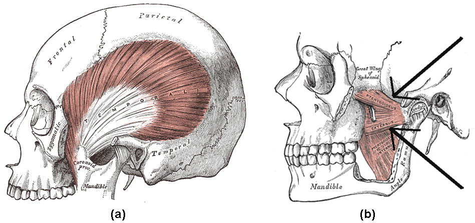
Location of the temporalis muscle and the lateral pterygoid: (a) Highlighted in orange, the temporalis muscle is a broad muscle extending from zygomatic bone. (b) Arrows indicate the location of the lateral pterygoid, highlighted with the medial pterygoid in orange.
KEY MOVEMENTS
- Elevation of the Jaw: Produced by the masseter, temporalis and medial pterygoid.
- Depression of the Jaw: Produced by the lateral pterygoid, assisted by the digastric, mylohyoid and geniohyoid muscles found in the neck.
- Protraction of the Jaw: Produced by the lateral pterygoid.
- Retraction of the Jaw: Produced by the temporalis.
- Lateral Movement of the Jaw: Produced by the lateral and medial pterygoid.
Neck Muscles
Cervical muscles are those associated with the front of the neck; vertebral muscles are associated with the vertebral column.
LEARNING OBJECTIVES
Outline the neck muscles and their movements
KEY TAKEAWAYS
Key Points
- Numerous muscles contribute to the processes of speaking and swallowing.
- These muscles can be divided into suprahyoid and ingrahyoid groups based on their locations relative to the hyoid bone.
- The hyoid bone, located beneath the mandible, acts as a key attachment point for muscles involved in speaking and swallowing.
- Numerous muscles contribute to both the stabilization and fine movements of the head and neck.
Key Terms
-
suprahyoid muscles: A group of muscles located above the hyoid bone, responsible for its elevation which widens the esophagus.
-
hyoid bone: A -shaped bone which sits below the mandibleand in front of the esophagus, facilitating the wide range of movements associated with speaking and swallowing.
-
infrahyoid muscles: A group of muscles located below the hyoid bone, responsible for its depression which narrows the esophagus.
Muscles of the neck play important roles in mastication (chewing), swallowing, speaking and supporting and moving the head. All muscles found in the neck are paired, meaning they exist to both the left and right side of the spine. The muscles involved in chewing have been discussed previously so only those involved in swallowing and support will be discussed below.
Muscles Involved in Swallowing and Speaking
Located to the anterior of the neck, these muscles are split into two based on their location relative to the hyoid bone. The U-shaped hyoid bone sits below the mandible and in front of the esophagus, providing a level of protection and facilitating the wide range of muscle activity required for speaking and swallowing.
Suprahyoid Muscles

Suprahyoid and infrahyoid muscles of the neck: Suprahyoid and infrahyoid muscle groups are named based on their location relative to the hyoid bone. The hyoid bone sits below the mandible and in front of the esophagus, providing a level of protection but also facilitating the wide range of muscle activity required for speaking and swallowing.
The four suprahyoid muscles found above the hyoid bone act in concert to elevate the hyoid bone, assisting with swallowing by widening the esophagus.
- Stylohyoid: The most superior of the suprahyoid muscles, the stylohyoid originates from the skull and attaches to the hyoid bone.
- Digastric: The digastric muscle is split into two parts that are connected by a tendon attached to the hyoid bone. The anterior section originates from the mandible and the posterior section from the skull.
- Mylohyoid: The mylohyoid is a broad flat muscle which forms the floor of the oral cavity. It originates from the mandible and attaches to the hyoid bone.
- Geniohyoid: The deepest of the suprahyoid muscles, the geniohyoid muscle originates from the mandible and attaches to the hyoid bone.
Infrahyoid Muscles
The four infrahyoid muscles found below the hyoid bone act in concert to depress the hyoid bone during swallowing and speaking, compressing the esophagus.
- Sternohyoid: A superficial muscle which originates from the sternum and attaches onto the hyoid bone.
- Omohyoid: Located laterally to the sternohyoid, the omohyoid muscle is split in two parts attached by a tendon. The inferior region originates from the scapula, joins the superior region, and attaches to the hyoid bone.
- Sternothyroid: Sitting deeper than the sternohyoid, the sternothyroid originates from the sternum and attaches to the thyroid cartilage associated with the hyoid bone.
- Thyrohyoid: A short continuation of the sternothyroid muscle, the thyrohyoid originates from the thyroid cartilage and attaches to the hyoid bone.
Muscles of the Back and Neck

Muscles of the back and neck: Muscles of the back and neck play an important role in maintaining posture and the movement of the head and neck.
The muscles of the back and neck are responsible for maintaining posture and facilitating movement of the head and neck. They are divided into three layers.
Superficial Layer
Two muscles in the superficial layer are responsible for rotation of the head.
- Splenius Capitis: A thick rectangular muscle, the most superior of the neck muscles.
- Attachments: Originates from the upper spine and attaches to the skull.
- Actions: Rotates and extend the head and neck.
- Splenius Cervicis: A small triangular-shaped muscle located immediately below the splenius capitis.
- Attachments: Originates from the spine and attaches several vertebrae higher.
- Actions: Rotate and extend the head and neck.
Intermediate Layer
Three columnar muscles in the intermediate layer are responsible for flexion and extension of the neck as well as posture maintenance. All three originate from a common tendon associated with the pelvis and can be divided into thoracic, cervicis, and capitis regions.
- Iliocostalis: The most laterally located of the three intermediate muscles.
- Attachments: Originates from the common tendon and attaches to the ribs and lower neck.
- Actions: Extends and controls abduction and adduction of the spine and neck.
- Longissimus: Located between the iliocostalis and spinalis muscles, this is the largest of the intermediate layer muscles.
- Attachments: Originates from the common tendon and attaches to the lower ribs, the spine, and the skull.
- Actions: Extends and controls abduction and adduction of the spine and neck.
- Spinalis: The most medially located and smallest of the three intermediate layer muscles.
- Attachments: Originates from the common tendon and attaches to the upper spine and skull.
- Actions: Extends and flexes to control abduction and adduction of the spine and neck.
Deep Layer
Two muscles in the deep layer are responsible for maintenance of posture and rotation of the neck.
- Semispinalis: The semispinalis is the most superficial of the deep muscles.
- Attachments: A broad origin on the upper regions of the spine, with each origin attaching several vertebrae higher or to the skull.
- Actions: Extends and rotates the head and maintains posture.
- Multifidus: The multifidus is located underneath the semispinalis muscle and is key in maintaining posture.
- Attachments: A broad origin up the length of the spine, with each origin attaching several vertebrae higher.
- Actions: Maintains posture through the spine.
Other Muscles That Act on the Neck
Several other muscles act on the head and neck. Below are three with a larger impact.
- Trapezius: The trapezius is the most superficial muscle of the back and forms a broad flat triangle.
- Attachments: The trapezius originates from the skull and spine of the upper back and neck. It attaches to the clavicle and scapula.
- Actions: The superior region supports the arm and elevates and rotates the scapula. It controls adduction, abduction and rotation of the head, the intermediate region retracts the scapula, and the inferior region rotates and depresses the scapula.
- Sternocleidomastoid: A thick rectangular muscle that is responsible for many movements within the neck.
- Attachments: Dual-headed, the sternocleidomastoid originates from the clavicle and the sternum and attaches to the mandible.
- Actions: Abduction, adduction, extension, flexion, and rotation of the neck depending on intra and inter-muscle contractions.
- Platysma: A broad sheet of muscle arising from the fascia covering the pectorals.
- Attachments: Originates from the fascia covering the pectorals and attaches to various locations within the mandible and dermis of the face and neck.
- Actions: Depresses the mandible and angles the lip and mouth, wrinkling the skin upon the neck flexing.
KEY MOVEMENTS
- Extension (tilting head backwards): Produced by the semispinalis, splenus capitis, longissimus, trapezius (superior fibers), and sternocleidomastoid (posterior fibers).
- Flexion (tilting head forwards): Produced by the sternocleidomastoid (anterior fibers).
- Abduction (tilting head towards shoulder): Produced by the sternocleidomastoid, longissimus, splenius capitis, semispinalis, and trapezius (superior fibers)
- Adduction (returning head to midline): Produced by the sternocleidomastoid, longissimus, splenius capitis, semispinalis, and trapezius (superior fibers)
- Rotation (rotation head to left or right): Produced by the sternocleidomastoid, longissimus, splenius capitis, semispinalis, and trapezius (superior fibers)

