
Coxa Saltans/Snapping hip syndrome also known as coxa saltans (or dancer’s hip) is a clinical condition characterized by an audible or palpable snapping sensation that is heard during movement of the hip joint. Snapping hip has multiple etiologies and is classified based on the anatomic structure that is the cause/source of the snapping sensation. Historically, two main snapping hip categories were recognized: The term intra-articular snapping hip has relatively fallen out of favor due to the increasing knowledge of intra-articular pathologies. These etiologies include loose bodies (e.g. secondary to synovial chondromatosis) or labral tears of the hip.
Snapping hip syndrome, also known as coxa saltans (or dancer’s hip), is a clinical condition characterized by an audible or palpable snapping sensation that is heard during movement of the hip joint. Snapping hip has multiple etiologies and is classified based on the anatomic structure that is the cause/source of the snapping sensation.
Snapping hip syndrome, also referred to as a dancer’s hip is a medical condition characterized by a snapping sensation felt when the hip is flexed and extended. This may be accompanied by a snapping or popping noise and pain or discomfort. Pain often decreases with rest and diminished activity. Snapping hip syndrome is commonly classified by the location of the snapping as either extra-articular or intra-articular.
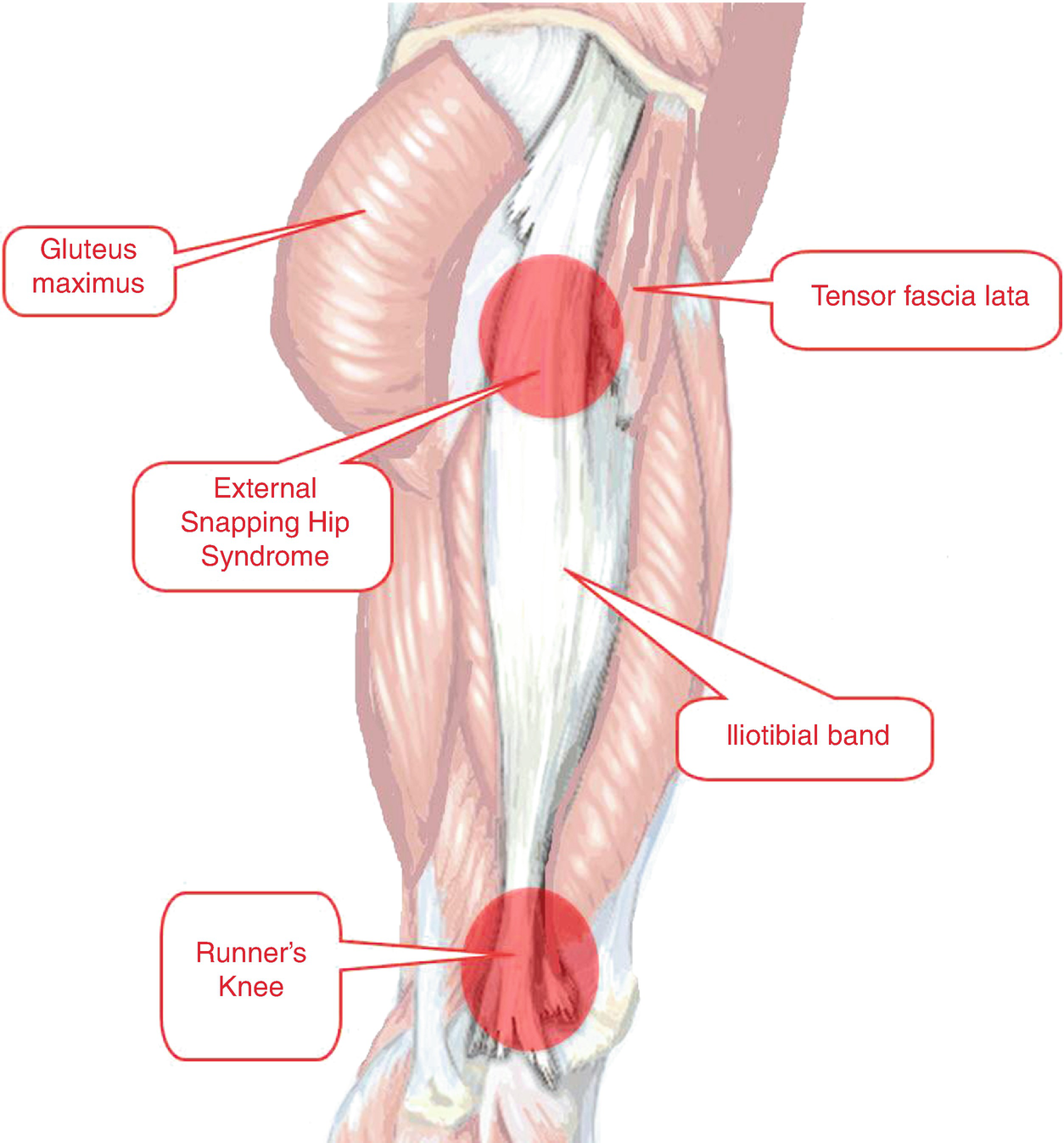
Types of Snapping Hip Syndrome
As the clinical terminology evolved, extra-articular snapping hip eventually became subclassified into:
- External snapping hip – is most commonly attributed to the iliotibial band moving over the greater trochanter of the femoral head during hip movements in flexion, extension, and external or internal rotation. Other causes include the proximal hamstring tendon rolling over the ischial tuberosity, either the fascia lata or the anterior aspect of the gluteus maximus rolling over the greater trochanter, and the psoas tendon rolling over the medial fibers of the iliacus muscle. A combination of defects is also possible; for example, the thickening of both the posterior iliotibial band and anterior gluteus maximus, which snap over the greater trochanter at the same time. [rx][rx]
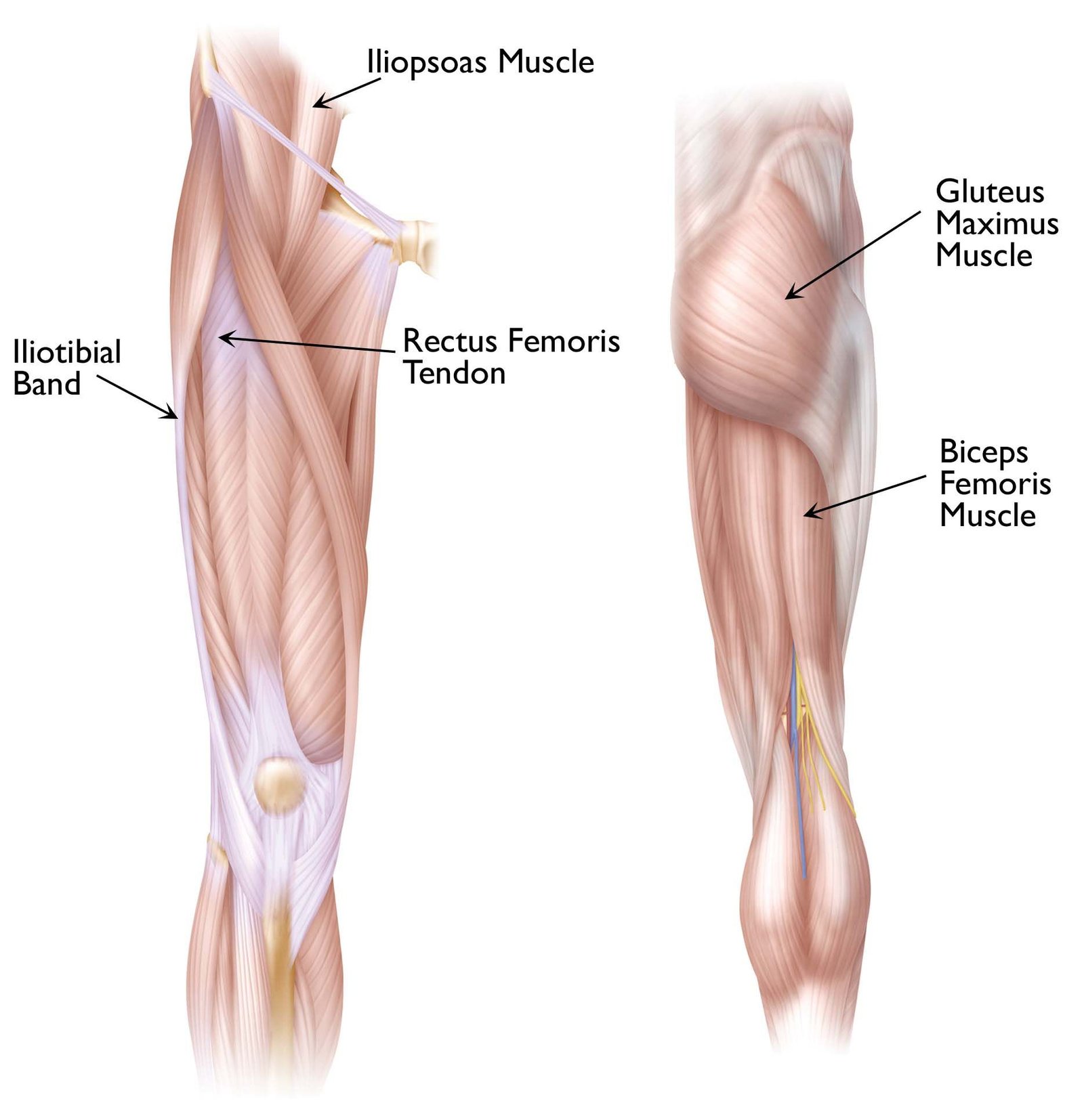
- Internal snapping hip – most commonly occurs as the iliopsoas tendon snaps over underlying bony prominences, such as the ilio pectinal eminence or the anterior aspect of the femoral head. Other causes include paralabral cysts and partial or complete bifurcation of the iliopsoas tendon. The snapping sensation can closely mimic intra-articular pathology since they both originate from the anterior hip area. Close physical exam and imaging can differentiate the two. It should be noted, however, that in approximately 50% of internal snapping hip cases, an additional intra-articular hip pathology is identified.
- Intra-articular – The term intra-articular snapping hip has relatively fallen out of favor due to the increasing knowledge of intra-articular pathologies. These etiologies include loose bodies (e.g. secondary to synovial chondromatosis) or labral tears of the hip.[rx][rx][rx] In this category, a snapping hip is caused by an actual hip joint issue or injury. Unlike external or internal SHS, intra-articular SFS isn’t caused by a tendon or muscle.
Causes of Snapping Hip Syndrome/Coxa Saltans
Snapping hip is most commonly
- The snapping muscle or tendon becomes irritated and inflamed – During snapping, the muscle or tendon is uncomfortably stretched and strained as it pulls over a bony protrusion and then snaps as the tension is released. Done repeatedly over time, this process can lead to muscle or tendon irritation, damage and pain.
- The snapping leads to painful hip bursitis – The snapping tendon or muscle can irritate a nearby bursa, a lubricating, fluid-filled structure that normally reduces friction between bone and soft tissue. This irritation can cause a bursa to become inflamed, a condition called hip bursitis.
- Overuse phenomenon – but may be precipitated by trauma, including intramuscular injection into the gluteus maximus and surgical procedures. For example, the smaller median femoral neck angle (i.e., coxa vera) following total hip arthroplasty specifically has been linked to the development of external snapping hip syndrome.
- Other anatomical variations – may predispose to coxa saltans including an increased distance between the greater trochanters, prominent greater trochanters, and narrow bi-iliac width.
- Additionally, iliotibial band tightness, shorter muscle or tendon lengths, muscle tightness, or inadequate relaxation of the muscles may contribute to snapping hip development. Sometimes no etiology is uncovered at all, resulting in an idiopathic classification.[rx]
- External snapping hip syndrome most commonly is caused by the iliotibial band snapping over the greater trochanter of the femoral head during movements such as flexion, extension, and external or internal rotation.
- Internal snapping hip is most commonly caused by the iliopsoas tendon snapping over underlying bony prominences, such as the iliopectinal eminence or the anterior aspect of the femoral head.
Symptoms of Snapping Hip Syndrome/Coxa Saltans
- The area over the greater trochanteric region may be painful due to greater trochanter bursitis, abductor tendon pathology, or inflammation of the iliotibial band.
- Pain
- Inflammation
- Leg muscle weakness when trying to lift your leg sideways or forward
- Swelling
- Difficulty with regular physical activity such as walking or rising from your chair
- Feeling your hip is coming out of place
Diagnosis of Snapping Hip Syndrome
History and Physical
History and physical exam often can help pinpoint the correct anatomic region of the snapping. The patient often can point with one finger to the area that is painful upon snapping and may even be able to recreate the snap for you. Symptoms develop and increase over a long period of time, typically months to years.
- External snapping hip – usually is more obvious on clinical exam, with the patient experiencing a snapping or sensation of subluxation of the hip (i.e., pseudo subluxation). Sometimes one can even visualize or palpate the snapping phenomenon under the patient’s skin.
- Ober test – Tests to provoke the snap usually include femoral rotation and or flexion. To examine the hip, the patient is placed in a lateral position, and the Ober test is done to test for iliotibial band tightness. While the patient is in this same position, the knee and hip can be cycled through flexion and extension to provoke the snapping.
- Internal snapping hip test – is usually described by the patient as a snapping or locking of the hip with an audible snap. Weakness in the gluteus medius is sometimes found as well. To examine the hip, the patient is placed in the supine position with the affected hip guided by the examiner into an externally rotated and flexed position. Then, from this position, the leg is extended into an anatomically neutral position next to the other resting leg. This test is positive if the snapping is reproduced at the anterior hip.
- Hula-Hoop test – Adduction with circumduction of the affected hip is done to test for external snapping hip syndrome and snap over greater trochanter is a positive sign.
- FABER test – can be used to differentiate between internal (iliopsoas) or external (ITB) generated snapping hip. By placing the affected hip into the FABER (flexion, abduction, external rotation) and passively moving the hip into an extended, adducted and internally rotated position, a palpable or audible snap may be heard.
- The Stinchfield test – The patient lies supine with the hip flexed at 30° and ask the patient to fully flex the hip, while the examines apply a resistance force. Pain in the anterior groin indicates a positive test.
Imaging
- Plain radiographs – usually are of little use to confirm the diagnosis; however, they should be done to rule out anatomical variations, developmental dysplasia, or other hip pathology. Additionally, a positive response to anesthetic joint injection in the affected area can help distinguish between external and internal snapping hip syndrome.[[rx][rx] Clinically visible external snapping hip syndrome can be confirmed on the T1 weighted axial as a thickened iliotibial band or thickened anterior edge of the gluteus maximus muscle.
- Dynamic ultrasonography – If the snapping is not visible on physical exam, dynamic ultrasonography can be used to demonstrate the snapping of the iliotibial band over the greater trochanter. Dynamic ultrasonography also can reveal associated tendonitis, iliopsoas bursitis, or muscle tears.
- MRI – A diagnosis of internal snapping hip syndrome can be confirmed using the iliopsoas biography combined with fluoroscopy, dynamic ultrasonography, magnetic resonance imaging, or magnetic resonance arthrography.
- Magnetic resonance arthrography – is preferred because it also can detect intra-articular hip pathology, which commonly accompanies internal snapping hip syndrome.
Treatment of Snapping Hip Syndrome
When pain is not present, treatment is not warranted. When pain is present upon snapping, treatment is conservative and consists of rest, stretching, steroid injections, oral anti-inflammatory medications, physical therapy, and activity modification. Most of the time, patients experience relief with these measures.
Nonsurgical Treatments
Below are non-surgical treatment typically recommended to alleviate pain caused by snapping hip syndrome:
- Protection – is meant to prevent further injury. For example, an injured leg or foot may be protected by limiting or avoiding weight-bearing through the use of crutches, a cane, or hiking poles. Partially immobilizing the injured area by using a sling, splint, or brace may also be a means of protection.
- Rest – People with snapping hip syndrome are advised to avoid the motions that cause the snapping, popping or clicking sensation. Resting limits joint irritation and allows the affected tendon, muscle or bursa to heal. If walking initiates inner snapping hip, the patient may be advised to walk with the affected leg rotated out a bit, as this may minimize hip snapping.
- Ice – refers to the use of cold treatments, also known as cryotherapy, to treat acute injuries. Ice is recommended with the intent to minimize and reduce swelling as well as to decrease pain. There are many ways to employ cryotherapy at home. The most common and most convenient is a simple plastic bag of crushed ice placed over a paper towel on the affected area. It is important to protect the skin and limit the cold exposure to 10 to 15 minutes. Cycles of 10 to 15 minutes on and 1 to 2 hours off are generally agreed upon as effective and safer than longer periods of continuous ice application.
Physical therapy
A licensed physical therapist can help loosen tension and encourage healing in the muscles and tendons that cause external and inner snapping hip. A physical therapist may employ:
- Stretching – People with external hip snapping may benefit from stretching the IT band. People with inner hip snapping may benefit from stretching the hip flexors.
- Massage – Just as stretching can help loosen tendons and muscles, a deep tissue massage or trigger-point massage can help reduce muscle tension, and reduce snapping hip symptoms.
- Retraining – Increasing an athlete’s physical awareness, improving posture, and modifying form can change the hip’s biomechanics and improve symptoms.
- Ultrasound – Tendon and muscle healing may get a boost from ultrasound, which uses high-frequency sound waves to stimulate soft tissues below the skin.
- Iontophoresis – This treatment uses a mild electrical current to administer an anti-inflammatory medicine (e.g. dexamethasone) through healthy skin and into the sore area. A doctor or physical therapist may recommend iontophoresis to a patient who does not tolerate injections or wants to avoid injections.
Snapping Hip Syndrome Stretches
Recommended exercises to treat snapping hip syndrome will vary depending on the type of snapping hip syndrome you have. Snapping hip syndrome stretches may include:
- Quadriceps stretch – Standing arm’s length from a wall, place the hand opposite the painful hip against the wall for support. With your other hand, take hold of the ankle of the painful leg and, keeping your knees together, pull your ankle up toward your buttocks. Hold for 30 to 60 seconds and release. Repeat three times.
- Hamstring stretch – Lie flat on your back on the floor in a doorway so that your upper body is on one side of the doorway and your lower body is on the other and the painful hip is against the door frame. Raise the painful leg and rest it against the wall next to the door frame. Hold for 30 to 60 seconds and then lower. Repeat three times.
- Iliotibial band stretch – Standing with your legs together, cross your uninjured leg in front of the painful leg, then bend down and touch your toes. Hold for 30 seconds, then return to starting position. Repeat three times.
- Iliotibial band stretch
- Stand next to a wall for support
- Cross the leg that is closest to the wall behind your other leg.
- Lean your hip toward the wall until you feel a stretch at the outside of your hip. Hold the stretch for 30 seconds.
- Repeat on the opposite side.
- Perform 2 to 3 sets of 4 repetitions on each side.
- Piriformis stretch – Lying on your back with both knees bent, place the foot of your uninjured leg flat on the floor and rest the ankle of your painful leg over the knee of your uninjured leg. Take hold of the thigh of the uninjured leg and pull the knee toward your chest. Hold for 30 to 60 seconds and release. Repeat three times.
- Lie on your back with bent knees and feet flat on the floor.
- Cross the foot of the affected hip over the opposite knee and clasp your hands behind your thigh.
- Pull your thigh toward you until you feel the stretch in your hip and buttocks. Hold the stretch for 30 seconds.
- Repeat on the opposite side.
- Perform 2 to 3 sets of 4 repetitions on each side.
Medication
If pain is severe and intolerable following medicine may be considered to prescribe to control pain and healing.
- COX-2 inhibitors – Taking non-steroidal anti-inflammatory drugs (NSAIDs) or COX-2 inhibitors for a limited period of time may ease inflammation and hip pain. or
- NSAIDs – Prescription-strength drugs that reduce both pain and inflammation. Pain medicines and anti-inflammatory drugs help to relieve pain and stiffness, allowing for increased mobility and exercise. There are many common over-the-counter medicines called non-steroidal anti-inflammatory drugs (NSAIDs). They include and Ketorolac, Aceclofenac, Naproxen, Etoricoxib.
- Corticosteroids – Also known as oral steroids, these medications reduce inflammation.
- Muscle Relaxants – These medications provide relief from associated muscle spasms.
- Neuropathic Agents – Drugs(pregabalin & gabapentin) that address neuropathic—or nerve-related—pain. This includes burning, numbness, and tingling.
- Opioids – Also known as narcotics, these medications are intense pain relievers that should only be used under a doctor’s careful supervision.
- Topical Medications – These prescription-strength creams, gels, ointments, patches, and sprays help relieve pain and inflammation through the skin.
- Calcium & vitamin D3 – to improve bone health and healing fracture. As a general rule, men and women age 50 and older should consume 1,200 milligrams of calcium a day, and 600 international units of vitamin D a day.
- Dietary supplement-to remove general weakness & improved health.
- Antidepressants – A drug that blocks pain messages from your brain and boosts the effects of endorphins (your body’s natural painkillers).
- Glucosamine & Diacerein, Chondroitin sulfate – can be used to tightening the loose tension, cartilage, ligament, and cartilage, ligament regenerates cartilage or inhabits the further degeneration of cartilage, ligament. They are structural components of articular cartilage, and the thought is that a supplement will aid in the health of articular cartilage.
- Steroid injections – A physician may recommend a steroid injection if the pain from the snapping hip syndrome interferes with daily living. Using medical imaging (e.g. ultrasound) a steroid injection can be made into the hip bursa or the thin sheath that surrounds the tendon. (Injections are not made directly into the tendon, because corticosteroids can weaken tendons).
Surgical Treatment
For external snapping hip syndrome, loosening of the iliotibial band is usually the goal and can be accomplished with either open or arthroscopic procedures. The iliotibial tendon is lengthened or completely released using various procedures including formal Z-lengthening, a cross-shaped release, a Z-shaped release, or a gluteus maximus release. Weakness in abduction may be a complication if the release is excessive or there is damage to the surrounding area.[rx]
- Iliotibial band release – This procedure involves lengthening your iliotibial band to reduce tension and hip snapping. It’s recommended for people experiencing pain from external SHS.
- Arthroscopic hip debridement – With this surgical procedure, your doctor will make a small incision at the hip joint cavity to remove debris. This procedure is most appropriate for those with intra-articular SHS.
- Iliopsoas tendon release – is sometimes recommended for people who have persistent and painful inner hip snapping syndrome. During surgery the physician makes strategic incisions in the iliopsoas tendon to lengthen it, reducing tension and the likelihood of hip snapping.
- Arthroscopic hip debridement – may be appropriate if the snapping hip is caused by a cartilage injury that has resulted in loose pieces of cartilage caught in the hip’s ball and socket joint. The surgeon makes a small incision to enter the hip joint cavity and remove debris.
- Acetabular labrum repair – may be recommended if x-rays and MRI show evidence of a tear to the ring of cartilage around the hip’s socket, or acetabulum. This surgery is typically done arthroscopically so that only small incisions are needed.
External
- Z-plastic of the iliotibial tract
- Resection of the posterior half of the iliotibial tract
- Elliptical resection of a portion of the iliotibial band
Internal
- Lengthening of the iliopsoas tendon
- Resection of the bony prominence of the lesser trochanter
- A complete release of the iliopsoas tendon
Intra-articular
- Cause not as clear due to possible involvement of many different intra-articular lesions that can cause symptoms
- Correction of intra-articular pathologies
Physical Therapy Exercise
Some common exercises to treat SHS include:
Iliotibial band stretch
- Lean sideways against a wall, standing on the leg with the affected hip. This leg should be closest to the wall.
- Cross your opposite leg in front of the affected leg.
- Lean away from the wall, gently stretching your hip.
- Hold this stretch for 15 to 30 seconds.
- Repeat two to three times.
Hip flexor stretch
- Kneel on your affected leg, with the opposite leg in front.
- With your back straight, push your hips forward.
- Stretch in this position until you feel slight tension in the upper thigh of your affected leg and your hip.
- Hold this stretch for 15 to 30 seconds.
- Repeat two to three times.
Lying-down hamstring stretch
- Lie flat on the floor.
- Lift your affected leg in the air perpendicular to your body.
- Gently pull your leg toward your body until you feel slight tension in the back of your thigh.
- Hold this stretch for 30 seconds.
- Repeat two to three times.
Bridges
- Lie on your back with both knees bent at 90 degrees.
- Lift your hips off of the floor until your shoulders, hips, and knees are in a straight line.
- Hold this stretch for about six seconds.
- Slowly lower your hips back down to the floor.
- Repeat 8 to 10 times.
Clamshell
- Lie on your side with the affected leg on top.
- Keep your legs together and bend your knees.
- Raise your top knee, keeping your feet together. Your legs should resemble a clamshell when opened.
- Hold this stretch for about six seconds.
- Slowly lower your knee back down.
- Repeat 8 to 10 times.
Rehabilitation and How to do the exercises
Iliotibial band stretch

- Lean sideways against a wall. If you are not steady on your feet, hold on to a chair or counter.
- Stand on the leg with the affected hip, with that leg close to the wall. Then cross your other leg in front of it.
- Let your affected hip drop out to the side of your body and against the wall. Then lean away from your affected hip until you feel a stretch.
- Hold the stretch for 15 to 30 seconds.
- Repeat 2 to 4 times.
Hip flexor stretch (kneeling)

- Kneel on your affected leg, and bend your good leg out in front of you, with that foot flat on the floor. If you feel discomfort in the front of your knee, place a towel under your knee.
- Keeping your back straight, slowly push your hips forward until you feel a stretch in the upper thigh of your back leg and hip.
- Hold the stretch for at least 15 to 30 seconds.
- Repeat 2 to 4 times.
Piriformis stretch

- Lie on your back with both knees bent and your feet flat on the floor.
- Put the ankle of your affected leg on your opposite thigh near your knee.
- Use your hands to gently lift the knee of your good leg off the floor. Gently pull that knee toward your chest until you feel a stretch in the buttock and hip of your affected leg.
- Hold the stretch for at least 15 to 30 seconds.
- Repeat 2 to 4 times.
Hamstring stretch (lying down)

- Lie flat on your back with your legs straight. If you feel discomfort in your back, place a small towel roll under your lower back.
- Holding the back of your affected leg for support, lift that leg straight up and toward your body until you feel a stretch at the back of your thigh.
- Hold the stretch for at least 30 seconds.
- Repeat 2 to 4 times.
Bridging

- Lie on your back with both knees bent. Your knees should be bent about 90 degrees.
- Then push your feet into the floor, squeeze your buttocks, and lift your hips off the floor until your shoulders, hips, and knees are all in a straight line.
- Hold for about 6 seconds as you continue to breathe normally, and then slowly lower your hips back down to the floor and rest for up to 10 seconds.
- Repeat 8 to 12 times.
Clamshell

- Lie on your side, with your affected leg on top and your head propped on a pillow. Keep your feet and knees together and your knees bent.
- Raise your top knee, but keep your feet together. Do not let your hips roll back. Your legs should open up like a clamshell.
- Hold for 6 seconds.
- Slowly lower your knee back down. Rest for 10 seconds.
- Repeat 8 to 12 times.
Alternate arm and leg (bird dog) exercise

- Start on the floor, on your hands and knees.
- Tighten your belly muscles by pulling your belly button in toward your spine. Be sure you continue to breathe normally and do not hold your breath.
- Raise one leg off the floor, and hold it straight out behind you. Be careful not to let your hip drop down, because that will twist your trunk.
- Hold for about 6 seconds, then lower your leg and switch to your other leg.
- Repeat 8 to 12 times on each leg.
- When you can do this exercise with ease and no pain, repeat steps 1 through 5 using your arms instead of your legs. Raise one arm off the floor, holding your arm straight out in front of you. Be careful not to let your shoulder drop down, because that will twist your trunk. Then switch to your other arm.
- When holding your arm straight out becomes easier, try raising your opposite leg at the same time, and repeat steps 1 through 5.
Lower abdominal strengthening

slide 8 of 8, Lower abdominal strengthening,
- Lie on your back with your knees bent and your feet flat on the floor.
- Tighten your belly muscles by pulling your belly button in toward your spine.
- Lift one foot off the floor and bring your knee toward your chest, so that your knee is straight above your hip and your leg is bent like the letter “L.”
- Lift the other knee up to the same position.
- Lower one leg at a time to the starting position.
- Keep alternating legs until you have lifted each leg 8 to 12 times.
- Be sure to keep your belly muscles tight and your back still as you are moving your legs. Be sure to breathe normally.
References
- https://www.ncbi.nlm.nih.gov/books/NBK448200/
- https://www.ncbi.nlm.nih.gov/pmc/articles/PMC4961351/
- https://www.ncbi.nlm.nih.gov/books/NBK448200/
- https://pubmed.ncbi.nlm.nih.gov/28846235/
- https://en.wikipedia.org/wiki/Snapping_hip_syndrome
- https://orthoinfo.aaos.org/en/diseases–conditions/snapping-hip/
- https://www.webmd.com/a-to-z-guides/snapping-hip-syndrome-causes-symptoms-treatments

