
Fourth Week of Development of Embryo (the embryonic disc) will grow from about 2 to about 6 mm. The processes of the third week (neural tube formation, development of blood circulation) continue in the fourth week. At the same time, the amnion increases enormously in size, while the yolk sac does not. This is the cause of a great change in the form.
This is primarily due to the folding of the germinal disk, to the differentiation of the somites and to the rapid development of the nervous system. In the fourth week the embryo has a length of 1.5 to 3.5 mm. At the beginning of the week it is almost straight.
Fourth Week of Development of Embryo
The fourth week of gestation is characterized by the flexion of the superior portion of the neural tube to create the mesencephalon.
Key Points
Neural tube flexion patterns determine neural development, including the positioning and differentiation of the prosencephalon and rhombencephalon, as well as the optical vesicle.
Important changes occur to the embryonic heart as well, including development of the pharyngeal arches.
At the end of the fourth week, the yolk sac presents the appearance of a small pear-shaped vesicle (the umbilical vesicle) that opens into the digestive tube by a long, narrow tube—the vitelline duct.
Key Terms
- mesencephalon: A part of the brain located rostral to the pons and caudal to the thalamus and the basal ganglia, composed of the tectum (dorsal portion) and the tegmentum (ventral portion).
- mandibular arch: The first pharyngeal arch, also called the mandibular arch, is the first of six aortic arches that develops in fetal life during the fourth week of development.
- optical vesicle: The eyes begin to develop as a pair of diverticula from the lateral aspects of the forebrain. These diverticula make their appearance before the closure of the anterior end of the neural tube; after the closure of the tube they are known as the optic vesicles.
Placentation
Trophoblast cells surrounding the embryonic cells proliferate and invade deeper into the uterine lining. They will eventually form the placenta and embryonic membranes. At the end of the fourth week the yolk sac presents the appearance of a small pear-shaped vesicle (the umbilical vesicle) opening into the digestive tube by a long narrow tube, the vitelline duct.
The chorion undergoes rapid proliferation and forms numerous processes. The chorionic villi, which invade and destroy the uterine decidua and at the same time absorb from it nutritive materials for the growth of the embryo. Until about the end of the second month of pregnancy the villi cover the entire chorion, and are almost uniform in size, but after this they develop unequally.
Mesencephalon Development
Late in the fourth week of gestation, the superior part of the neural tube flexes at the level of the future midbrain, the mesencephalon. Superior to the mesencephalon is the prosencephalon (future forebrain) and inferior to it is the rhombencephalon (future hindbrain). The optical vesicle (which will eventually become the optic nerve, retina, and iris) forms at the basal plate of the prosencephalon.

Embryo brain at four weeks: At four weeks the embryo’s brain can be differentiated into the proscephalon, mesencephalon, and rhombencephalon. A white circle represents the area of the optical vesicle.
Pharyngeal Arch Development
The embryonic heart attains functionality and starts beating at 22 days after conception (about five weeks after the last menstrual period). It can sometimes be seen as flickering in the embryonic chest by an ultrasound performed during the fourth week after conception.
During the fourth week of development, the first pharyngeal arch, also called the mandibular arch, is the first of six pharyngeal arches that begins to develop. It is located between the stomodeum and the first pharyngeal groove.

Developing fetus: A schematic of a developing fetus with the first, second, and third arches labeled.
The growth of the amnion
In the third week, the embryonic disc got thickness and a symmetry-axis. The tissues of the embryonic disc extend into the tissues of the peripheral organs: ectoderm into the amnion membrane, entoderm into the wall of the yolk sac and mesoderm into the extra-embryonic mesoderm and the connective stalk. The embryo is still an open disc on all sides, with ectoderm on the backside and entoderm on the breastside.
In order to let an independent body come into being, a skin has be formed around the entire embryo. This happens by a dramatic growth of the amnion, that is pushed to the outside through the pressure of the amniotic fluid. The yolk sac is not growing and its liquid does not give pressure, which results in a yolk sac that hangs loose. By these growth-movements, the amnion enfolds around the embryonic disc. This happens all around: at the head, the tail and on both lateral sides. It happens faster at the head side than at the tail side.
In figures 28 to 31 this enfolding process is shown. It is difficult to imagine this three-dimensional process. It is advisable to compare the drawn sections, copy them and draw other cross-sections (eg. through the heart) in order to understand the process better.
What happens?
- With the growth of the amnion the relatively large heart is moved from its cranial position above the head to its destination in the chest.
- A little later the connective stalk is pushed towards the belly, to a place somewhat caudal from the heart. The umbilical cord will eventually develop around it.
- The neural tube thickens quickly on the cranial end, causing the cranial end to fold inside.
- The amnion pushes the yolk sac to the inside on the cranial and caudal as well as the lateral sides. The solidity that the notochord gives to the embryonic disc causes the embryonic disc to stay more or less straight. Ectoderm and entoderm are joined together at the mouth membrane and at the cloacal membrane by hinging points. This leads to the formation of the digestive tract.
- The formation of the digestive tract goes from the tips to the centre.
- The umbilical cord is created in the centre. It attaches the embryo to the chorion and it transports nutrients and waste products.
- In the umbilical cord the connective stalk, the yolk sac and allantois are found.
- In the enclosing of the embryonic disc by the amnion, a part of the chorion is included in the embryo as the body cavity (the intra-embryonic coelom; .
- The embryo becomes more or less cylindrical, and later it makes a wrapping movement in longitudinal direction .
- Once it is formed, the digestive tract grows rapidly.
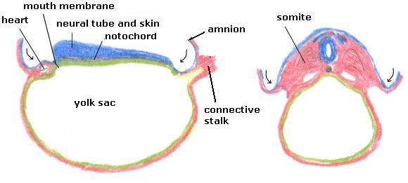
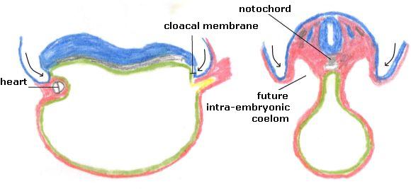
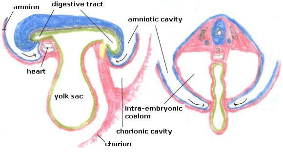

Shows two side views of the embryo in the fourth week. In the drawing from the middle of the fourth week, the 24th day the amnion surrounds the embryonic disc and the cylindrical embryo is created. Visible is the large hump of the heart that has been pushed from its cranial position above the head to a location in the chest. The neural tube is still open on both sides. The connective stalk and the yolk sac are visible.
At the end of the fourth week, the 28th day, the complete enclosure of the amnion and the umbilical cord (containing blood vessels, connective stalk, yolk sac and allantois) are visible. The heart has grown and descended a bit more and now lies on its final place adjacent to the umbilical cord. The embryo has a short tail and has become rounder. The head and tail are bent to the inside. The head is large; the headside develops faster then the tailside. At the head folds have appeared, caused by the fast thickening of the tissue of the neural tube that thereby bends the head downward. The first arch is the mandibulary arch, from which the upper jaw grows. The last two folds are called branchial (gill) arches, similar to the folds in fish. The beginning of an eye and an ear are visible. The limbs grow, the first beginnings of an arm and a leg can be seen (see the next page for the development of the limbs).
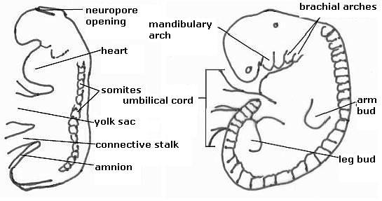
Figure 32. Left: an embryo on day 24, right on 28 days, respectively. 2 and 5 mm long.
Neural tube and digestive tract
The neural tube arises from the centre, the digestive tract from the extremities. In the neural tube there is clear amniotic fluid, which exerts pressure on the wall. In the digestive tract is turbid yolk liquid, which does not. The neural tube is mainly a straight tube, the digestive tract is a tube that winds in and out (the intestines). The neural tube consists of ectoderm, the digestive tract of entoderm. Ectoderm comes from the back, which may be called the antipathy-side of the body (to turn your back on someone). Entoderm comes from the front side, the sympathy-side of our body. With ectoderm we demarcate ourselves and create distance (observing), with entoderm we create connections (digestion of food).
| neural tube | digestive tract | |
| arises from | the middle | the extremities |
| fluid | clear amniotic fluid, exerts pressure | trouble yolk fluid, no pressure |
| tissue | ectoderm | entoderm |
| form | straight | winding |
| where | backside = antipathy-side | frontside = sympathy-side |
Differences between the neural tube and the digestive tract
Characteristics
The processes of the third week gave the embryo volume, but did not give the embryonic disc a boundary. At the beginning of the fourth week, the embryonic disc extends into the enclosing tissues of amnion, chorion and yolk sac. The enclosing movement of the amnion separates the embryo from the enclosing tissues and emancipates the embryonic disc. It still takes a long time before the embryo is independent, but the beginning is there, now that the body gets its first form and is connected to the nourishing tissues by the umbilical cord. Steiner called this stage “Paradise Man”, by which he meant to say that this is the first separation of man from his environment.
Now the body is apart and three-dimensional. The body bends and is directed towards a centre (Fig. 32). Hartmann uses the name “Animal Man”.
The enveloping movement of the amnion has as a result, that the back-side is now on the outside and what was on the ventral side now lies within. It is an enveloping gesture.
The umbilical cord is on the ventral side of the embryo. It started as the connective stalk at the backside, then it moved to the tail and now it shifts to the abdomen. The nourishment came from behind in the stage of the “Plant Man”; from the front in the stage of the “Animal Man”.
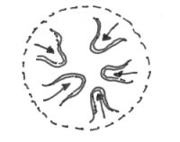
Figure 33. Animal Man (from Van der Wal, by Hartmann)
Indicated is that an animal is an organism with content or volume. It is focused inwardly on a centre.
The three tissues of the embryonic disc and what arises from them
The embryonic disc consists of three germ layers:
- Ectoderm
- Entoderm
- Mesoderm
From the ectoderm arise:
- The skin
- The nervous system
- The senses
From the entoderm arise:
- The digestive tract and digestive organs (liver, pancreas)
- The lungs
- The bladder (from the allantois)
The mesoderm can create three types of tissues:
- It can concentrate and grow inward to form muscles, tendons, ligaments and bones;
- And the kidneys, spleen and reproductive organs.
- It can go to the periphery to form body cavities, such as the pericardium, the lung cavity, the abdominal cavity, etc.
- It can do both at once and form blood cells, blood vessels and the heart.
Stages of Growth Month-by-Month in Pregnancy
The fetus will change a lot throughout a typical pregnancy. This time is divided into three stages, called trimesters. Each trimester is a set of about three months. Your healthcare provider will probably talk to you about fetal development in terms of weeks. So, if you’re three months pregnant, you’re about 12 weeks.
You’ll see distinct changes in the fetus, and yourself, during each trimester of pregnancy.
First trimester
The first trimester will span from conception to 12 weeks. This is generally the first three months of pregnancy. During this trimester, the fertilized egg will change from a small grouping of cells to a fetus that begins to have human features. The first trimester is exciting, but also when most people develop unpleasant symptoms like morning sickness and fatigue.
Month 1 (weeks 1 through 4)
Although it’s strange, the first two weeks of pregnancy are a “getting ready” period. Your body slowly releases more hormones and your uterus prepares for a potential pregnancy. At the end of the second week, your ovary releases an egg (ovulation). If sperm meets an egg just after ovulation, the process to pregnancy continues.
- Week 3: Fertilization occurs during the third week. A sperm and egg join and create a zygote.
- Week 4: The tiny bundle of cells turns into a blastocyst and implants into your uterine lining. The beginning of what will become the placenta forms. A water-tight sac forms around the blastocyst. This is the amniotic sac, and it provides cushioning to a fetus during pregnancy.
By the end of the fourth week, the blastocyst is about 2 millimeters (mm) long — the size of a poppy seed.
Month 2 (weeks 5 through 8)
The second month of pregnancy is when most people realize they’re pregnant. Pregnancy hormones go into overdrive, and by about the fifth week, an at-home pregnancy test will show as positive. This is when many people begin to feel symptoms of pregnancy.
- Week 5: The neural tube (brain, spinal cord and other neural tissue of the central nervous system) forms. The tiny “heart” tube will pulse 110 times a minute by the end of the fifth week.
- Week 6: Tiny buds that become arms and legs also develop. Blood cells are taking shape, and circulation will begin. Structures that’ll become the ears, eyes and mouth take form. Your healthcare provider can probably detect pulses in the cluster of cells that will form the heart on a vaginal ultrasound.
- Week 7: Bones begin replacing soft cartilage and genitals begin to form. The embryo’s head is large in proportion to the rest of its body. Some people think the embryo resembles a small tadpole or seahorse due to its prominent tail (which recedes) and large head.
- Week 8: All of the major organs and body systems are developing. The embryo has web-like hands and feet. Eyes become visible and ears begin to form. The umbilical cord is fully developed and helps to transport oxygen and blood to the embryo.
After the 8th week, healthcare providers refer to the embryo as a fetus. It will remain a fetus until birth.
By the end of the second month, the fetus is about 0.5 to 1 inch (in) long — about the size of a black bean.
Month 3 (weeks 9 through 12)
The third month of pregnancy is when an embryo becomes a fetus. It’s a period of rapid growth and development. The fetus develops distinct facial features, limbs, organs, bones and muscles. By the end of the 12th week, the fetus has an assigned sex, but it won’t be visible on ultrasound for several more weeks.
- Week 9: The beginnings of teeth and taste buds are forming. Its muscles are forming and its body shape takes on more of a human appearance. But, its head is still 50% of its length. Your provider may be able to hear its heartbeat with a Doppler ultrasound.
- Week 10: The arms, hands, fingers, feet and toes are fully formed (no more webbed fingers). Fingernails and toenails are beginning to develop and the external ears form. The external genitals also begin to form, but it’s too soon to see them on an ultrasound.
- Week 11: The fetus is starting to explore a bit by doing things like opening and closing its fists and mouth. Its knees, elbows and ankles are working, but it’s too soon to feel any kicks. The bones are hardening, but its skin is still see-through. Facial features are more prominent.
- Week 12: All the organs, limbs, bones and muscles are present and will continue to develop in order to become fully functional. The circulatory, digestive and urinary systems are also working and the liver produces bile. The fetus is drinking and peeing amniotic fluid.
Since the most critical development has taken place, your chance of miscarriage drops considerably after 12 weeks (the end of the first trimester). Most people begin feeling some relief from morning sickness now, too.
At the end of the third month, the fetus is about 2.5 to 3 inches long — about the size of a plum.
Second trimester
The second trimester of pregnancy is often thought of as the best part of the experience. By this time, any morning sickness is probably gone and the discomfort of early pregnancy has faded. You may also start to feel movement as the fetus flips and turns in your uterus. During this trimester, many people find out about the fetus’s assigned sex. This is typically done during an anatomy scan (an ultrasound that checks physical development) at around 20 weeks.
Month 4 (weeks 13 through 16)
Many people begin showing signs of being pregnant at this point in pregnancy, especially if you’ve been pregnant before. Your pregnancy care provider can hear the fetal heartbeat loud and clear on a Doppler ultrasound. The fetus can even suck its thumb, yawn, stretch and make faces.
- Week 13: Vocal cords form and the fetus’s large head begins to grow proportionate to its body.
- Week 14: The fetus’s skin starts to thicken and fine hair begins to grow. It can start bringing its fingers to its mouth and turn its head. External genitals are fully developed and fingerprints begin to form.
- Week 15: Some organs, like intestines and ears, are moving to their permanent location. The fetus still uses amniotic fluid to practice breathing, but its lungs are beginning to develop. The fetus begins to make more purposeful movements, like sucking its thumb or smiling.
- Week 16: The fetus has lips and its ears are developed enough that it can hear you talk. Even though its eyes are closed, the fetus can react to light by turning away from it.
By the end of the fourth month, the fetus is about 5 inches long and weighs about 4 ounces. For reference, that’s about as big as an avocado.
Month 5 (weeks 17 through 20)
By the end of the fifth month of pregnancy, most people begin to feel the fetus moving around. The first movements are called quickening and can feel like a flutter. If your pregnancy has been healthy to this point, you’ll finally get your first ultrasound. You may even get to find out the fetus’s assigned sex.
- Week 17: The fetus still has thin skin, but will start to put on fat. Its skin is covered with a whitish coating called vernix. This “cheesy” substance is thought to protect fetal skin from long-term exposure to amniotic fluid.
- Week 18: The fetus is covered in lanugo, a peach fuzz-like hair. It helps keep the fetus warm and provides another layer of protection. The fetus may have a sleep-wake cycle, and loud noises may wake the fetus if it’s asleep.
- Week 19: The fetus is getting stronger and most people begin to feel kicks and punches. The fetus also has its own unique set of fingerprints and can hiccup.
- Week 20: The fetus’s nails grow towards the end of its fingers. The area of the brain responsible for its five senses begins to develop.
By the end of the fifth month, the fetus is about 9 to 10 inches long and weighs about 1 pound.
Month 6 (weeks 21 through 24)
If you could look inside your uterus right now, you’d see that the fetus’s skin is reddish in color, wrinkled and veins are visible through translucent skin. In the sixth month of pregnancy, its eyelids begin to part and you may notice regular, jerky movements. The fetus responds to sounds by moving or increasing its pulse.
- Week 21: Limb movements are coordinated and frequent. The fetus has bone marrow that helps it produce blood cells.
- Week 22: The fetus’s grasp is getting stronger and it can touch its ears and the umbilical cord. It can hear your heartbeat, your stomach rumble and your breathing.
- Week 23: If born prematurely, the fetus may survive after the 23rd week with intensive care. It will begin rapidly adding fat to its body.
- Week 24: The fetus’s lungs are fully developed, but not well enough to work outside your uterus.
By the end of the sixth month, the fetus is about 12 inches long and weighs about 2 pounds.
Month 7 (weeks 25 through 28)
The fetus continues to mature and develop reserves of body fat. The fetus changes position frequently and responds to stimuli, including sound, pain and light. The amniotic fluid begins to diminish.
- Week 25: More body fat makes the fetus’s skin less wrinkled and plumper. Its nervous system is quickly maturing.
- Week 26: The fetus makes melanin, the substance that gives skin and eyes their color. The fetus’s lungs start to make surfactant, a substance that helps it breathe after birth.
- Week 27: The fetus can open its eyes and blink. It also has eyelashes.
- Week 28: The fetus may begin turning head-down in your uterus as it gets ready for birth. At the end of the seventh month, the fetus is about 14 to 15 inches long and weighs between 2 and 3 pounds.
Third trimester
This is the final part of your pregnancy. You may be tempted to start counting down the days to your due date and hope that it comes early, but each week of this final stage of development helps the fetus prepare for birth. Throughout the third trimester, the fetus gains weight quickly, adding body fat that’ll help after birth.
Your healthcare provider will monitor you closely as you approach your due date. You’ll visit your provider biweekly and then weekly. Make sure to ask your provider any questions you have about labor and delivery.
Month 8 (weeks 29 through 32)
The fetus continues to mature and develop reserves of body fat. The brain develops most rapidly during this time. The fetus can see and hear most stimuli. Most internal systems are well-developed, but the lungs may still be immature.
- Week 29: You may notice the kicks and jabs feel more like pokes now that the fetus is getting cramped in the amniotic sac.
- Week 30: The fetus can control its own body heat. Its brain is maturing and growing rapidly.
- Week 31: The fetus can process more information and stimuli. You can probably notice more distinct patterns in when it’s awake and when it’s asleep.
- Week 32: The fetus’s skin isn’t translucent anymore. Other than the lungs and brain, most other organs are well-formed and ready for birth.
The fetus is about 17 to 18 inches long and weighs as much as 5 pounds.
Month 9 (weeks 33 through 36)
During this stage, the fetus continues to grow and mature. The lungs are close to being fully developed at this point in pregnancy. The ninth month is mostly about putting the finishing touch on growth and brain development.
- Week 33: The fetus’s bones are hardening, with exception of the cranial bones around its brain, which needs to be soft to descend the birth canal.
- Week 34: The vernix that protects the fetus’s skin starts to get thicker.
- Week 35: The fetus’s brain continues to grow, but still only weighs two-thirds of what it should at birth.
- Week 36: The fetus loses its lanugo and it has hair on its head.
The fetus is about 17 to 19 inches long and weighs from 6 to 7 pounds.
Month 10 (Weeks 37 through 40)
In this final month, you could go into labor at any time. At this point, the fetus’s position may have changed to prepare for birth. Ideally, it’s head-down in your uterus. You may feel very uncomfortable in this final stretch of time as the fetus drops down into your pelvis and prepares for birth. Your provider may encourage you to perform kick counts, which is a way to track how much the fetus moves.
- Week 37: The fetus’s toenails reach the end of its toes. You may start to feel the fetus drop into your pelvis.
- Week 38: The fetus is packing on 0.5 pounds per week to get to its final size.
- Week 39: The fetus is full-term and ready to meet the world!
- Week 40: It’s your due date week. Call your pregnancy care provider if you notice any signs of labor.
The fetus is about 18 to 20 inches long and weighs about 7 to 9 pounds.
References
- https://www.ncbi.nlm.nih.gov/books/NBK279385/
- https://www.ncbi.nlm.nih.gov/books/NBK535404/
- https://www.ncbi.nlm.nih.gov/books/NBK482385/
- https://www.ncbi.nlm.nih.gov/books/NBK499861/
- https://www.ncbi.nlm.nih.gov/books/NBK441896/
- https://www.ncbi.nlm.nih.gov/books/NBK554544/
- https://www.ncbi.nlm.nih.gov/books/NBK544228/
- https://www.ncbi.nlm.nih.gov/books/NBK441859/
- https://www.ncbi.nlm.nih.gov/books/NBK532934/
- https://www.ncbi.nlm.nih.gov/books/NBK499952/
- https://www.ncbi.nlm.nih.gov/books/NBK507815/
- https://www.ncbi.nlm.nih.gov/books/NBK431105/
- https://www.ncbi.nlm.nih.gov/books/NBK459218/
- https://www.ncbi.nlm.nih.gov/books/NBK470390/
- https://www.ncbi.nlm.nih.gov/books/NBK536971/
- https://www.sciencedirect.com/book/9781437709872/
- https://medlineplus.gov/ency/article/000500.htm
- https://www.nice.org.uk/guidance/ng148
- https://www.nhs.uk/conditions/acute-kidney-injury/
- https://www.mayoclinic.org/diseases-conditions/kidney-failure/diagnosis-treatment/drc-20369053
- https://en.wikipedia.org/wiki/Acute_kidney_injury
- https://www.ncbi.nlm.nih.gov/pmc/articles/PMC5198510/
- https://www.aafp.org/afp/2012/1001/p631.html
- https://www.kidney.org/atoz/content/what-creatinine
- https://www.nuh.nhs.uk/creatinine-clearance-calculator
- https://www.sciencedirect.com/topics/medicine-and-dentistry/uremia
- https://en.wikipedia.org/wiki/Uremia
- https://www.mayoclinic.org/diseases-conditions/polycystic-kidney-disease/symptoms-causes/syc-20352820
- https://medlineplus.gov/genetics/condition/polycystic-kidney-disease/
- https://www.kidney.org/atoz/content/AcuteKidneyInjury
- https://tomvangelder.antrovista.com/the-fourth-week-the-form-emerges-158m77.html

